
Enchodelus brevidentatus Thorne, 1939
|
publication ID |
https://doi.org/10.1163/156854107779969646 |
|
DOI |
https://doi.org/10.5281/zenodo.8111776 |
|
persistent identifier |
https://treatment.plazi.org/id/8C029544-A91F-FFD3-FFA9-FDD0FE17CE2A |
|
treatment provided by |
Carolina (2023-07-04 01:08:12, last updated 2024-11-27 08:37:23) |
|
scientific name |
Enchodelus brevidentatus Thorne, 1939 |
| status |
|
Enchodelus brevidentatus Thorne, 1939
( Figs 4 View Fig , 5 View Fig )
MATERIAL EXAMINED
Nineteen females, 18 of them mounted on one slide labelled Enchodelus brevidens, and collected from foothill soil in Lehi , Utah ( USA) on March 8, 1926; a few specimens in acceptable condition, but most of them becoming flattened. The remaining female, mounted on slide labelled Enchodelus macrodoroides 2a, and collected from Wellsville Hill, Utah ( USA) on March 28, 1925, is in quite bad condition.
MEASUREMENTS
See Table 2. View Table 2
DESCRIPTION
Female
Moderately slender nematodes of medium size, 1.6- 1.9 mm long. Body cylindrical, tapering towards both ends but more so towards posterior. Cuticle 2.0-3.0 µ m thick at anterior region, 2.0-4.0 µ m at midbody and 3.5- 6.0 µ m at tail; its outer layer thinner than inner, bearing fine but distinct transverse striations that are more visible at level of anterior body region and caudal region. Lateral chord occupying 18-30% of midbody diam, with granular appearance. Lateral pores obscure. Lip region rather angular, offset by distinct depression, 2.4-2.9 times as broad as high and ca one-third (some specimens ca one-fifth, but obviously flattened) of body diam. at neck base; lips mostly amalgamated; labial and cephalic papillae protruding, in particular the inner labial ones. Amphid fovea cup-shaped, opening at level of cephalic depression and occupying one-third to one-half of corresponding body diam. Cheilostom a truncate cone to practically cylindrical, with no particular differentiation. Odontostyle relatively slen- der, practically straight; 7.7-11.3 times as long as wide or 1.1-1.4 times as long as lip region diam.; aperture small, one-tenth to one-fifth of total length. Odontophore rod-like, lacking any specialisation, 1.2-1.5 times odontostyle length long. Guiding ring simple, at 8.5-10.5 µ m or 0.7- 0.8 lip region diam. from anterior end. Pharynx consisting of a slender but well muscular portion expanding gradually into basal expansion at 54-65% of total neck length, reaching full diam. at 62-70%; pharyngeal expansion occupying 33-42% of total neck length, and ca three-fifths of corresponding body diam. Pharyngeal gland nucleilocatedas follows: DN = 67-72 (n = 10); S 1 N 1 = 22; S 1 N 2 = 31 (n = 1); S 2 N 1 = 49-55; S 2 N 2 = 50- 57 (n = 7). Base of pharyngeal expansion surrounded by faint membrane-like structure. Cardia rounded conoid, wider than long, 16 × 11 µ m. Genital system didelphic-amphidelphic, with both branches equally and well developed. Ovaries relatively large, 109-320 µ m long, usually reaching and surpassing sphincter level; oocytes first in two or more rows, then in one row. Oviduct 120-273 µ m long or 2-3 times corresponding body diam., consisting of a slender portion with prismatic cells and a well developed pars dilatata with distinct lumen. Sphincter present between oviduct and uterus. Uterus bipartite, i.e., with a wider proximal portion with distinct lumen, and a narrower distal part with narrow lumen and refractive inner lining; length 65-126 µ m or 0.9-2.0 times body diam. at its level; a group of hyaline cells observed surrounding distal part of uterus. Sperm not observed in genital tract. Only one uterine egg observed in female from Wellsville, measuring 91 × 35 µ m. Vagina extending inwards one-third (flattened specimens) to two-fifths of body diam.; pars proximalis vaginae 11-16 × 13-25 µ m, with almost straight or slightly convergent walls and enveloped by moderately developed circular musculature; pars refringens vaginae with (in lateral view) two trapezoidal sclerotisations which have a combined width of 9.5-15.5 µ m and are somewhat separated by a weakly sclerotised intermediate area; pars distalis vaginae short, 2.5-5.0 µ m long. Vulva a transverse slit. Prerectum 1.8-3.2, rectum 0.8-1.2 anal body diam. long. Tail conical, regularly ventrad curved, with rounded tip; hyaline terminal portion 10-15 µ m or 15-21% of total length. Two pairs of caudal pores at the middle of the tail: one subdorsal, another practically lateral.
Male Unknown.
Enchodelus brevidentatus is distinguished by its body 1.6-1.9 mm long, lip region 12-13.5 µ m diam., odontostyle 14.5-17 µ m long or 1.1-1.4 times the lip region diam., odontophore rod-like, neck length 264-308 µ m long, pharyngeal expansion 91-125 µ m long or occupying 33-42% of total neck length, female genital system amphidelphic, uterus bipartite, pars refringens vaginae with two trapezoidal sclerotisations, vulva in form of a transverse slit (V = 47-53), tail conical and regularly ventrad curved (55-71 µ m, c = 24-33, cļ = 1.3-2.1), and males unknown.
REMARKS
The material examined is certainly the original described by Thorne (1939) and, as a consequence, the above description fits that by the American author, although ratio a is lower (vs a = 30-35) due to flattening of the specimens.
Since its original description, E. brevidentatus has been recorded from Italy ( Vinciguerra, 1972), Hungary ( Andrássy, 1996) and Spain ( Liébanas et al., 2004). The material described by Vinciguerra differs from type population in several relevant features: shorter body (L = 1.2-1.5) and odontostyle (10-12 µ m) and males as frequent as females. Some doubts therefore exist as to the true identity of the Italian material.
ANDRASSY, I. (1996). Free-living nematodes in the Bukk Mountains, Hungary. The Fauna of the Bukk National Park, 1996, pp. 33 - 63.
LIEBANAS, G., GUERRERO, P., MARTIN- GARCIA, J. - M. & PENA- SANTIAGO, R. (2004). Spatial distribution of dorylaimid and mononchid nematodes from the southeast Iberian peninsula: environmental characterization of chorotypes. Journal of Nematology 36, 114 - 122.
THORNE, G. (1939). A monograph of the nematodes of the superfamily Dorylaimoidea. Capita Zoologica 8, 1 - 261.
VINCIGUERRA, M. T. (1972). Nematodi di Sicilia. Nota I. Bolletino delle Setude dell'Accademia Gioenia di Scienze Naturali in Catania 11, 3 - 35.
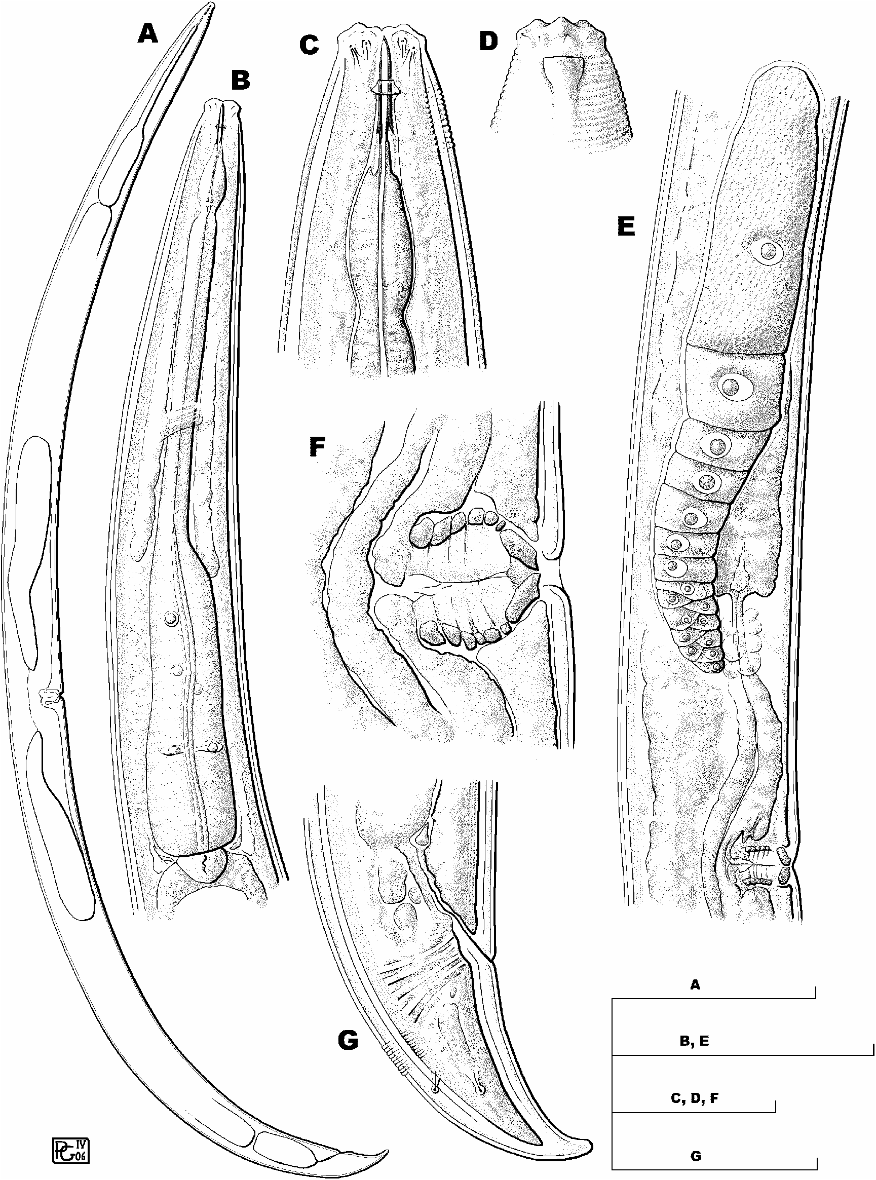
Fig. 4. Enchodelus brevidentatus Thorne, 1939 (female). A: Entire body; B: Pharyngeal region; C: Anterior region; D: Reconstruction of superficial appearance of cephalic region; E: Anterior genital branch; F: Vulval region; G: Tail. (Scale bars: A = 250 µm; B, E = 100 µm; C, D, F = 25 µm; G = 50 µm.)
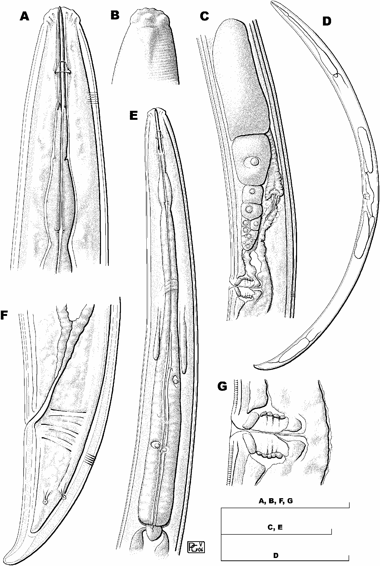
Fig. 5. Enchodelus brevidentatus Thorne, 1939 (female). A: Variability of anterior region; B: Posterior genital branch showing sphincter between uterus and oviduct (arrow); C: Expanded part of pharynx showing dorsal nucleus (large arrow) and one of posterior subventral nuclei (small arrow); D: Sphincter at junction of uterus-oviduct; E: Egg; F: Lateral chord; G: Variability of vulval region; H: Variability of tail. (Scale bars: A, D, F-H = 25 µm; B = 100 µm; C, E = 50 µm.)
No known copyright restrictions apply. See Agosti, D., Egloff, W., 2009. Taxonomic information exchange and copyright: the Plazi approach. BMC Research Notes 2009, 2:53 for further explanation.
|
Kingdom |
|
|
Phylum |
|
|
Class |
|
|
Order |
|
|
Family |
|
|
Genus |
1 (by carolina, 2023-07-04 01:08:12)
2 (by carolina, 2023-07-04 01:18:56)
3 (by ExternalLinkService, 2023-07-04 01:19:01)
4 (by ExternalLinkService, 2023-07-04 01:27:07)
5 (by carolina, 2023-07-04 17:49:44)
6 (by ExternalLinkService, 2023-07-04 18:00:02)
7 (by ExternalLinkService, 2023-07-04 19:25:18)
8 (by plazi, 2023-11-09 12:41:14)