
Hansenomysis sorbei, Vicente, Carlos San, 2009
|
publication ID |
https://doi.org/ 10.5281/zenodo.190521 |
|
DOI |
https://doi.org/10.5281/zenodo.6213384 |
|
persistent identifier |
https://treatment.plazi.org/id/8A3F879F-D04D-147F-8F96-FB5CFCAF2076 |
|
treatment provided by |
Plazi |
|
scientific name |
Hansenomysis sorbei |
| status |
sp. nov. |
Hansenomysis sorbei n. sp.
( Figs.1–5 View FIGURE 1 View FIGURE 2 View FIGURE 3 View FIGURE 4 View FIGURE 5 )
Material examined. Holotype ( ICMM 1/2009). Mature female, 24.3 mm TL. Stn. 26TA, Bellingshausen Sea, 70º 14’14”S – 81º46’07”W, 1869 m depth, 10–50 cm water layer above bottom, sediment with a low organic matter content (2.0%) and a high percentage of fine sand (49.4%), 20 January 2006.
Paratypes ( ICMM 2/2009). Five immature females (18.5 mm TL, 6.6 mm CL, 14.5 mm TL, 12.1 mm TL and 4.3 mm CL), same locality.
Diagnosis. Hepatic region of the carapace with a strong bollard-like process. Antennal scale lanceolate, armed with a row of 5–8 evenly graduated spines. Posterior margins of abdominal somites 4 to 6 produced into five strong spines. Exopod and endopod of uropod sub-equal in length and extending to the apex of telson.
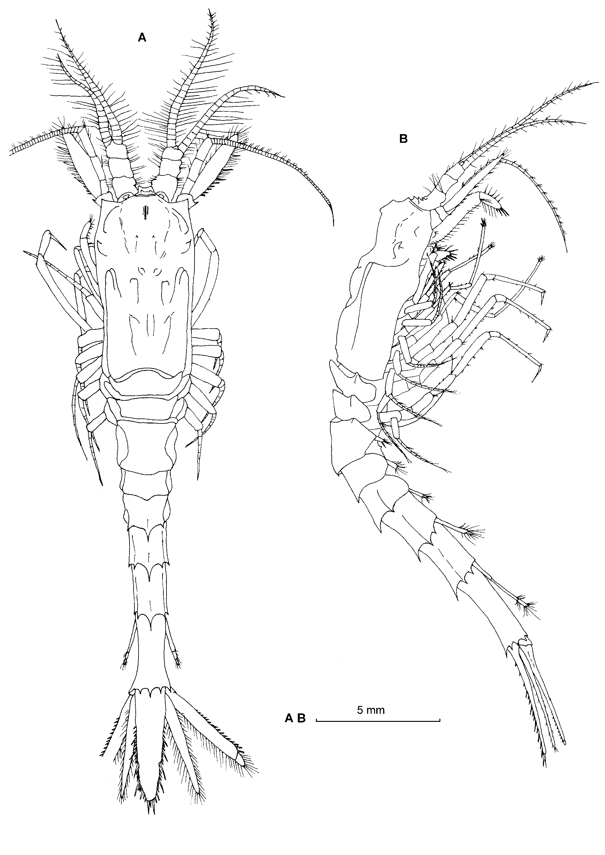
Description. The morphological characteristics refer only to females, male is unknown. Body robust, elongate ( Figs. 1 View FIGURE 1 A, B). Carapace without spines, leaving the two posterior thoracic somites exposed in dorsal view; anterior margin rounded without rostrum; antero-lateral corners slightly produced, not projecting beyond the carapace anterior margin. In the median line, one small keel near the anterior margin, rectangular in shape in lateral view. Two strong bollard-like process projects forward from the hepatic region. Posterior to the sulcus, carapace almost parallel and raised into two keels that converge posteriorly and run forward on each side ( Figs. 1 View FIGURE 1 A, B).
Tergum of the anterior margin of thoracic somites 7 and 8 produced forward and overlapping the preceding somite. Tergum of abdominal somite 1 thickened and produced backward, more or less covering the second abdominal somite. Abdominal somites 2–5 increasing regularly in length distally; somite 6 twice as long as the preceding one. The posterior margins of abdominal somite 3 produced into three strong spines: one dorsal and one pair on lateral angles. The posterior margins of abdominal somites 4 to 6 produced into five strong spines: one dorsal, one pair on lateral angles and other pair between them ( Figs. 1 View FIGURE 1 A, B).
Eyes fused in a single plate that does not reach the first half of the basal joint of the antennular peduncle.
Antennular peduncle short and robust. Inner margin of articles 1, 2 and 3 armed with a row of long setae; well-developed Tattersall organ which appear at the eyeplate sides as two oval conical plates in dorsal view ( Fig. 1 View FIGURE 1 A).
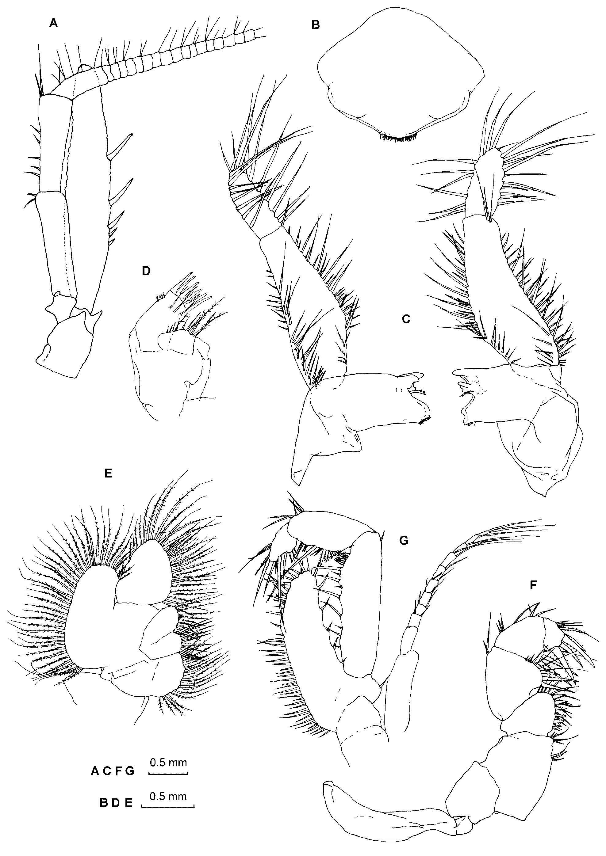
Sympod of antenna relatively slender, with outer distal angle produced into two spines. Peduncle relatively long and slender, extending beyond the antennular peduncle by more than half the length of article 3; middle curved protuberance on the inner margin of article 1. Antennal scale lanceolate, nearly six times as long as maximum width; extending for nearly one third of its length beyond the antennular peduncle. Outer margin armed with five (immature female) to eight (mature female) spinose setae, gradually increasing in length distally; distal half of outer margin setose, setae extending around the apex and along the entire inner margin ( Figs. 1 View FIGURE 1 A, 2A).
Labrum symmetrical, pentagonal, wider than long, with the posterior margin convex and armed with a row of 23 small setae ( Fig. 2 View FIGURE 2 B).
Mandibles with strong lacinia mobilis; palp large and slender, 3-jointed, with numerous long setae, first article shortest, second article about twice as long as third ( Fig. 2 View FIGURE 2 C). Maxillule basis (outer lobe) apex armed with eight cuspidate setae and three long setae on ventral surface; coxal endite (inner lobe) with ten setae, the four apical setae are large and plumose ( Fig. 2 View FIGURE 2 D). Maxilla with distal article of endopod a little longer than wide, densely setose on inner and outer margins; endites rounded distally, armed with plumose setae on inner margins; exopod large, with 38 plumose setae on outer margin ( Fig. 2 View FIGURE 2 E).
First and second thoracic appendages formed as maxillipeds. First thoracopod small and robust; exopod absent, epipod well developed; endopod preischium long, dactylus short with four setulated setae on distal margin; carpopropodus bearing two pappose setae on inner margin; merus with three pappose setae on inner margin; ischium with seven shorter pappose setae on inner margin; preischium with eight setae on inner distal margin ( Fig. 2 View FIGURE 2 F). Second thoracopod robust, endopod with one seta on outer margin of ischium, inner margin produced into a conspicucous lamellar lobe armed with many simple setae; preischium short; merus long; carpopropodus with six short and pappose setae on its proximal inner margin; dactylus with four long setae ( Fig. 2 View FIGURE 2 G).
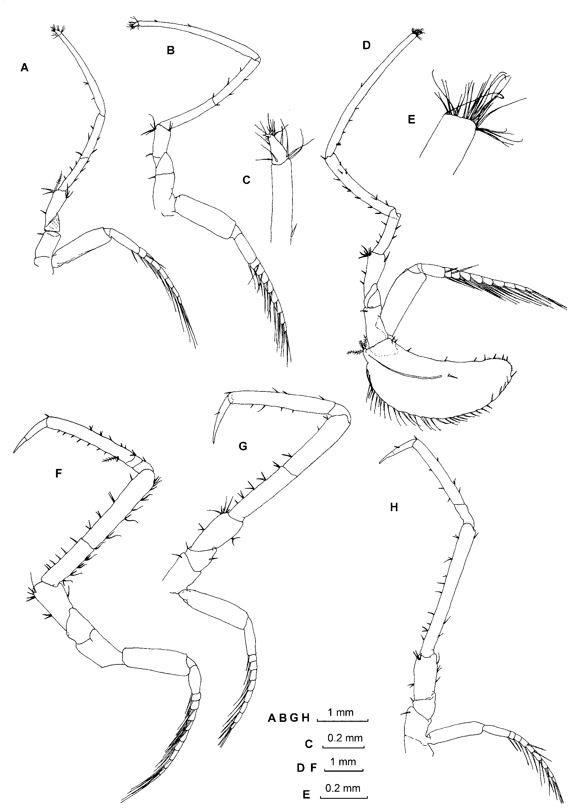
Third to fifth thoracic appendages with endopods long and slender, forming minute chelate structure terminally, surrounded by crown of long setae ( Figs. 3 View FIGURE 3 A–E). Sixth to eighth thoracic appendages with endopod long and slender; dactylus and nail together forming a long claw-like termination of the endopod. Endopod of sixth and seventh thoracopods with carpopropodus and separated into two subsegments, merus subdivided in two by a transverse suture ( Figs. 3 View FIGURE 3 F–G). Endopod of eigth thoracopod with carpopropodus separated into two subsegments, merus entire ( Fig. 3 View FIGURE 3 H). Thoracic exopods annulated, proximal segment takes the form of a flattened plate followed by one long segment and several small segments distally, 10 segments in second thoracopod and 9–12 segments in thoracopods 3–8. Female with seven pairs of developed oostegites ( Figs. 1 View FIGURE 1 B, 3D).
Pleopods of the female uniramous increasing in length posteriorly ( Fig. 4 View FIGURE 4 A–E). First to second pleopod rami 1–segmented, third and fourth pleopod 2–segmented, fifth pleopod 4–segmented and about twice as long as the fourth pleopod.
Uropods long and slender ( Fig. 4 View FIGURE 4 F), exopod and endopod sub–equal in length, extending to the apex of telson. Exopod with distal diaeresis, outer margin unarmed on the proximal third of its length, distal two thirds armed with a row of 26 setae, the distal one the longest. Endopod undivided, without statocyst, setose all round, without spines.
Telson long and narrow, almost 1.5 times longer than sixth abdominal somite and about 4 times as long as broad ( Fig. 4 View FIGURE 4 F). Lateral margins almost parallel on proximal two thirds and then converging evenly to the apex; irregularly distributed setose setae on the proximal third lateral margins; series of three to six spinose setae between five large ones on distal lateral margins. Apex with five spinose setae. Distal setae pappose ( Fig. 4 View FIGURE 4 G).
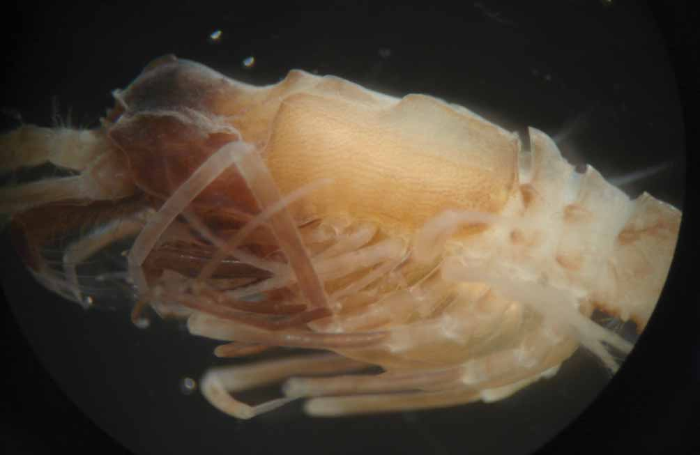
Colour in live specimens: white tegument with brown pigmentation irregularly distributed on the body and appendages ( Fig. 5 View FIGURE 5 ).
Etymology. This species is named in honour of Professor Jean Claude Sorbe in recognition of his contribution to the knowledge of world seas suprabenthos.
Remarks. The main distinguishing feature of Hansenomysis sorbei is the presence of strong dorsal projections taking on the form of spine-like processes on the posterior margins of the abdominal somites. Similar spines are seen in H. menziesi from Peru-Chile trench, H. nouveli from the Bay of Biscay and H. carinata from New Caledonian. However, H. sorbei differs from H. menziesi and H. nouveli in the number of these dorsal projections (present only in the posterolateral angle of abdominal somites in the latter two species), the relative size of uropods (shorter than the telson in the latter two species) and in the armature of the carapace.
The new species can be distinguished from H. carinata by the form of the carapace (hepatic region with a strong bollard-like process, absent in H. carinata ), the shape of the sixth abdominal somite (posterior margin with five strong projections versus three in H. carinata ) and the relative size of the uropods (exopod and endopod sub-equal in length and extending to the apex of telson versus both rami longer than the telson in H. carinata ).
Hansenomysis sorbei is the fifth species to be described from the Southern Ocean, after H. antarctica , H. falklandica , H. angusticauda and H. anaramosa e. H. sorbei is easily distinguished from these southern species by the strong projections of the abdominal somites.

No known copyright restrictions apply. See Agosti, D., Egloff, W., 2009. Taxonomic information exchange and copyright: the Plazi approach. BMC Research Notes 2009, 2:53 for further explanation.
|
Kingdom |
|
|
Phylum |
|
|
Class |
|
|
Order |
|
|
Family |
|
|
Genus |