
Diplectrona aiensis Kobayashi 1987
|
publication ID |
https://doi.org/10.11646/zootaxa.5082.3.3 |
|
publication LSID |
lsid:zoobank.org:pub:99F75ED7-13CE-402E-8621-8F8534C8C08E |
|
DOI |
https://doi.org/10.5281/zenodo.5789477 |
|
persistent identifier |
https://treatment.plazi.org/id/854F87B5-FFFF-681B-FF0A-B96DFDE8AAB9 |
|
treatment provided by |
Plazi (2021-12-17 08:49:55, last updated 2024-11-27 08:38:31) |
|
scientific name |
Diplectrona aiensis Kobayashi 1987 |
| status |
|
Diplectrona aiensis Kobayashi 1987 View in CoL
( Figs 3 View FIGURE 3 , 7 View FIGURE 7 )
Diplectrona aiensis Kobayashi 1987 View in CoL , 19, 32, male; Morita 2013, 62–63; Nozaki 2016, 348, male; Tanaka 2016, 2, 5, male.
Diagnosis. Male genitalia of this species are similar to those of Diplectrona guangxiensis Sun 2017 described from China but are distinguished by the length of the lateral filaments of the abdominal segment V: Each lateral filament is about 1.7 times longer than the segment V in D. aiensis , but slightly shorter than the segment in D. guangxiensis . Furthermore, the pair of internal glands of the abdominal segment VIII of D. aiensis are larger than those of D. guangxiensis .
Adult ( Figs 3A–3J View FIGURE 3 ). General appearance similar to D. kibuneana ( Fig. 3A View FIGURE 3 ). On head ( Fig. 3B View FIGURE 3 ), frontal setal wart (f.s.w.) small oval; each anterior setal wart (a.s.w.) divided into 3 warts, 2 anterior ones each small oval, posterior one triangular; each posterior setal wart (p.s.w.) large oval. Forewings each 7.0–9.5 mm long in male (n = 10), 9.3–11.5 mm in female (n = 10), venation similar to that of D. kibuneana ( Fig. 1C View FIGURE 1 ). Pair of lateral filaments (l.f.) of abdominal segment V (V) long, each about 1.7 times as long as segment V in male ( Fig. 3C View FIGURE 3 ), 1.4 times in female. Segment V with small internal gland (i.g.) in both male and female. Segment VIII (VIII) with large internal gland in male ( Fig. 3C View FIGURE 3 ), slightly shorter than segment VII (VII), lacking in female.
Male genitalia ( Figs 3D–3G View FIGURE 3 ). Segment IX longitudinally short in lateral aspect ( Fig. 3D View FIGURE 3 ), anterior margins angled 50° in lateral aspect. Segment X (X) triangular in lateral aspect ( Fig. 3D View FIGURE 3 ), with pair of mesocaudal lobes (m.c.l.) V-shaped in dorsal aspect ( Fig. 3E View FIGURE 3 ); pair of lateral lobes (l.l) rectangular in dorsal aspect ( Fig. 3E View FIGURE 3 ), each with apex directed dorsad and with short setae ventrally ( Fig. 3D View FIGURE 3 ); preanal appendages indistinct, forming only large setose areas. Inferior appendages each with basal segment long club-like, extending beyond apex of segment X ( Fig. 3D View FIGURE 3 ); distal segment slightly shorter than 1/3 of basal segment, tapering to apex, weakly curved mesad ( Fig. 3E View FIGURE 3 ). Phallic apparatus thick basally and with apical 2/3 almost straight in lateral aspect (3F), thick apex with two pairs of endothecal processes dorsally, each apex somewhat sharp ( Fig. 3F View FIGURE 3 ).
Female genitalia ( Figs 3H–3J View FIGURE 3 ). Sternum VIII cleft from base, lateral lobes (l.l.) widely separated from each other posteriorly in ventral aspect ( Fig. 3J View FIGURE 3 ). Segment IX (IX) semicircular in dorsal aspect ( Fig. 3I View FIGURE 3 ), anterior margins gently convex anteriorly about 1/3 from base in lateral aspect ( Fig. 3H View FIGURE 3 ); with pair of shallow slits posterodorsally; pair of short external sclerotized ribs (s.r.) ventrally ( Figs 3H, 3J View FIGURE 3 ). Vulval scale (v.s.) with pair of short protrusions basolaterally in ventral aspect (marked with arrow in Fig. 3J View FIGURE 3 ), apical part broadly membranous. Segment X (X) rhomboid in lateral aspect ( Fig. 3H View FIGURE 3 ).
Immature stage. Morphology and biology of this species will be reported in a future work.
Specimens examined. Holotype: Male (in alcohol, M-8232), Ai (200 m), Nita-gun , Shimane, 22.vii.1985, M. Kobayashi ( KPM-NK). Honshu , Akita: 2 males, Sashimaki, Tazawa-ko, Semboku-shi, 29.vi.2012, M. Tanaka. Ibaraki: 3 males, Uwaso, Ishioka-shi, 7.vi.2008, N. Katsuma; 1 female, same locality, 15.vii. 2008, N. Katsuma; 5 males, 4 females, same locality, 4.vi.2011, N. Katsuma; 6 males, 5 female, same locality, 11.vi.2011, N. Katsuma. Aichi: 2 males, Hashigoda, Kikko, Moriyama-ku, Nagoya-shi, 16.v.2011, T. Nozaki. Mie: 1 male, Tabika, Komono-cho, alt. 85 m, 1–8.x.2000, H. Morita; 4 males, 1 female, same locality, 6.v.2006, H. Morita; 2 males, same locality, 31.v.2011, H. Morita; 3 males, 4 females, Okubano, Iga-shi, 9–22.vi.2012, H. Morita ( NK). Shiga: 1 male, Shishitobi-bashi, Seta-gawa, Oishi-higashi, Otsu-shi, 2.ix.2014, S. Kobayashi. Okayama: 1 male, Okutsu-kawanishi, Kagamino-cho, 26.vi.2013, K. Nojima ( KN). Kyushu, Saga: 1 male, Fuji-cho, Saga-shi, 6.v.2011, T. Iwai et al.
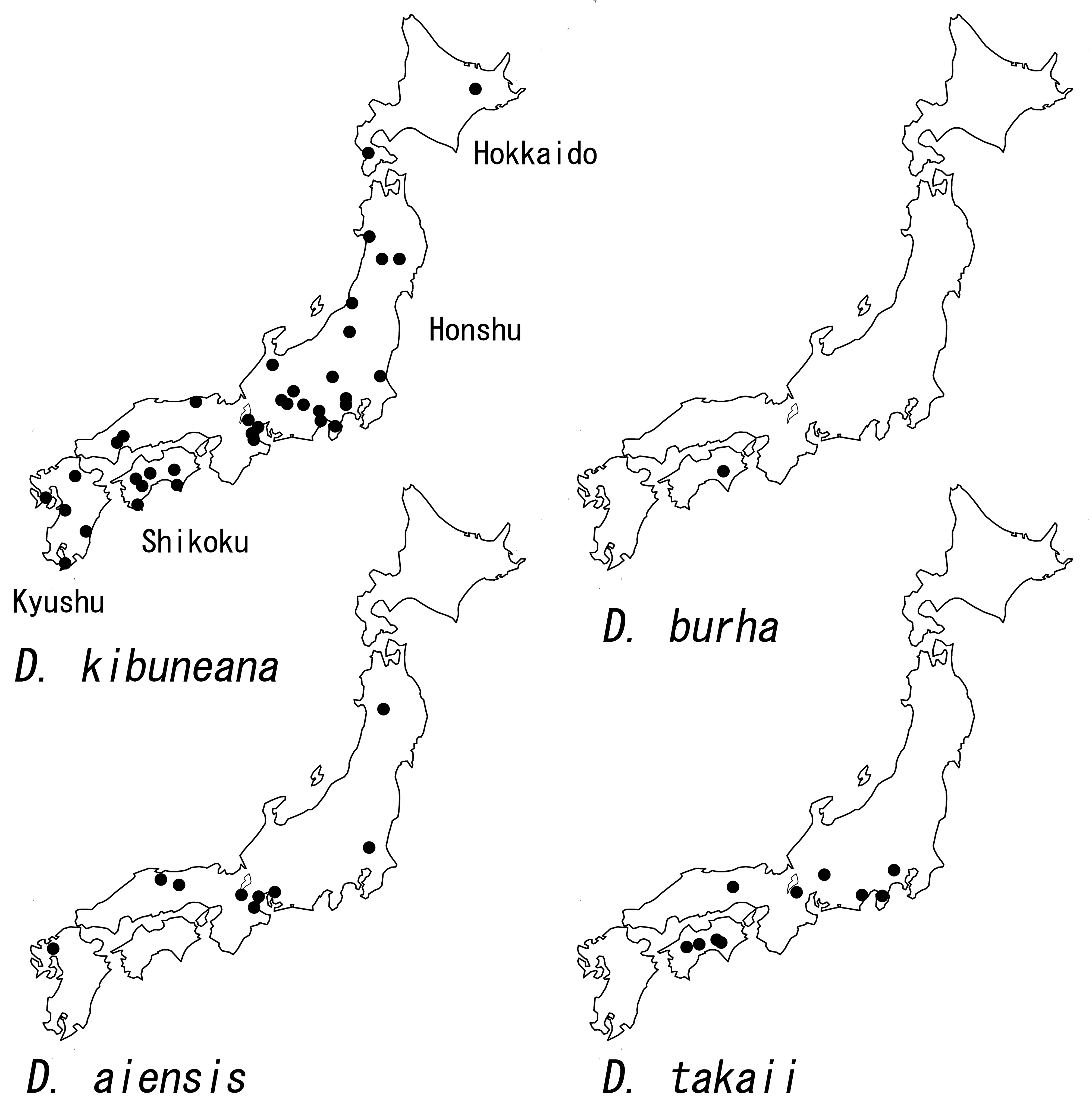
Distribution. Japan: Honshu, Kyushu.
Japanese name. Ai-shima-tobikera.
Kobayashi, M. (1987) Caddisflies or Trichoptera from Shimane Prefecture in Japan (Insecta). Bulletin of the Kanagawa prefectural Museum (Natural Science), 17, 13 - 35.
Morita, H. (2013) Aoyama-kogen no tobikera-so. Hirakura, 57 (3), 60 - 64. [in Japanese]
Nozaki, T. (2016) Trichoptera. In: Maruyama, H. & Hanada, S. (Ed.), A Field Guide to Japanese Aquatic Insects: Adults of Mayflies, Stoneflies and Caddisflies. Zenkoku Noson Kyoiku Kyokai, Tokyo, pp. 69 - 87 + 294 - 410. [in Japanese]
Sun, C-h. (2017) Eight new species of Diplectrona (Trichoptera: Hydropsychidae) from China. Journal of the Kansas Entomological Society, 90 (2), 146 - 161. https: // doi. org / 10.2317 / 0022 - 8567 - 90.2.146
Tanaka, M. (2016) Akita-ken no tobikera-moku bunpushiryo 7 [Caddisflies of Akita Prefecture 7]. Akita Nature Study, 70, 1 - 5. [in Japanese]
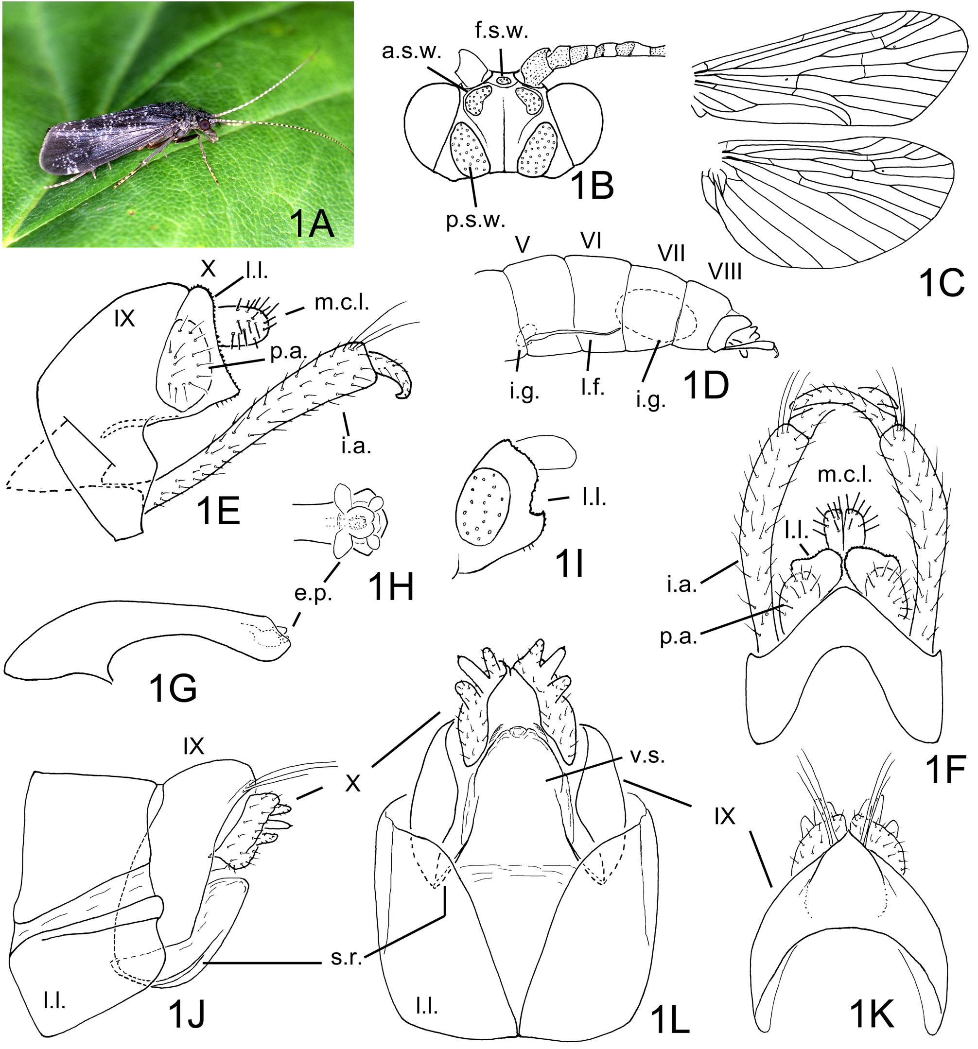
FIGURE 1. Diplectrona kibuneana Tsuda 1940. 1A–1I, male: 1A, habitus (Kochi, photographed by M. Takai), right lateral; 1B, head, dorsal; 1C, right wings, dorsal; 1D, abdominal segments V–X, left lateral; 1E, genitalia, left lateral; 1F, same, dorsal; 1G, phallic apparatus, left lateral; 1H, same, apical part, dorsal; 1I, segment X, left lateral, type series male of Hydropsyche difficultata Kobayashi. 1J–1L, female genitalia: 1J, left lateral; 1K, dorsal; 1L, ventral. Abbreviations: a.s.w. = anterior setal wart (paired), e.p. = endothecal process (paired), f.s.w. = frontal setal wart, i.a. = inferior appendage (paired), i.g. = internal gland (paired) of segment V or VIII, m.c.l. = mesocaudal lobe of segment X (paired), l.f. = lateral filament (paired), l.l. = lateral lobe of segment VIII in female or X in male (paired), p.a. = preanal appendage of segment X (paired), p.s.w. = posterior setal wart (paired), s.r. = sclerotized rib (paired), V–X = abdominal segments V–X, v.s. = vulval scale.
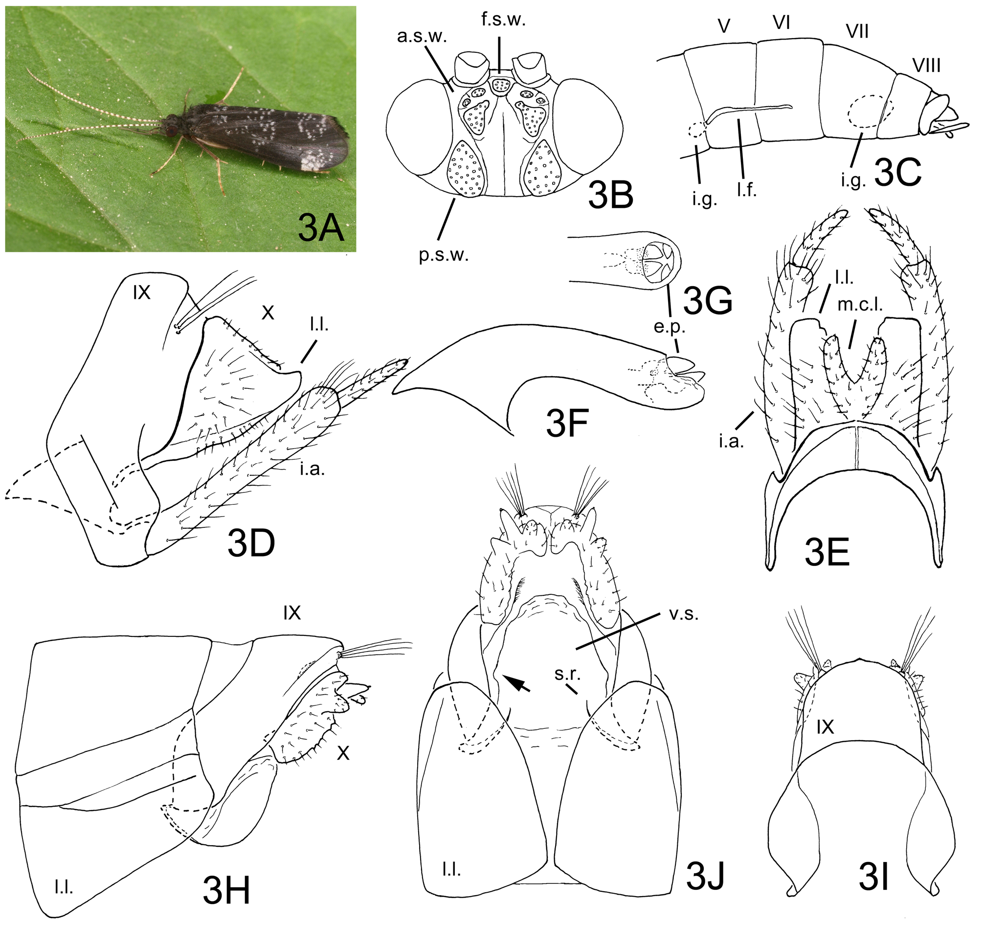
FIGURE 3. Diplectrona aiensis Kobayashi 1987. 3A–3G, male: 3A, habitus (Aichi), left lateral; 3B, head, dorsal; 3C, abdominal segments V–X, left lateral; 3D, genitalia, left lateral; 3E, same, dorsal; 3F, phallic apparatus, left lateral; 3G, same, apical part, dorsal. 3H–3J, female genitalia: 3H, left lateral; 3I, dorsal; 3J, ventral. Abbreviations: a.s.w. = anterior setal wart (paired), e.p. = endothecal process (paired), f.s.w. = frontal setal wart, i.a. = inferior appendage (paired), i.g. = internal gland (paired) of segment V or VIII, m.c.l. = mesocaudal lobe of segment X (paired), l.f. = lateral filament (paired), l.l. = lateral lobe of segment VIII in female or X in male (paired), p.s.w. = posterior setal wart (paired), s.r. = sclerotized rib (paired), V–X = abdominal segments V–X, v.s. = vulval scale. Arrow: see text.
| T |
Tavera, Department of Geology and Geophysics |
No known copyright restrictions apply. See Agosti, D., Egloff, W., 2009. Taxonomic information exchange and copyright: the Plazi approach. BMC Research Notes 2009, 2:53 for further explanation.
|
Kingdom |
|
|
Phylum |
|
|
Class |
|
|
Order |
|
|
Family |
|
|
Genus |
Diplectrona aiensis Kobayashi 1987
| Nozaki, Takao 2021 |
Diplectrona aiensis
| Kobayashi 1987 |