
Xizangiana linzhiensis ( Hu, 2001 )
|
publication ID |
https://doi.org/10.11646/zootaxa.5346.4.5 |
|
publication LSID |
lsid:zoobank.org:pub:E4FD4F32-49FF-4E81-A9B8-AD9F0361DC69 |
|
DOI |
https://doi.org/10.5281/zenodo.8407520 |
|
persistent identifier |
https://treatment.plazi.org/id/803A87D0-0663-8D2F-FF01-BCC7FD332EB5 |
|
treatment provided by |
Plazi (2023-09-29 11:10:17, last updated 2024-11-27 02:24:41) |
|
scientific name |
Xizangiana linzhiensis ( Hu, 2001 ) |
| status |
|
Xizangiana linzhiensis ( Hu, 2001) View in CoL View at ENA
(Hĭqǥ)
Figs 2A–B View FIGURE 2 , 3A View FIGURE 3 , 4–5 View FIGURE 4 View FIGURE 5 , 6A–B View FIGURE 6
Callilepis linzhiensis Hu, 2001: 229 , fig. 123. 1–4 (♀).
Scotophaeus himalayaensis Hu, 2001: 271 , fig. 155. 1–5 (♁).
Xizangia linzhiensis Song et al., 2004: 244 View in CoL , 337, fig. 142A–I (♁ ♀, transfer from Callilepis View in CoL , synonymy of S. himalayaensis ).
Xizangiana linzhiensis Sherwood et al., 2022: 64 View in CoL (transfer from Xizangia View in CoL View at ENA ).
Type material. Holotype ♀ (MHBU-ARA-005319), CHINA: Xizang Autonomous Region, Linzhi City , 3000 m elev., 30 July 1988, leg. Fuping Zhang. Paratype: 2♀ (MHBU-ARA-005320), same data as holotype; 2♁ (MHBU-ARA-005604 to 5), CHINA: Xizang Autonomous Region, Nyalam County, Zhangmu Town , 25 June 1985, leg. Aihua Li, examined .
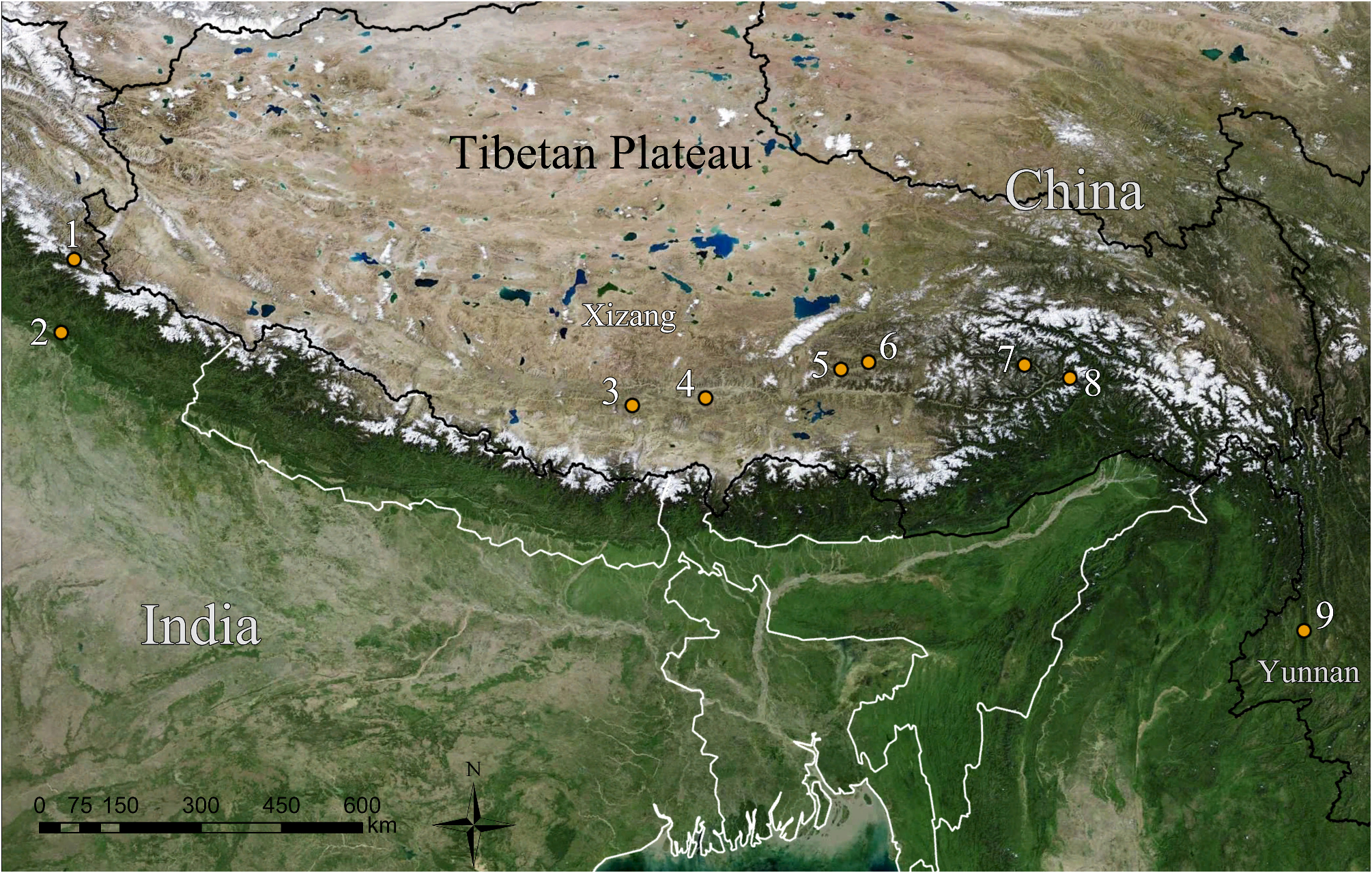
Other material examined. 1♁ (MHBU-ARA- 2019-31-1), CHINA: Xizang Autonomous Region, Linzhi City, Bayi District , Bayi Town , Duobu Village , 29°44.061′N, 94°07.546′E, 3099 m elev., 21 May 2019, leg. Luyu Wang; 1♀ (MHBU-ARA-2018-97-1) GoogleMaps , same data as the male, except 4 July 2018, leg. Luyu Wang; 1♀ (MHBU-ARA-2018-95-1) GoogleMaps , CHINA: Xizang Autonomous Region, Linzhi City , Mainling County, 29°27.147'N, 94°44.900'E, 2929 m elev., 3 July 2018, leg. Luyu Wang. GoogleMaps
Diagnosis. Males differ from all the congeners except X. xiangbi by having retrolateral tibial apophysis with arc-shaped membranous projection ( Fig. 3 View FIGURE 3 ). It can be distinguished from X. xiangbi by having an arc-shaped embolar base projection distolaterally, the absence of radix projection, and the absence of bump on tegulum ( Fig. 5 View FIGURE 5 ; vs. having a hook-like embolar base projection retrolaterally, radix projection present, tegulum with a bump ventrally, Fig. 10 View FIGURE 10 ). Females can be distinguished from all the congeners by the almost triangular atrium, with five curved, partly sclerotized transversal folds, the heart-shaped, well sclerotized lateral folds, and the presence of a small scape, almost length of primary spermathecae ( Fig. 6A–B View FIGURE 6 ).
Redescription. Male. Total length 3.66–5.68. One paratype ( Figs 4C–D View FIGURE 4 ; MHBU-ARA-005604): total length 4.12; carapace 1.91 long, 1.43 wide; abdomen 2.21 long, 1.24 wide. Eye sizes and interdistances: AME 0.06, ALE 0.09, PME 0.07, PLE 0.08; AME‒AME 0.04, AME‒ALE 0.01, PME‒PME 0.06, PME‒PLE 0.07, ALE‒PLE 0.07. Leg measurements: I 5.18 (1.45, 0.77, 1.11, 0.99, 0.86), II 4.09 (1.21, 0.64, 0.80, 0.81, 0.63), III 3.93 (1.11, 0.54, 0.86, 0.82, 0.60), IV 5.72 (1.46, 0.62, 1.17, 1.65, 0.82). Leg formula: 4123. Cheliceral promargin with four teeth, retromargin with two ( Fig. 2B View FIGURE 2 ). Color in alcohol ( Fig. 4C–D View FIGURE 4 ): carapace dark brown; legs yellow-brown.
Palp ( Figs 3A View FIGURE 3 , 5 View FIGURE 5 ). Retrolateral tibial apophysis broad, finger-like, dorsally with an arc-shaped membranous projection; embolar radix fused with embolar base; embolar base distolaterally with an arc-shaped embolar base projection; embolus slightly curved, with pinhead-shaped apex; median apophysis wide at base, narrowest at median part, almost two times as thin as base.
Female. Total length 6.94–8.86. Holotype ( Fig. 4A–B View FIGURE 4 ; MHBU-ARA-005319): total length 7.86; carapace 3.05 long, 2.34 wide; abdomen 4.81 long, 2.60 wide. Eye sizes and interdistances:AME 0.08, ALE 0.13, PME 0.09, PLE 0.11; AME‒AME 0.08, AME‒ALE 0.03, PME‒PME 0.11, PME‒PLE 0.13, ALE‒PLE 0.10. Leg measurements: I 7.26 (2.16, 1.04, 1.81, 1.26, 0.99), II 6.17 (1.71, 0.73, 1.62, 1.26, 0.85), III 5.45 (1.44, 0.61, 1.37, 1.22, 0.81), IV 8.39 (2.26, 0.95, 1.90, 2.16, 1.12). Leg formula: 4123. Cheliceral promargin with six teeth, retromargin with three ( Fig. 2A View FIGURE 2 ). Color in alcohol ( Fig. 4A–B View FIGURE 4 ) as in male.
Epigyne ( Fig. 6A–B View FIGURE 6 ). Atrium almost triangular, with five transversal folds, partly sclerotized; anterior fold strongly sclerotized; scape oval, small, almost length of primary spermathecae; lateral folds heart-shaped, strongly sclerotized; secondary spermathecae oval, small.
Distribution. China (Xizang) ( Fig. 23 View FIGURE 23 ).
Hu, J. L. (2001) s. n. In: Spiders in Qinghai-Tibet Plateau of China. Henan Science and Technology Publishing House, Zhengzhou, pp. 229 - 230.
Sherwood, D., Li, S. Q. & Zhang, F. (2022) A replacement name for Xizangia Song, Zhu & Zhang, 2004 (Araneae: Gnaphosidae). Acta Arachnologica Sinica, 31 (1), 64 - 65. https: // doi. org / 10.3969 / j. issn. 1005 - 9628.2022.01.011
Song, D. X., Zhu, M. S. & Zhang, F. (2004) s. n. In: Fauna Sinica: Invertebrata Vol. 39. Arachnida: Araneae: Gnaphosidae. Science Press, Beijing, pp. 243 - 246.

FIGURE 2. Chelicerae, ventral view. A–B. X. linzhiensis, female (A) and male (B); C–D. X. namchabarwa sp. n., female (C) and male (D); E. X. rigaze, male. F–H. X. shenxian sp. n., female (F–G) and male (H); I–J. X. benae sp. n., female (I) and male (J); K–L. X. xiangbi sp. n., female (K) and male (L); M–O. X. longlin sp. n., male (M–N) and female (O); Scale bars = 0.1 mm.
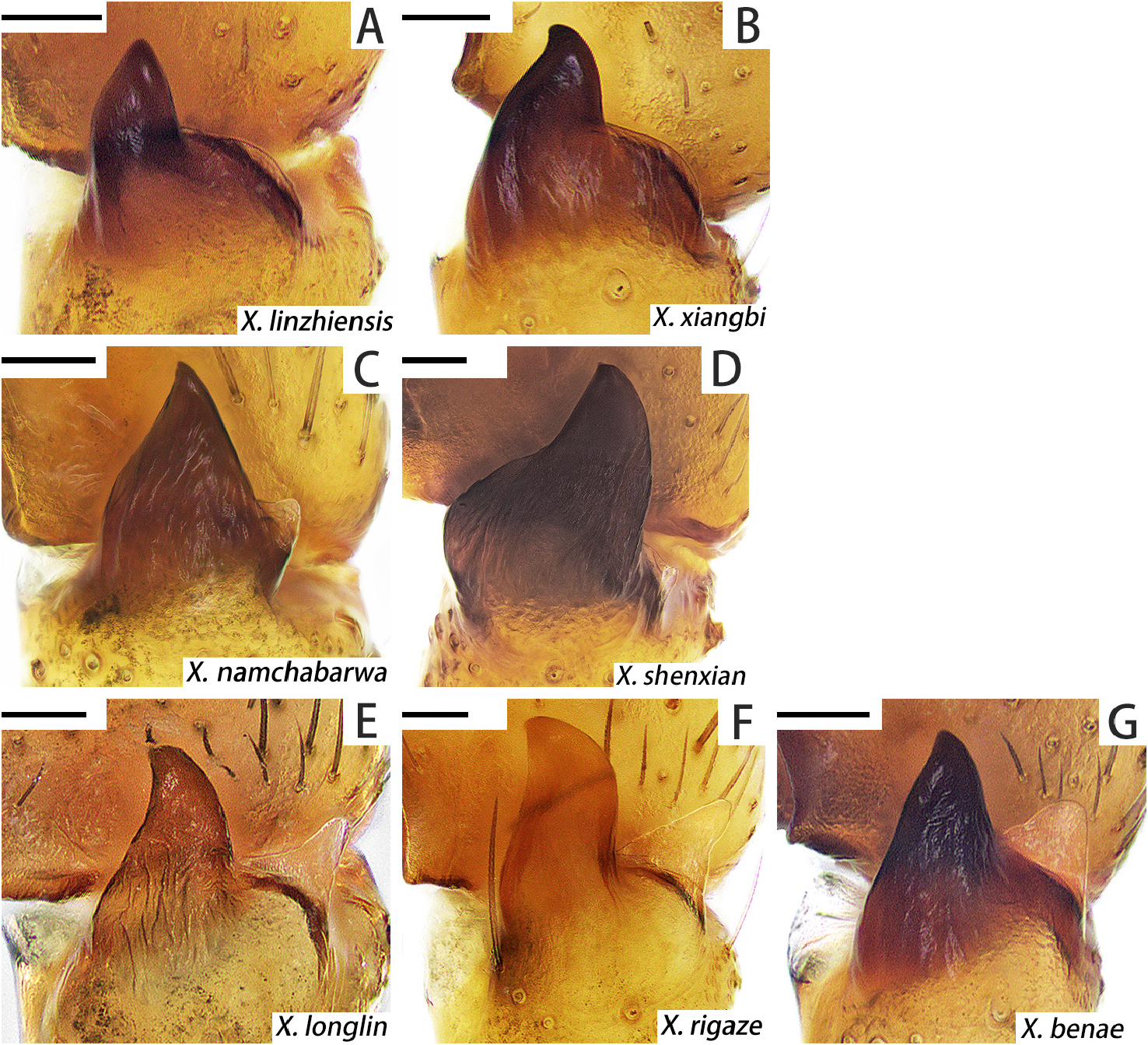
FIGURE 3. RTA, retrolateral view. A. X. linzhiensis; B. X. xiangbi sp. n.; C. X. namchabarwa sp. n.; D. X. shenxian sp. n.; E. X. longlin sp. n.; F. X. rigaze; G. X. benae sp. n.. Scale bars = 0.05 mm.

FIGURE 4. Xizangiana linzhiensis. A–B. Female, habitus, in dorsal (A) and ventral (B) view; C–D. Male, habitus, in dorsal (C) and ventral (D) view; E. Female, metatarsus IV, preening brush, ventral view; F. Female, carapace, frontal view; G–H. Female, eye region, in frontal (G) and dorsal (H) view.
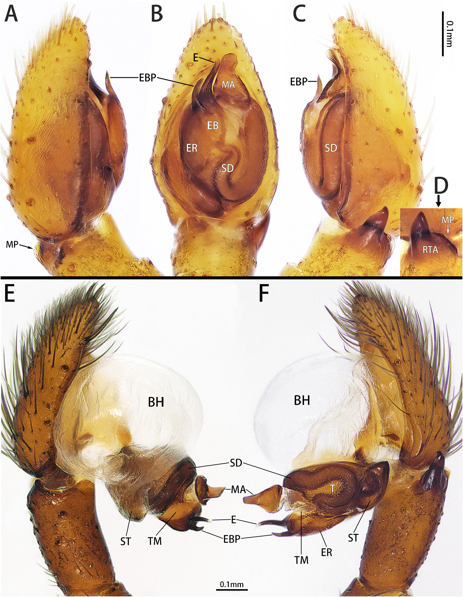
FIGURE 5. Palp of Xizangiana linzhiensis, in prolateral (A), ventral (B) and retrolateral (C) view. D. RTA, retrolateral view; E–F. Expanded palp, in prolateral (E) and retrolateral (F) view.
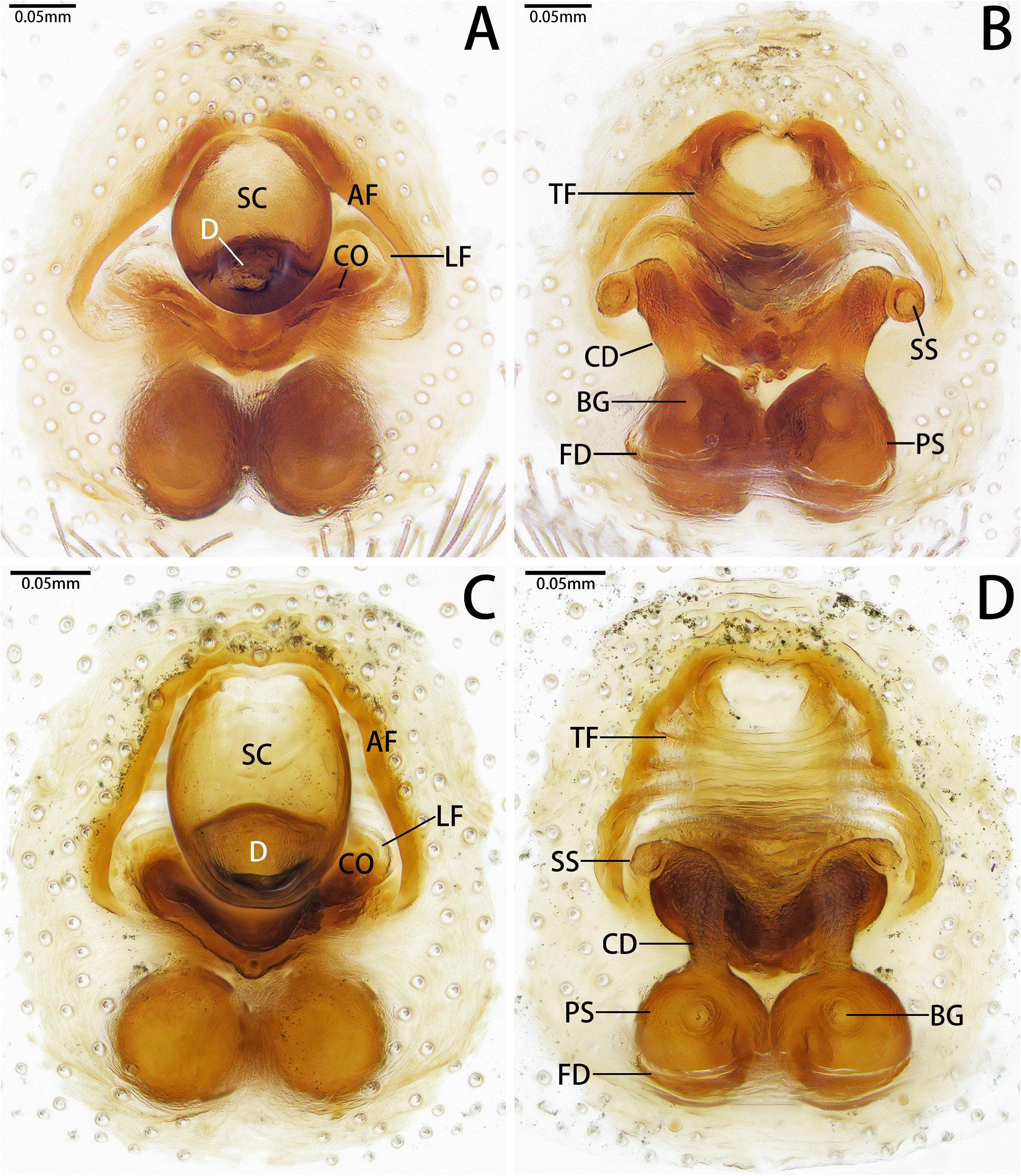
FIGURE 6. Female genitalia, in ventral (A, C) and dorsal (B, D) view. A–B. Xizangiana linzhiensis; C–D. X. namchabarwa sp. n..
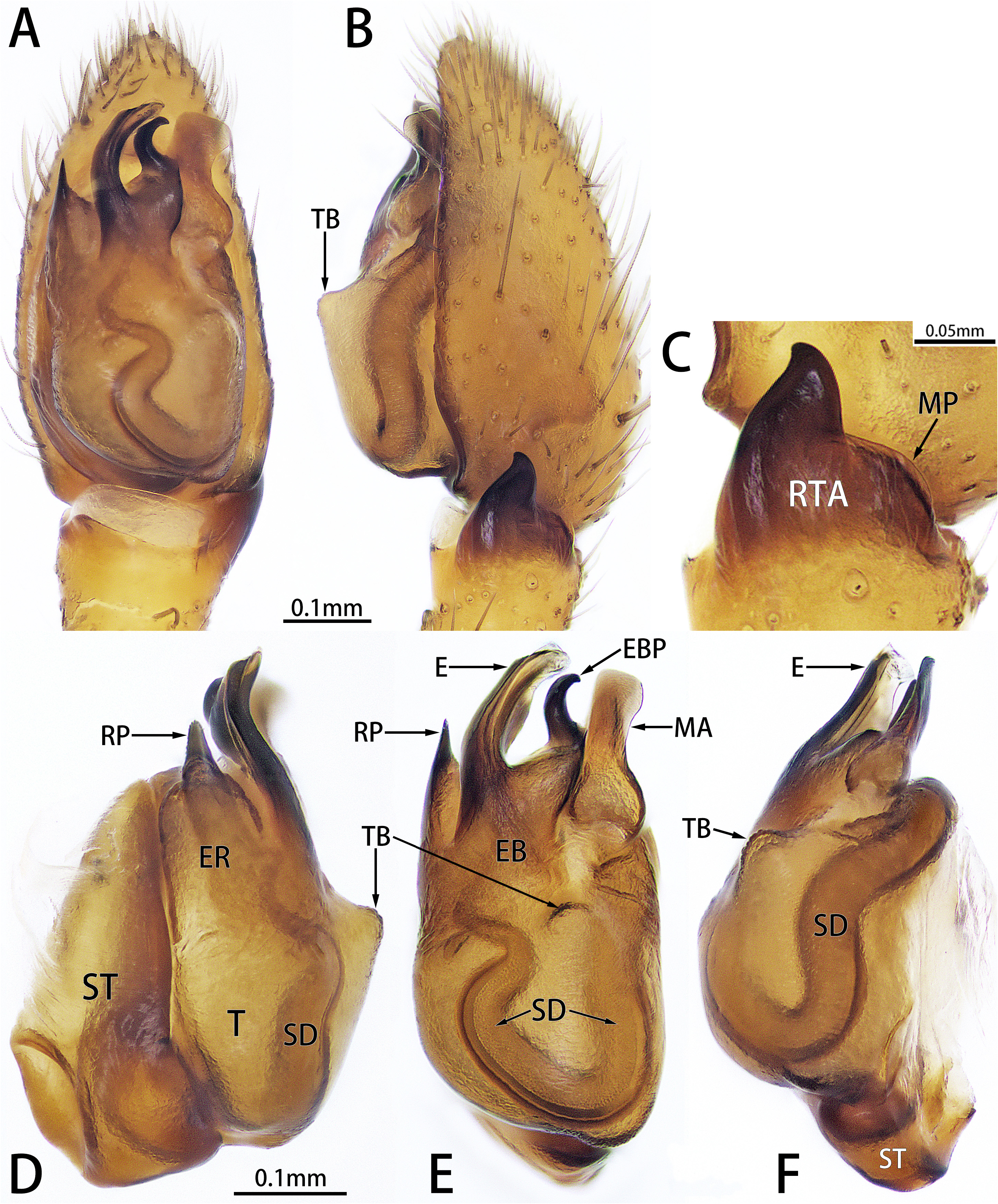
FIGURE 10. Xizangiana xiangbi sp. n.. A–B. Palp, in ventral (A) and retrolateral (B) view; C. RTA, retrolateral view; D–F. Bulb, in prolateral (D), ventral (E) and retrolateral (F) view.
No known copyright restrictions apply. See Agosti, D., Egloff, W., 2009. Taxonomic information exchange and copyright: the Plazi approach. BMC Research Notes 2009, 2:53 for further explanation.
|
Kingdom |
|
|
Phylum |
|
|
Class |
|
|
Order |
|
|
Family |
|
|
Genus |
Xizangiana linzhiensis ( Hu, 2001 )
| Liu, Bo, Wang, Luyu & Zhang, Feng 2023 |
Xizangiana linzhiensis
| Sherwood, D. & Li, S. Q. & Zhang, F. 2022: 64 |
Xizangia linzhiensis
| Song, D. X. & Zhu, M. S. & Zhang, F. 2004: 244 |
Callilepis linzhiensis
| Hu, J. L. 2001: 229 |
Scotophaeus himalayaensis
| Hu, J. L. 2001: 271 |