
Carnarvoncochlea cf. carnarvonensis ( Ponder & Clark, 1990 ), 2019
|
publication ID |
https://doi.org/10.11646/zootaxa.4583.1.1 |
|
publication LSID |
lsid:zoobank.org:pub:27F24995-359E-46F6-AB22-75568BACFDCF |
|
persistent identifier |
https://treatment.plazi.org/id/724987F6-FFA9-242C-FF7E-BEEEFA843E64 |
|
treatment provided by |
Plazi (2019-04-12 07:30:42, last updated 2025-02-11 16:15:37) |
|
scientific name |
Carnarvoncochlea cf. carnarvonensis ( Ponder & Clark, 1990 ) |
| status |
sp. nov. |
Carnarvoncochlea cf. carnarvonensis ( Ponder & Clark, 1990) View in CoL
Jardinella n. sp. (C.400126) Perez et al., 2005: 547.
Material examined. Figured specimen: Queensland, Carnarvon NP, Carnarvon Gorge, small creek on side of track emerging from N side of gorge, 25° 02' 37" S, 148° 12' 15" E, on leaves, dead ferns and roots etc., W.F. Ponder & C. Lydeard, 7 May 2001, C.479955. Other material examined: Same data, C.400126, 20+; QM MO85772 , 5 .
Shell ( Fig. 21A, B View FIGURE 21 ). Ovate, spire outline moderately convex, normally coiled, transparent. Length 2.1–2.7 mm (mean 2.4 mm), width 1.9–2.3 mm (mean 2.1 mm). Protoconch of about 1.3–1.4 whorls, sometimes a few very weak irregular transverse folds near suture on first half whorl, very weak, minute pits over whole surface; one specimen with few spiral lines near outer edge. Teleoconch whorls moderately convex, evenly rounded, total number 3.7–4.0 (mean 3.9). Umbilicus moderately wide. Aperture ovate or pyriform, inner lip narrow, thin or medium, slightly separated along whole length of parietal wall, outer lip thin or medium. Periostracum very thin and inconspicuous, white or pale yellow.
Operculum ( Fig. 21 View FIGURE 21 C–E). Transparent, yellow-brown, flat. Inner side lacking white smear, simple or with few very weak protuberances ( Fig. 21D, E View FIGURE 21 ).
Head-foot and external body. Snout, tentacles, dorsal and lateral foot and opercular lobes unpigmented, neck unpigmented or pigmented, mantle roof and visceral coil unpigmented or weakly pigmented.
Mantle cavity. Ctenidium well-developed, filaments 17–20, broadly triangular, apex right edge. Osphradium narrowly oval, towards posterior end of ctenidium, length relative to gill 0.25–0.31. Hypobranchial gland not distinguishable (absent). Rectum with U-shaped bend, faecal pellets longitudinally orientated, anus at or very near mantle collar. Kidney half or more in roof of mantle cavity. Renal gland longitudinal, or transverse. Pericardium half or more in roof of mantle cavity, overlapping posterior end of ctenidium.
Radula ( Fig. 22 View FIGURE 22 ). Central teeth with cusp formula 4+1+4, basal cusps 2+2; median cusp sharp, about twice as long as adjacent cusps. Lateral teeth with cusp formula 2–3+1+2 (–3), main cusp blunt to pointed, about twice as long as adjacent cusps. Inner marginal teeth with about 17–19 cusps. Outer marginal teeth with about 22–24 cusps.
Female reproductive system ( Fig. 23C, D View FIGURE 23 ). Ovary simple sac or weakly lobed. Renal oviduct thick, forming simple tight near vertical U-shaped fold. Seminal receptacle near anterior edge of bursa, oval, duct intermediate; orientated sperm also located in renal oviduct and/or bursa duct. Bursa copulatrix behind albumen gland, elongately-oval or triangular, shorter than albumen gland, bursal duct enters bursa mid anteriorly or anteroventrally, bursal duct joins coiled oviduct little behind posterior mantle cavity wall. Albumen gland partly in mantle cavity. Capsule gland with two distinct glandular zones, medium thickness in cross section, markedly indented by rectum. Anterior vestibule large, opening subterminal, intermediate in size, no associated gutter.
Male reproductive system ( Fig. 23A, B View FIGURE 23 ). Prostate gland mostly in mantle roof, oval, medium in cross section. Posterior pallial vas deferens coiled, anteriorly coiled. Penis towards middle of head, intermediate, distal end narrow, blunt, terminal papilla small.
Distribution and habitat. This material was found in a small stream on the north side of the main Carnarvon Gorge ( Fig. 1 View FIGURE 1 ).
Remarks. The shell of this material differs from typical C. carnarvonensis in its slightly larger size (up to 2.7 mm in length compared with a maximum of 2.5 mm in the type material of C. carnarvonensis and 1.7 mm in C. exigua ) and in its slightly broader shape. There is also a greater separation of the inner lip from the parietal wall than in other material. These differences are not sufficient to recognise more than one species, although a more detailed analysis of the populations in the Carnarvon Gorge area may possibly reveal that more than one species can be recognised.
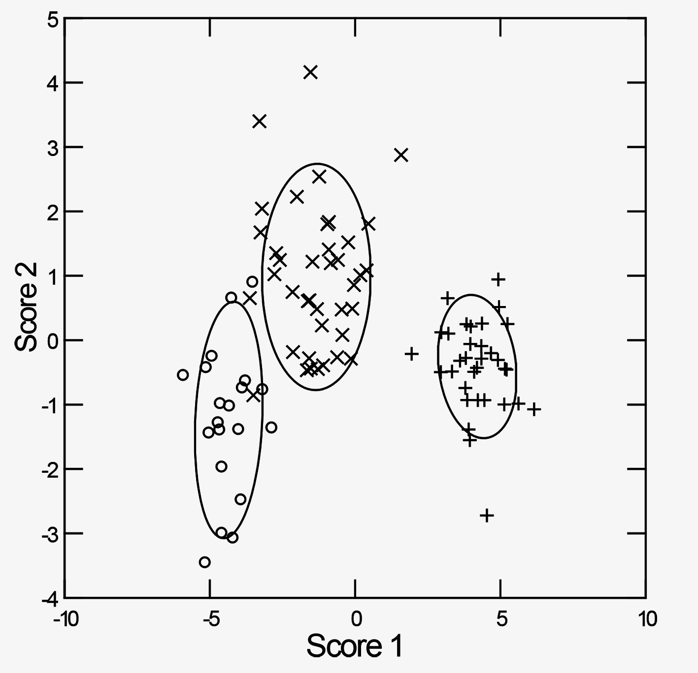
A discriminant function analysis identified 98% of the specimens of this form correctly (Wilks’s Lambda 0.049 p<0.000) ( Fig. 24 View FIGURE 24 ). One specimen of C. carnarvonensis was misidentified as C. cf. carnarvonensis and one of C. cf. carnarvonensis was misidentified as C. carnarvonensis while all C. exigua were correctly identified.
Perez, K. E., Ponder, W. F., Colgan, D. J., Clark, S. A. & Lydeard, C. (2005) Molecular phylogeny and biogeography of springassociated hydrobiid snails of the Great Artesian Basin, Australia. Molecular Phylogenetics and Evolution, 34, 545 - 556. https: // doi. org / 10.1016 / j. ympev. 2004.11.020
Ponder, W. F. & Clark, G. A. (1990) A radiation of hydrobiid snails in threatened artesian springs in Western Queensland. Records of the Australian Museum, 42, 301 - 363. https: // doi. org / 10.3853 / j. 0067 - 1975.42.1990.119
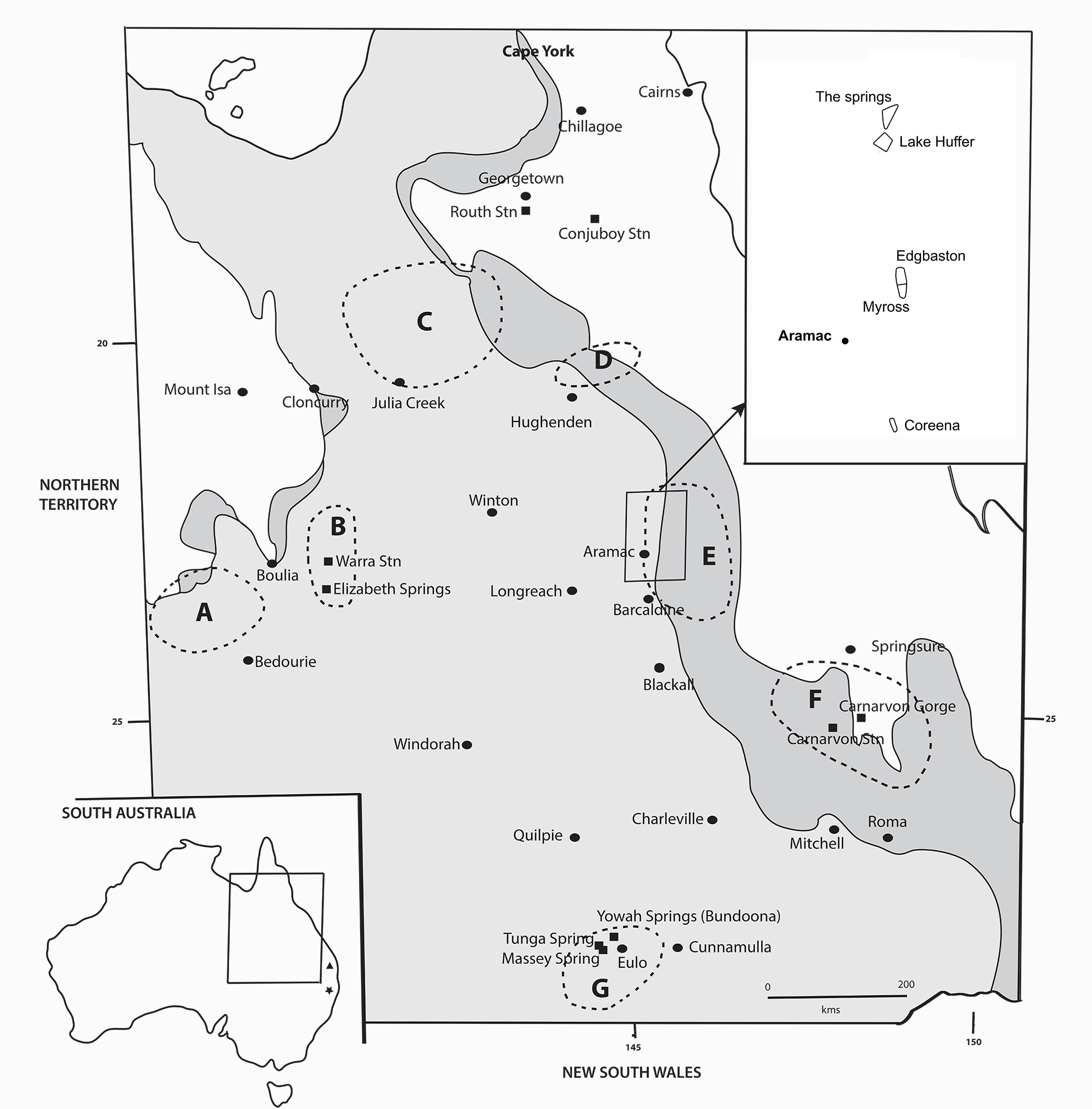
FIGURE 1. Map of the spring supergroups associated with the Great Artesian Basin (GAB) in Queensland, showing the main non-town locations mentioned in the text (squares) and some of the towns in the area (ovals). The pale grey shading is the discharge part of the GAB, the dark grey the recharge part. The inset map of Australia shows the area illustrated. The approximate location of Conondale is indicated by a triangle and Nundle by a star. The spring supergroups are outlined with dotted lines and are as follows: A, Mulligan River; B, Springvale; C, Flinders River; D, Hughenden; E, Barcaldine (see inset at upper right for details); F, Springsure; G, Eulo.
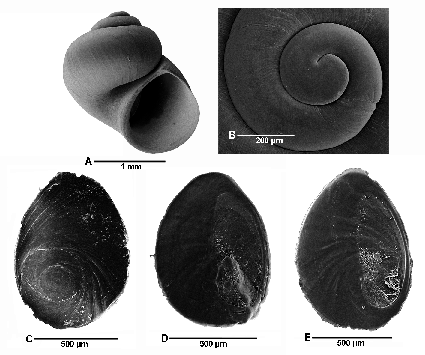
FIGURE 21. Shell and operculum of Carnarvoncochlea cf. carnarvonensis (Ponder & Clark, 1990). A. Shell (C.479955). B. Protoconch. C–E. Operculum, outer (C), and inner (D, E) sides (C.400126).
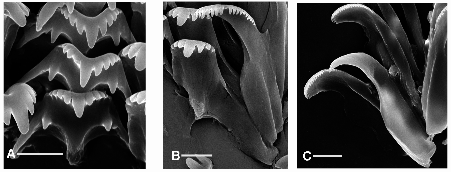
FIGURE 22. Radula of Carnarvoncochlea cf. carnarvonensis (Ponder & Clark, 1990) (C.400126). A. Detail of central teeth. B. Detail of lateral and inner marginal teeth. C. Detail of outer marginal teeth. Scale bars 10 µm.

FIGURE 23. Male and female reproductive anatomy of Carnarvoncochlea cf. carnarvonensis (Ponder & Clark, 1990) (C.400126). A. Penis, showing penial duct and vas deferens. B. Prostate, with associated vas deferens and vas efferens. C. Female system shown from left side. D. Bursa copulatrix and seminal receptacle. Abbreviations: ag—albumen gland; avdanterior vas deferens; bc—bursa copulatrix; cg—capsule gland; o—opening of oviduct; pd—penial duct; pw—position of posterior wall of pallial cavity; ro—renal oviduct; sr—seminal receptacle; vc—ventral channel; vd—vas deferens; ve—vas efferens. Scale bar 200 µm.
| QM |
Queensland Museum |
No known copyright restrictions apply. See Agosti, D., Egloff, W., 2009. Taxonomic information exchange and copyright: the Plazi approach. BMC Research Notes 2009, 2:53 for further explanation.
|
Kingdom |
|
|
Phylum |
|
|
Class |
|
|
Order |
|
|
Family |
|
|
Genus |
1 (by plazi, 2019-04-12 07:30:42)
2 (by ImsDioSync, 2019-04-12 07:47:43)
3 (by ImsDioSync, 2019-07-24 12:26:28)
4 (by ImsDioSync, 2019-07-25 10:20:41)
5 (by ImsDioSync, 2019-07-26 12:32:21)
6 (by ImsDioSync, 2019-07-30 09:35:43)
7 (by ExternalLinkService, 2019-09-25 22:54:12)
8 (by felipe, 2020-04-20 15:21:29)
9 (by ExternalLinkService, 2021-10-20 02:45:54)
10 (by ExternalLinkService, 2021-10-20 04:16:13)
11 (by ExternalLinkService, 2022-11-22 21:42:57)
12 (by plazi, 2023-10-30 13:13:40)
13 (by ExternalLinkService, 2023-10-31 02:47:01)
14 (by ExternalLinkService, 2024-11-26 02:35:24)