
Hydrobiosella arcuata Kimmins
|
publication ID |
https://doi.org/10.24199/j.mmv.2010.67.01 |
|
DOI |
https://doi.org/10.5281/zenodo.8064963 |
|
persistent identifier |
https://treatment.plazi.org/id/704FCC34-FFDB-E60A-BEA2-F9798C05A8C3 |
|
treatment provided by |
Felipe (2023-06-21 14:02:09, last updated 2024-11-26 22:45:36) |
|
scientific name |
Hydrobiosella arcuata Kimmins |
| status |
|
Hydrobiosella arcuata Kimmins View in CoL
Figures 1 View Figures 1–7 , 23–25 View Figures 17–25 , 44–45 View Figures 35–49
Hydrobiosella arcuata Kimmins in Mosely and Kimmins, 1953:
397, fig. 271. — Neboiss, 1986: 103.
Type material (not seen). Holotype. Male, Queensland, Montville , 3 Oct 1912, R. J. Tillyard ( BMNH).
Material examined. Queensland. 1 male, Booloumba Ck , 8 km SW Kenilworth, 26°39'S, 152°39'E, 12 Dec 1984, G. Theischinger GoogleMaps ; 1 male (specimen CT-573 figured), Booloumba Ck, Mary R . catchment, 26°41'S, 152°37'E, 26 Oct 1993, collector unknown; 1 male, 1 female, Branch Ck, Brisbane R . catchment, 26°52'S, 152°41'E, 26 Apr 1993; collector unknown; 1 male, 2 females (specimen CT-603 figured), Stony Ck, Brisbane R . catchment, 26°52'S, 152°43'E, 18 Aug 1992; collector unknown ( NMV) GoogleMaps .
Diagnosis. Hydrobiosella arcuata can be separated from other species in the group by the shape of the harpago, where the ventral margin is curved or arched strongly so that the apex points downwards.
Description. (Revised after Kimmins in Mosely and Kimmins, 1953). Wings similar to other species in the group (fig. 1), length of forewing: male 6.3–7.3 mm, female 6.1–8.0 mm. Forewing fork 2 long, length fork 2 about 1.6 times length of fork 1; length fork 3 about twice length footstalk; fork 4 length about 4.5 times length footstalk. Hind wing fork 1 sessile or with very short footstalk; fork 3 length about 1.0–1.3 times length of footstalk.
Male. Segment IX with shallow notch medially on distal margin in between a pair of small knobs (fig. 25). Segment X with a slender mesal lobe, with a pair of short hairs/bristles subapically, in lateral view slightly downturned distally (fig. 24); in dorsal view not narrowed subapically (fig. 23), with a pair of more pigmented lateral lobes, which terminate in small, slightly backward and outward projecting hooks (figs 23–24). Phallus truncate apically, with a pair of robust parameres arising from the phallus subapically (fig. 24). Inferior appendages in lateral view, with basal segment length about 1.7 times maximum width, broad basally, rounded distally; harpago more slender, with ventral margin sharply angled near middle, curved in distal half with downward pointing acute apex (fig. 24).
Female. Genitalia typical of genus, with a small, shallow, rounded projection on sternite VIII mesodistally (figs 44–45).
Remarks. Males and females of Hydrobiosella arcuata have been collected from five sites in addition to the type locality, all in southeastern Queensland (latitudinal range 26°39'– 26°52'S).
Kimmins’ (in Mosely and Kimmins 1953) and Neboiss’ (1986) figures have been redrawn to allow direct comparisons and to accompany the description that is revised in light of new interpretations of Hydrobiosella genitalic structures.
Mosely, M. E. and Kimmins D. E. 1953. The Trichoptera (caddis-flies) of Australia and New Zealand. London: British Museum (Natural History). 550 pp.
Neboiss, A. 1986. Atlas of Trichoptera of the SW Pacific-Australian Region. Dr W. Junk Publishers, Dordrecht. 286 pp.
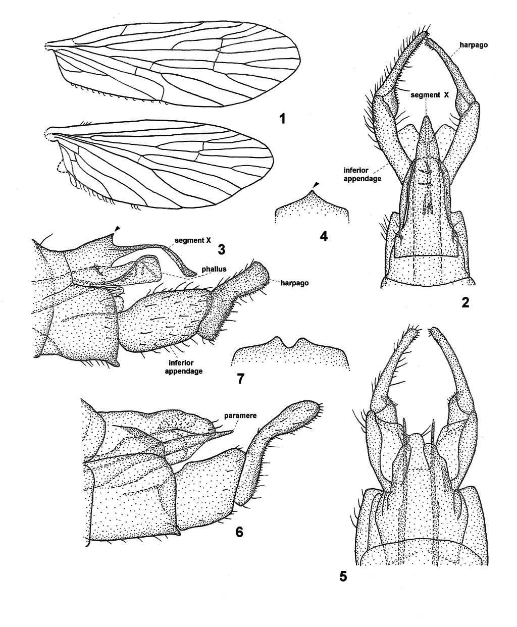
Figures 1–7. Hydrobiosella spp.; 1, Hydrobiosella arcuata Kimmins, wings; 2–7, Hydrobiosella spp., male genitalia in dorsal, lateral and part ventral views; 2–4, Hydrobiosella unispina sp. nov.; 2, dorsal; 3, lateral; 4, ventral, mesodistal margin of segment IX; 5–7, Hydrobiosella gurara sp. nov.; 5, dorsal; 6, lateral; 7, ventral, mesodistal margin of segment IX.
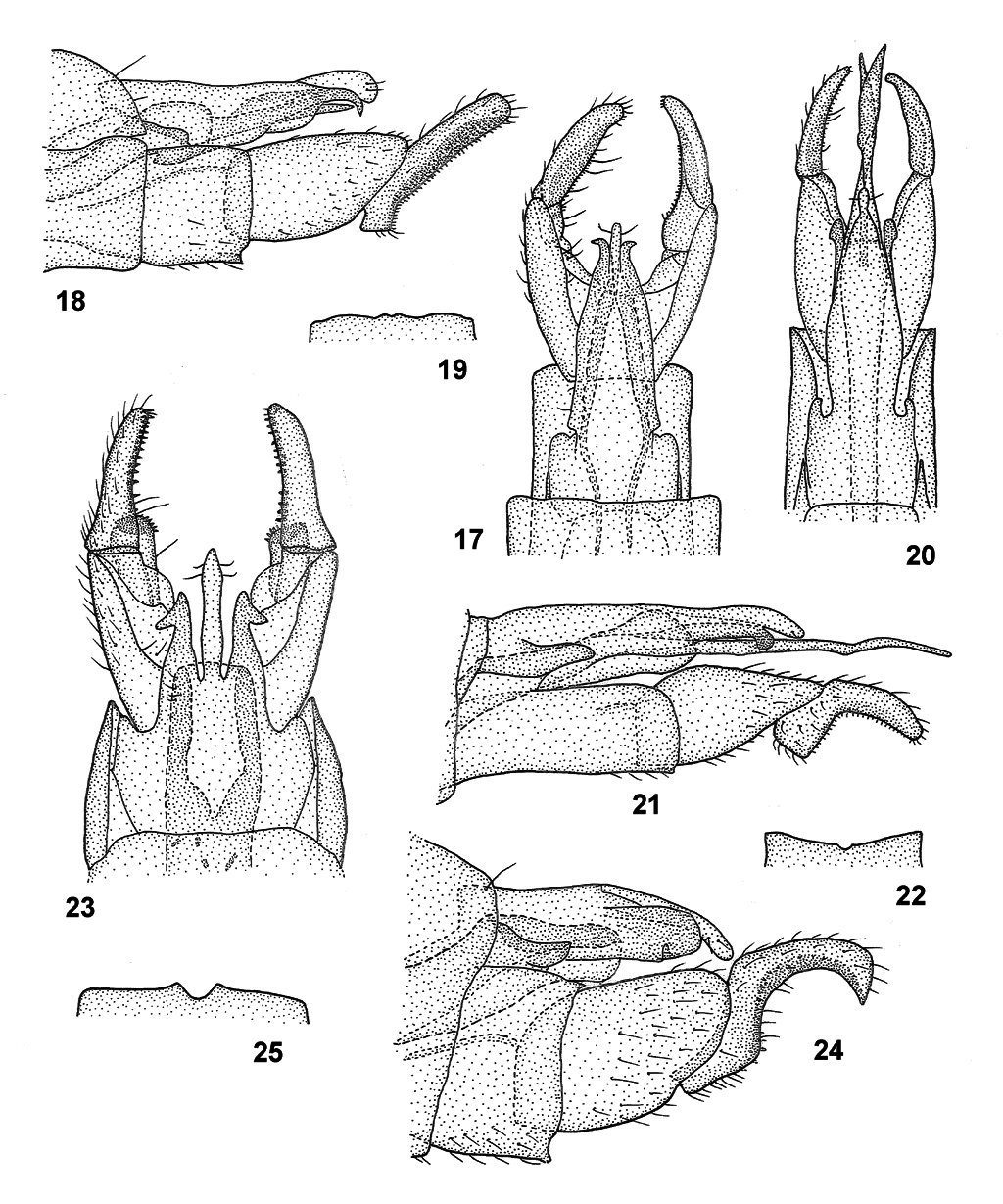
Figures 17–25. Hydrobiosella spp. male genitalia in dorsal, lateral and part ventral views; 17–19, Hydrobiosella mundagurra sp. nov.; 17, dorsal; 18, lateral; 19, ventral, mesodistal margin of segment IX; 20–22, Hydrobiosella bispina Kimmins.; 20, dorsal; 21, lateral; 22, ventral, mesodistal margin of segment IX; 23–25, Hydrobiosella arcuata Kimmins; 23, dorsal; 24, lateral; 25, ventral, mesodistal margin of segment IX.
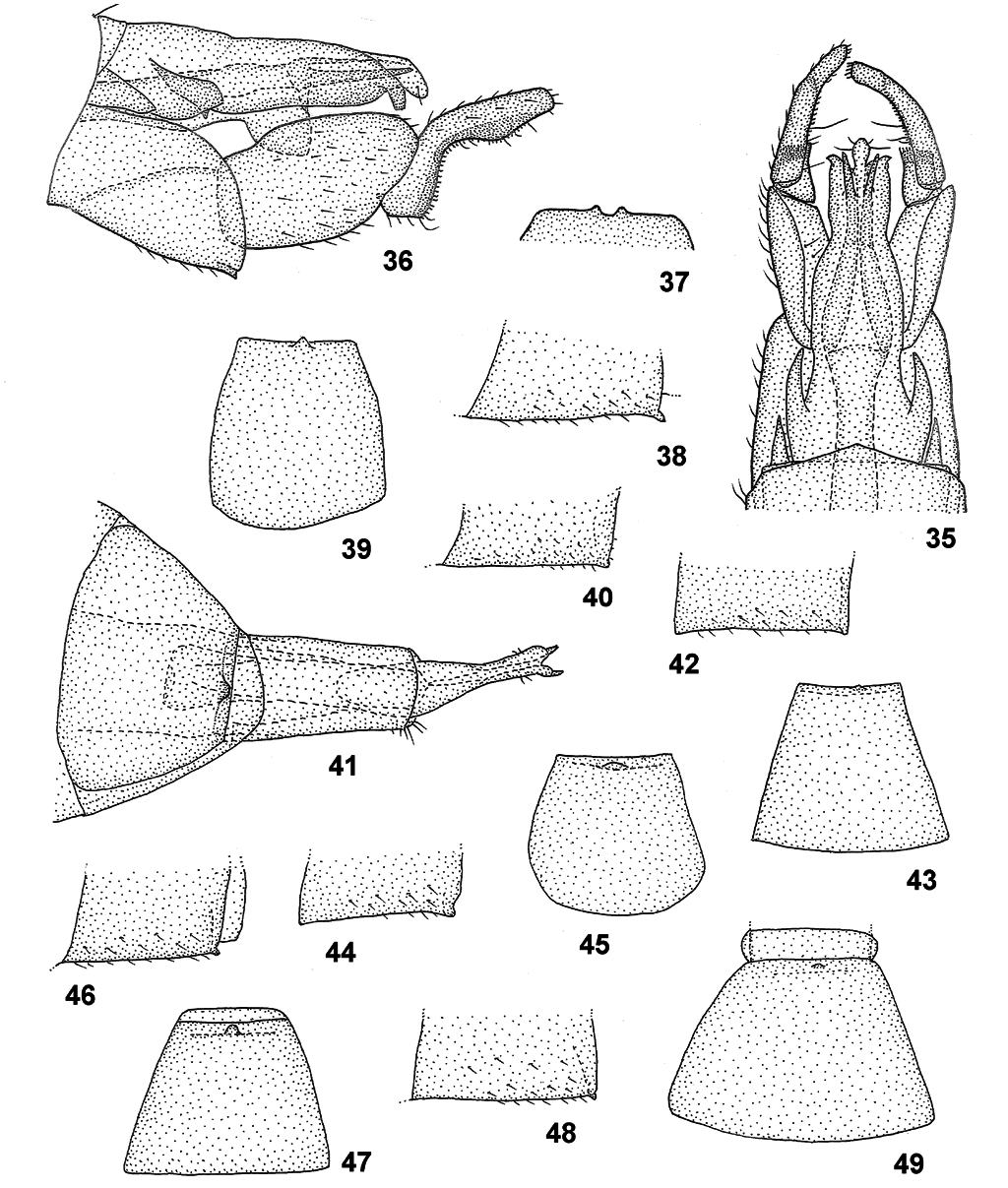
Figures 35–49. Hydrobiosella spp.; 35–37, Hydrobiosella yokunna sp. nov. male genitalia in dorsal, lateral and part ventral views; 35, dorsal; 36, lateral; 37, ventral, mesodistal margin of segment IX; 38–49, Hydrobiosella spp. female genitalia (part segment VIII) in lateral and (segment VIII) ventral view; 38–39, Hydrobiosella unispina sp. nov.; 38, lateral; 39, ventral; 40–41, Hydrobiosella mundagurra sp. nov.; 40, lateral; 41, female genitalia, ventral; 42–43, Hydrobiosella bispina Kimmins; 42, lateral; 43, ventral; 44–45, Hydrobiosella arcuata Kimmins; 44, lateral; 45, ventral; 46–47, Hydrobiosella woonoongoora sp. nov.; 46, lateral; 47, ventral; 48–49, Hydrobiosella thurawal sp. nov.; 48, lateral; 49, ventral.
| R |
Departamento de Geologia, Universidad de Chile |
| NMV |
Museum Victoria |
No known copyright restrictions apply. See Agosti, D., Egloff, W., 2009. Taxonomic information exchange and copyright: the Plazi approach. BMC Research Notes 2009, 2:53 for further explanation.
|
Kingdom |
|
|
Phylum |
|
|
Class |
|
|
Order |
|
|
Family |
|
|
Genus |