
Hydrobiosella bispina Kimmins
|
publication ID |
https://doi.org/10.24199/j.mmv.2010.67.01 |
|
DOI |
https://doi.org/10.5281/zenodo.8070782 |
|
persistent identifier |
https://treatment.plazi.org/id/704FCC34-FFD5-E60A-BD07-F9F18BA7ABD3 |
|
treatment provided by |
Felipe (2023-06-21 14:02:09, last updated 2024-11-26 22:45:36) |
|
scientific name |
Hydrobiosella bispina Kimmins |
| status |
|
Hydrobiosella bispina Kimmins View in CoL View at ENA
Figures 20–22 View Figures 17–25 , 42–43 View Figures 35–49
Hydrobiosella bispina Kimmins in Mosely and Kimmins, 1953:
394, fig. 270.— Neboiss, 1986: 102.
Type material (not seen). Holotype. Male, New South Wales, Stanwell Park , 23 Apr 1916, R. J. Tillyard ( BMNH).
Paratype (not seen). New South Wales. 1 male, collected with holotype ( BMNH) .
Material examined. New South Wales. 3 males, 6 females, Wilson R ., Wilson R . Reserve , 11 Feb 2008, R . St Clair; 1 male, Wilson R ., Bobs Ridge Rd , 31°15'S, 152°31'E, 4 Dec 2007, A. Glaister, J. Dean and R. St Clair GoogleMaps ; 1 male (specimen PT-579 figured), Tubrabucca, Barrington Tops , 15 Nov 1953, A. Neboiss ( NMV) ; 2 males, 16 females, Dilgry R ., Banksia camp ground, 31°53'S, 151°32'E, 2 Dec 2007, A. Glaister, J. Dean and R GoogleMaps . St Clair; 3 males, 4 females, Gloucester R ., Gloucester R . camping area, 32°03'S, 151°41'E, 1 Dec 2007, A. Glaister, J. Dean and R GoogleMaps . St Clair; 1 male, 1 female (specimen CT-605 figured), Gloucester Tops, el. 1280 m, Malaise , 19 Nov to 4 Dec 1988, D. Bickle ; 3 males, 1 female, Gloucester Tops , 32°04'S, 151°34'E, el. 1300 m, 2–3 Dec 1988, Theischinger and Mueller GoogleMaps ; 1 male, 1 female, Jerusalem Falls near Karuah , 6 Dec 1988, G. Theischinger ; 1 male, 1 female, Wilson R . near Bellangry , 5 Dec 1988, G. Theischinger ; 1 male, Wollomi Brook, The Basin, Olney State Forest , 33°06'S, 151°14'E, 26 Nov 2007, A. Glaister, J. Dean and R GoogleMaps . St Clair ( NMV) .
Diagnosis. Hydrobiosella bispina can be separated from other species in the group by the very long parameres, which reach the tip of the inferior appendages.
Description. (Revised after Kimmins in Mosely and Kimmins, 1953.) Wings similar to those of H.arcuata (fig. 1), length of forewing: male 6.3–8.3 mm, female 7.2–8.6 mm. Forewing fork 2 long, length fork 2 about 1.5 times length of fork 1; length fork 3 about twice length footstalk; fork 4 length about 8 times length footstalk. Hind wing fork 1 sessile; fork 3 length about 1.5 times length of footstalk.
Male. Segment IX with small, shallow notch medially on distal margin (fig. 22). Segment X with a slender mesal lobe, with a pair of short hairs/bristles subapically, in lateral view slightly downturned distally (fig. 21); in dorsal view slightly narrowed subapically (fig. 20); with a pair of pigmented lateral lobes, which end in small, slightly downward and outward projecting rounded hooks (figs 20–21). Phallus generally slender, slightly dilated subapically; with a pair of very slender and elongate parameres arising from the phallus near the apex (fig. 21). Inferior appendages in lateral view, with basal segment length about twice maximum width, broadest near middle, tapered strongly distally; harpago more slender, with ventral margin sharply angled at about 90 degrees near middle, tapered slightly distally (fig. 21).
Female. Genitalia typical of genus, with a small, shallow, triangular projection on sternite VIII mesodistally (figs 42–43).
Remarks. Males and females of Hydrobiosella bispina have been collected from nine sites in addition to the type locality, all in eastern New South Wales (latitudinal range 31°15'– 33°06'S).
Kimmins’ (in Mosely and Kimmins 1953) and Neboiss’ (1986) figures have been redrawn to allow direct comparisons and to accompany the description that is revised in light of new interpretations of Hydrobiosella genitalic structures.
Mosely, M. E. and Kimmins D. E. 1953. The Trichoptera (caddis-flies) of Australia and New Zealand. London: British Museum (Natural History). 550 pp.
Neboiss, A. 1986. Atlas of Trichoptera of the SW Pacific-Australian Region. Dr W. Junk Publishers, Dordrecht. 286 pp.
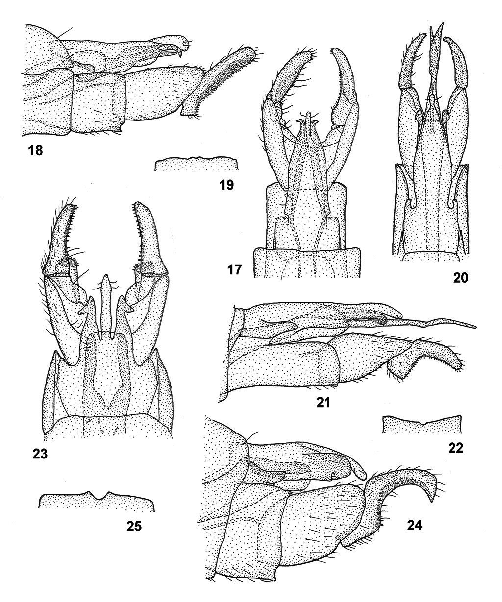
Figures 17–25. Hydrobiosella spp. male genitalia in dorsal, lateral and part ventral views; 17–19, Hydrobiosella mundagurra sp. nov.; 17, dorsal; 18, lateral; 19, ventral, mesodistal margin of segment IX; 20–22, Hydrobiosella bispina Kimmins.; 20, dorsal; 21, lateral; 22, ventral, mesodistal margin of segment IX; 23–25, Hydrobiosella arcuata Kimmins; 23, dorsal; 24, lateral; 25, ventral, mesodistal margin of segment IX.
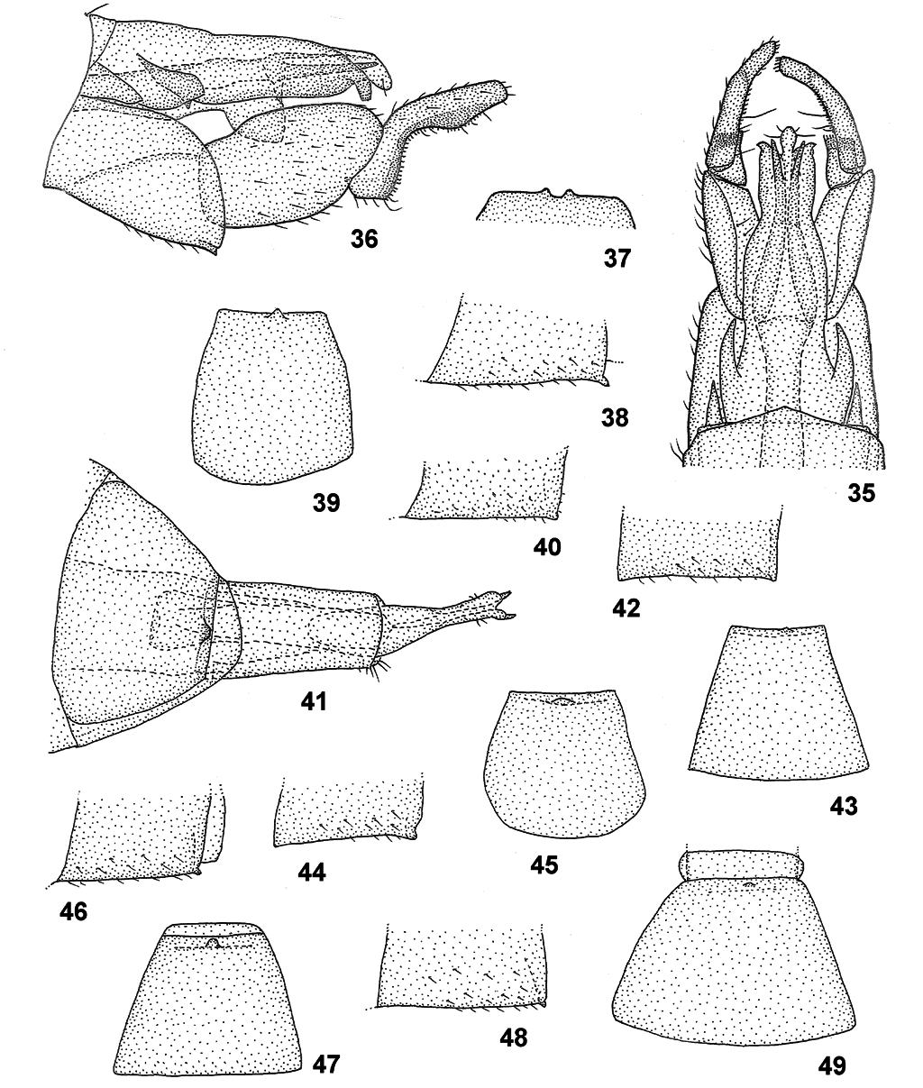
Figures 35–49. Hydrobiosella spp.; 35–37, Hydrobiosella yokunna sp. nov. male genitalia in dorsal, lateral and part ventral views; 35, dorsal; 36, lateral; 37, ventral, mesodistal margin of segment IX; 38–49, Hydrobiosella spp. female genitalia (part segment VIII) in lateral and (segment VIII) ventral view; 38–39, Hydrobiosella unispina sp. nov.; 38, lateral; 39, ventral; 40–41, Hydrobiosella mundagurra sp. nov.; 40, lateral; 41, female genitalia, ventral; 42–43, Hydrobiosella bispina Kimmins; 42, lateral; 43, ventral; 44–45, Hydrobiosella arcuata Kimmins; 44, lateral; 45, ventral; 46–47, Hydrobiosella woonoongoora sp. nov.; 46, lateral; 47, ventral; 48–49, Hydrobiosella thurawal sp. nov.; 48, lateral; 49, ventral.
| R |
Departamento de Geologia, Universidad de Chile |
| NMV |
Museum Victoria |
No known copyright restrictions apply. See Agosti, D., Egloff, W., 2009. Taxonomic information exchange and copyright: the Plazi approach. BMC Research Notes 2009, 2:53 for further explanation.
|
Kingdom |
|
|
Phylum |
|
|
Class |
|
|
Order |
|
|
Family |
|
|
Genus |