
Ponera, Latreille, 1804
|
publication ID |
https://doi.org/10.11646/zootaxa.4594.1.1 |
|
publication LSID |
lsid:zoobank.org:pub:4308D364-BCD7-473D-83BC-5B4130C32287 |
|
persistent identifier |
https://treatment.plazi.org/id/6E6AB616-6F7A-FF82-FF06-5FFDFA2ACC2A |
|
treatment provided by |
Plazi (2019-05-04 09:51:11, last updated 2024-11-26 07:16:12) |
|
scientific name |
Ponera |
| status |
|
Revised worker key to the East Asian species of Ponera View in CoL
The worker key to East Asian Ponera species is modified and updated based on the previous keys and species descriptions provided in Wheeler (1928a, b), Taylor (1967); Xu (2001a, b), Zhou (2001), Terayama (1986, 1996, 2009), Wang et al. (2009) and Yoshimura et al. (2009).
The key provides general directions for the identification of Ponera species in Eastern Asia. However, due to a potentially high number of undescribed species in the region, the readers should refer to specific species diagnoses to confirm their identification.
Head width (HW), which has been widely used for diagnosis in past studies, and which is positively correlated with body size (e.g. Taylor 1967, Terayama 1996), is an important identification aid. We plotted mean and range of HW for all the species included in this study ( Figure 5 View FIGURE 5 ). The simultaneous use of the key and figure 5 is suggested.
1a. In lateral view, petiolar node very thick with convex posterior margin; with upper portion of posterior margin bulging ( Fig. 6A View FIGURE 6 ).................................................................................................... 2
1b. In lateral view, petiolar node not as developed and presenting straight to convex posterior margin; with upper portion of poste- rior margin not bulging ( Fig. 6B, C View FIGURE 6 )...................................................................... 3
2a. Petiolar node in dorsal view with slightly convex anterior margin and slightly concave posterior margin ( Fig. 7A View FIGURE 7 ). Smaller species, HW: 0.54–0.60 mm. Body color dark.............................................. P. rishen Terayama, 2009
2b. Petiolar node in dorsal view with strongly convex anterior margin and strongly concave posterior margin ( Fig. 7B View FIGURE 7 ). Larger species, HW: 0.65–0.75 mm. Body color reddish brown.................................... P. takaminei Terayama, 1996
3a. Eye large, each consisting of 20 or more facets ( Fig. 8A View FIGURE 8 ). Larger species, HW: ca. 0.68 mm ..... P. kohmoku Terayama, 1996 3b. Eye small, each consisting of 10 or fewer facets ( Fig. 8B View FIGURE 8 ). Size variable.......................................... 4
4a. Masticatory margin of mandible with 5 subequal large teeth ( Fig. 9A View FIGURE 9 ). Smaller species, HW: 0.53–0.55 mm ............................................................................................... P. pentodontos Xu, 2001a
4b. Masticatory margin of mandible with 3 enlarged apical teeth followed by a series of small to indistinct denticles ( Fig. 9B View FIGURE 9 ). Size variable............................................................................................. 5 5a. Antennal scape, when laid backward, exceeding posterior margin of head in full-face view ( Fig. 10A View FIGURE 10 ).................. 6 5b. Antennal scape, when laid backward, not exceeding posterior margin of head in full-face view ( Fig. 10B View FIGURE 10 )............... 8 6a. Scape exceeding posterior border of head by nearly 34% of scape length............... P. hubeiensis Wang & Zhou, 2009 6b. Scape exceeding posterior border of head by <10% of scape length.............................................. 7 7a. Petiolar node in lateral view with straight posterior margin ( Fig. 11A View FIGURE 11 ). HW: 0.58–0.65 mm ....... P. alisana Terayama, 1986 View in CoL 7b. Petiolar node in lateral view with convex posterior margin ( Fig. 11B View FIGURE 11 ). HW: 0.61–0.70 mm ....... P. chapmani Taylor, 1967 View in CoL
8a. Petiolar node in dorsal view subrectangular or subtrapezoidal, anterior and lateral margins not forming a single arch ( Fig. 12A, B View FIGURE 12 : the dotted line not forming a continuously circular arc; lateral margins present), with slightly convex or almost straight anterior margin........................................................................................... 9
8b. Petiolar node in dorsal view semi-circular or oval, anterior and lateral margins constituting a single arch ( Fig. 12C, D View FIGURE 12 : the dotted line forming a continuously circular arc), with distinctly convex anterior margin................................ 12
9a. Dorsum of petiolar node in lateral view convex and relatively acute, with broadly convex posterior margin ( Fig. 13A View FIGURE 13 )............................................................................................. P. terayamai sp. n.
9b. Dorsum of petiolar node in lateral view slightly convex, blunt, with straight and sloping posterior margin ( Fig. 13B, C View FIGURE 13 ).... 10
10a. Subpetiolar process with large posteroventral teeth and large circular fenestra ( Fig. 13B View FIGURE 13 ). Larger species, HW: 0.39–0.43 mm. Body color dark brown......................................................... P. chiponensis Terayama, 1986 View in CoL
10b. Subpetiolar process with small or indistinct posteroventral teeth, and small circular fenestra ( Fig. 13C View FIGURE 13 ). Smaller species, HW: Ĺ 0.35 mm. Body color yellowish brown.................................................................. 11
11a. Petiolar node in dorsal view rectangular, broader than long, with slightly convex anterior margin ( Fig. 14A View FIGURE 14 ). Larger species, HW: ca. 0.33 mm ........................................................................ P. baka Xu, 2001a
11b. Petiolar node in dorsal view subquadrate, about as wide as long, with straight anterior margin ( Fig. 14B View FIGURE 14 ). Smaller species, HW: 0.27–0.28 mm .................................................................. P. shennong Terayama, 2009 12a. Subpetiolar process without developed posteroventral teeth ( Fig. 15A View FIGURE 15 ).......................................... 13 12b. Subpetiolar process with developed posteroventral teeth ( Fig. 15B View FIGURE 15 )............................................. 16
13a. Metanotal groove in lateral view indistinctly or weakly incised ( Fig. 16A View FIGURE 16 ). Petiolar node in lateral view anterior and posterior margins converging, not parallel......................................................................... 14
13b. Metanotal groove in lateral view deeply and broadly incised ( Fig. 16B View FIGURE 16 ). Petiolar node in lateral view with parallel anterior and posterior margins................................................................. P. nangongshan Xu, 2001a
14a. In full-face view, posterior margin of head distinctly concave ( Fig. 17A View FIGURE 17 ). Petiolar node in lateral view thick (LPeI: 63), with sloping and straight posterior margin. Smaller species, HW: ca. 0.35 mm. Body yellowish brown...... P. xantha Xu, 2001b
14b. In full-face view, posterior margin of head slightly concave or straight ( Fig. 17B, C View FIGURE 17 ). Petiolar node in lateral view relatively thin (LPeI: ca. 50), with almost vertical and slightly convex posterior margin. Larger species, HW ± 0.40 mm. Body brown to dark brown.......................................................................................... 15
15a. Petiolar node in dorsal view oval, with slightly convex posterior margin ( Fig. 18A View FIGURE 18 ). Larger species, HW: ca. 0.53 mm ........................................................................................... P. bawana Xu, 2001b
15b. Petiolar node in dorsal view arched, with straight posterior margin ( Fig. 18B View FIGURE 18 ). Smaller species, HW: ca. 0.40 mm ......................................................................................... P. yuhuang Terayama, 2009
16a. Length of 8 th antennal segment as long as 7 th antennal segment ( Fig. 19A View FIGURE 19 ) Metanotal groove indistinctly incised. Smaller spe- cies, HW: 0.30–0.31 mm ............................................................... P. szaboi Wilson, 1957 View in CoL
16b. Length of 8 th antennal segment ± 1.5 times length of 7 th antennal segment ( Fig. 19B View FIGURE 19 )............................... 17 17a. Metanotal groove in dorsal view indistinctly incised or almost obliterated ( Fig. 20A View FIGURE 20 )............................... 18 17b. Metanotal groove in dorsal view weakly incised ( Fig. 20B View FIGURE 20 )................................................... 22
18a. Petiolar node in dorsal view oval, with convex posterior margin ( Fig. 21A View FIGURE 21 ). Smaller species, HW: ca. 0.45 mm ............................................................................................... P. longlina Xu, 2001a
18b. Petiolar node in dorsal view not oval, with straight to slightly concave posterior margin ( Fig. 21B, C, D View FIGURE 21 ). Larger species, HW ± 0.49 mm .......................................................................................... 19 19a. Side of propodeum with weak to strong striae and hairs in lateral view ( Fig. 22A, B, C View FIGURE 22 ). Body color variable. Larger species, HW ± 0.55 mm ...................................................................................... 20
19b. Side of propodeum shining and smooth ( Fig. 22D View FIGURE 22 ). Body color reddish brown. Smaller species, HW: 0.49–0.52 mm ................................................................................................... P. wui sp. n.
20a. Lateral faces of metapleuron and propodeum weakly striate ( Fig. 22 View FIGURE 22 AC)......................................... 21
20b. Lateral faces of metapleuron and propodeum strongly striate ( Fig. 22B View FIGURE 22 )........................ P. menglana Xu, 2001a
21a. Apex of antennal scape, when laid backward, with a remaining distance to the posterolateral corner of ca. 10% of the scape length. Lateral faces of metapleuron and propodeum weakly striate ( Fig. 22C View FIGURE 22 )................ P. paedericera Zhou, 2001
21b. Apex of antennal scape, when laid backward, with a remaining distance to the posterolateral corner of Ĺ 5% of the scape length. Metapleuron and propodeum with relatively strong striae and abundant hairs in lateral view ( Fig. 22A View FIGURE 22 )............................................................................................. P. scabra Wheeler, 1928b View in CoL
22a. Anterodorsal corner of petiolar node in lateral view protruding forward ( Fig. 23A View FIGURE 23 )................. P. diodonta Xu, 2001b
22b. Anterodorsal corner of petiolar node in lateral view not protruding forward ( Fig. 23B, C, D View FIGURE 23 )......................... 23
23a. Apex of antennal scape, when laid backward, with a remaining distance to the posterolateral corner of Ĺ 5% of the scape length................................................................................................... 29
23b. Apex of antennal scape, when laid backward, with a remaining distance to the posterolateral corner of ± 10% of the scape length.............................................................................................. 24
24a. Dorsum of petiolar node in lateral view convex without forming distinct angle on posterodorsal corner; anterodorsal corner clearly much higher than posterodorsal corner ( Fig. 24 View FIGURE 24 AB).................................................... 25
24b. Dorsum of petiolar node in lateral view slightly convex without forming distinct angle on posterodorsal corner; anterodorsal corner slightly higher, or as high as posterodorsal corner ( Fig. 24 View FIGURE 24 CDEF)......................................... 26
25a. Third abdominal tergum distinctly longer than broad in dorsal view (ATI ± 105).............. P. bishamon Terayama, 1996 25b. Third abdominal tergum broader than long in dorsal view (ATI Ĺ 96).......................... P. pianmana Xu, 2001b 26a. Petiolar node in dorsal view relatively thicker ( Fig. 25A View FIGURE 25 )..................................................... 27 26b. Petiolar node in dorsal view relatively thin ( Fig. 25B View FIGURE 25 )........................................................ 28
27a. Petiolar node in dorsal view relatively thin ( Fig. 24E View FIGURE 24 ). Posterior margin of head slightly concave ( Fig. 26A View FIGURE 26 ). Clypeus without distinct median tooth............................................................... P. incerta (Wheeler, 1933) View in CoL
27b. Petiolar node in lateral view relatively thicker ( Fig. 24F View FIGURE 24 ). Posterior margin of head more concave ( Fig. 26B View FIGURE 26 ). Clypeus with distinct median tooth present (observation of the specimen using various angles needed)........ P. taiyangshen Terayama, 2009
28a. Posterior margin of head concave ( Fig. 27A View FIGURE 27 ). Anterior margin of petiolar node in dorsal view well convex ( Fig. 25C View FIGURE 25 ). Larger species, HW: 0.41–0.50 mm. Body color brown......................................... P. japonica Wheeler, 1906 View in CoL 28b. Posterior margin of head almost straight ( Fig. 27B View FIGURE 27 ). Anterior margin of petiolar node in dorsal view well convex ( Fig. 25D View FIGURE 25 ). Smaller species, HW: 0.32–0.34 mm. Body color yellowish brown......................... P. swezeyi (Wheeler, 1933) View in CoL
29a. Petiolar node in lateral view with strongly convex posterior margin ( Fig. 28A, B View FIGURE 28 ). Petiolar node in dorsal view with concave posterior margin, and large portion of the declivity clearly visible ( Fig. 29A, B View FIGURE 29 )................................... 30
29b. Petiolar node in lateral view with straight to slightly convex posterior margin ( Fig. 28C, D View FIGURE 28 ). Petiolar node in dorsal view with straight or slightly concave posterior margin, and small portion of the declivity visible ( Fig. 29C, D View FIGURE 29 )................... 31
30a. Petiolar node in lateral view with broadly convex posterior margin ( Fig. 28A View FIGURE 28 ). Petiolar node in dorsal view thin ( Fig. 29A View FIGURE 29 ). Body color dark brown............................................................ P. sinensis Wheeler, 1928a View in CoL
30b. Petiolar node in lateral view with convex posterior margin ( Fig. 28B View FIGURE 28 ). Petiolar node in dorsal view relatively thicker ( Fig. 29B View FIGURE 29 ). Body color lighter brown...................................................... P. tamon Terayama, 1996
31a. Posterodorsal corner of petiolar node forming a blunt angle in lateral view ( Fig. 30A View FIGURE 30 specimen collected in Sarawak, Malaysia; also see Taylor 1967, Fig. 71 View FIGURE 71 ). Subpetiolar process with small posteroventral teeth.............. P. oreas (Wheeler, 1933) View in CoL
31b. Posterodorsal corner of petiolar node convex in lateral view ( Fig. 30B View FIGURE 30 specimen collected in Vietnam; also see Zhou 2001, Fig. 74). Subpetiolar process with larger posteroventral teeth.................................. P. guangxiensis Zhou, 2001
Taylor, R. W. (1967) A monographic revision of the ant genus Ponera Latreille (Hymenoptera: Formicidae). Pacific Insects Monograph, 13, 1 - 112.
Terayama, M. (1986) Two new ants of the genus Ponera (Hymenoptera, Formicidae) from Taiwan. Kontyu, 54, 591 - 595.
Terayama, M. (1996) Taxonomic Studies on the Japanese Formicidae, Part 2 Seven Genera of Ponerinae, Cerapachyinae and Myrmicinae. Nature and Human Activities, 1, 9 - 32.
Terayama, M. (2009) A synopsis of the family Formicidae of Taiwan (Insecta, Hymenoptera). Research Bulletin of Kanto Gakuen University, Liberal Arts, 17, 81 - 266.
Wang, W., Shen, Z. K. & Zhao, Y. H. (2009) A taxonomic study on the family Formicidae from Hubei Province (Insecta: Hymenoptera: Formicidae). Huayu Nature Book Trade, China, 210 pp. [In Chinese]
Wilson, E. O. (1957) The tenuis and selenophora groups of the ant genus Ponera (Hymenoptera: Formicidae). Bulletin of the Museum of Comparative Zoology, 116, 355 - 386.
Yoshimura, M., Hosoishi, S., Kuboki, Y., Onoyama, K. & Ogata, K. (2009) New synonym and new Japanese record of the ant genus Ponera (Hymenoptera: Formicidae). Entomological Science, 12, 194 - 201. https: // doi. org / 10.1111 / j. 1479 - 8298.2009.00325. x
Zhou, S. Y. (2001) Ants of Guangxi. Guangxi Normal University Press, Guilin, China, 255 pp. [In Chinese]

FIGURE 5. Average head width of 32 Ponera species from the East Asia. Error bars are the range (Taylor 1967; Terayama 1986; Xu 2001a, b; Zhou 2001; Terayama 2004; Wong 2009; Yoshimura et al. 2009). Color of point refers to known distribution, extracted from antmaps.org, 2017. The zoogeographic definition refers to Holt et al. (2013).
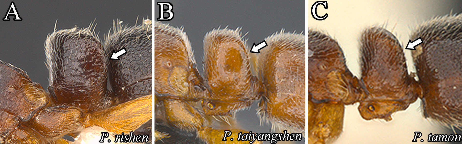
FIGURE 6. Lateral view of petiolar node (the arrow indicates the upper portion of posterior margin of petiolar node), A: P. rishen, B: P. taiyangshen, C: P. tamon.

FIGURE 7. Dorsal view of petiolar node (the arrow indicates the posterior margin of petiolar node), A: P. rishen, B: P. takaminei.

FIGURE 8. Lateral view of head (the arrow indicates the compound eye), A: P. kohmoku, B: P. alisana.
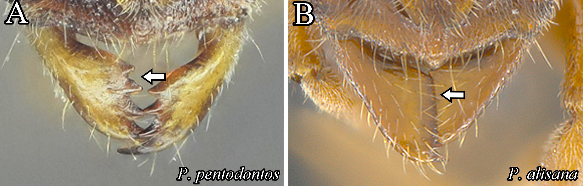
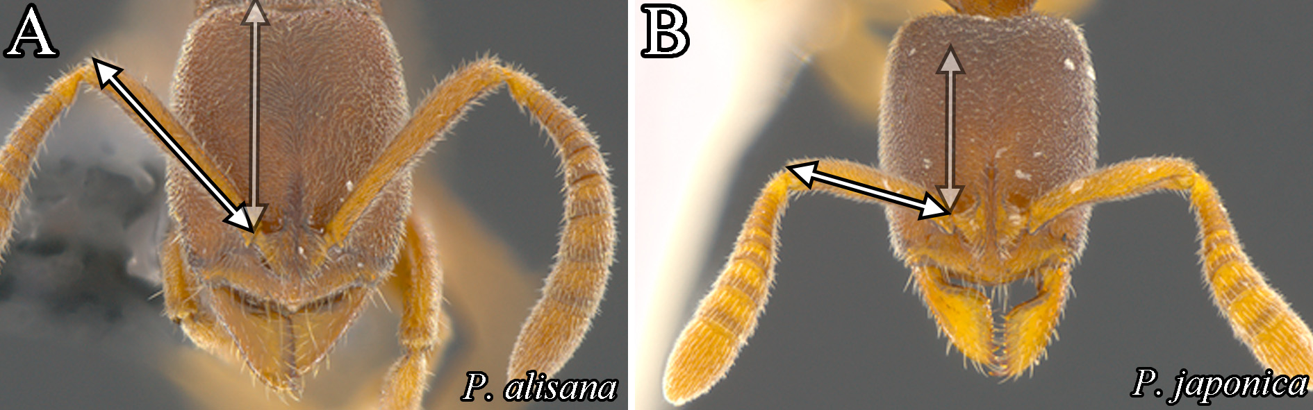
FIGURE 10. Full-face view of head (the line indicates the scape length), A: P. alisana, B: P. japonica.

FIGURE 11. Lateral view of petiole node (the arrow indicates the posterior margin of petiolar node), A: P. alisana, B: syntype of P. chapmani (MCZ-ENT00030919, © The Museum of Comparative Zoology).
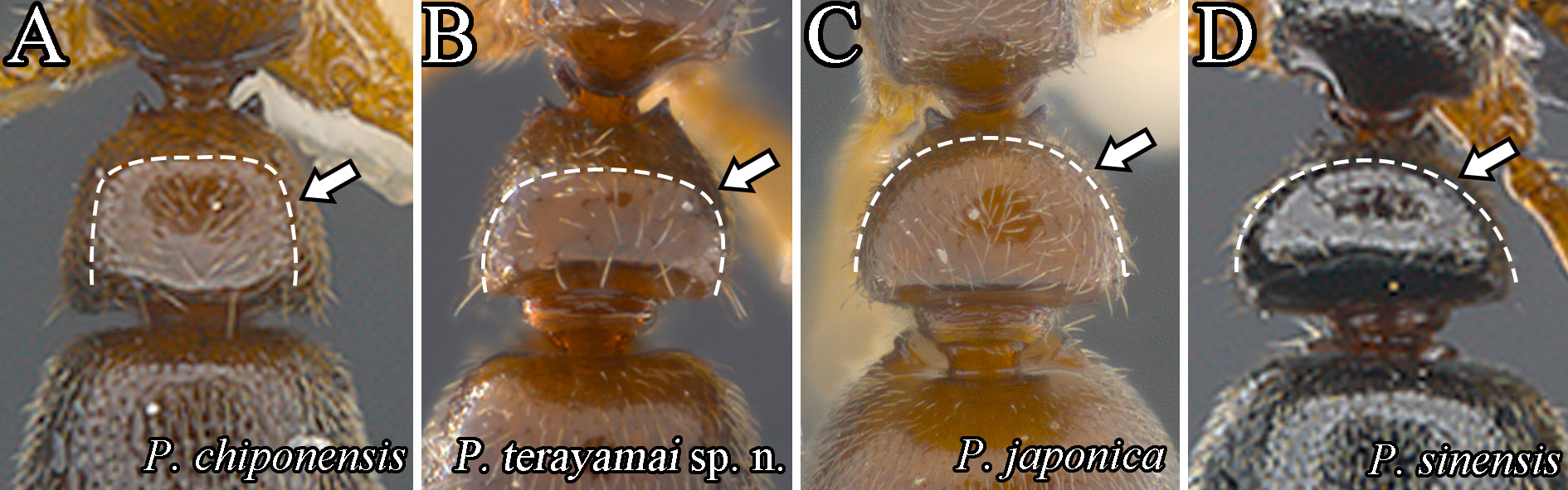
FIGURE 12. Dorsal view of petiole node (the dotted line and arrow indicate the anterior and lateral margins of petiolar node), A: P. chiponensis, B: P. terayamai sp. n., C: P. japonica, D: P. sinensis.

FIGURE 13. Lateral view of petiolar node (the arrow indicates the dorsal margin of petiolar node), A: P. terayamai sp. n., B: P. chiponensis, C: P. baka.
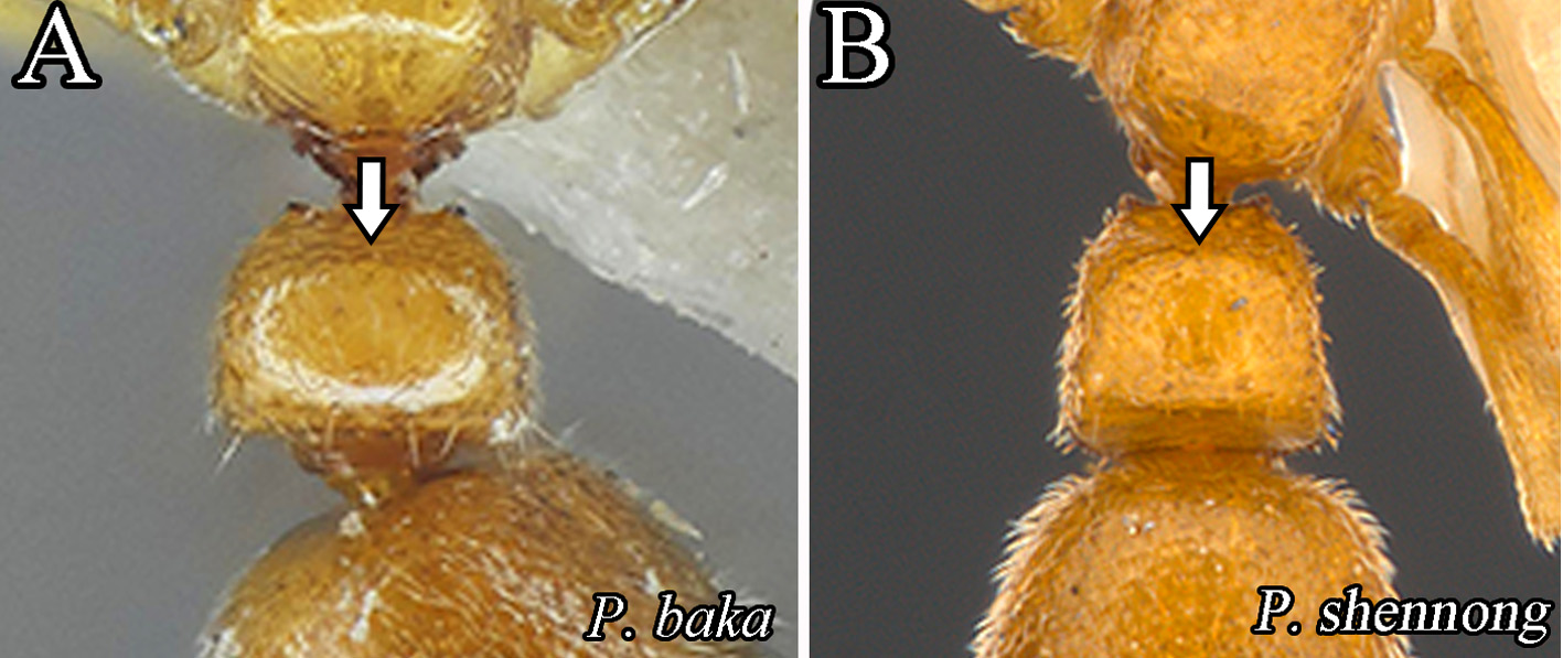
FIGURE 14. Dorsal view of petiolar node (the arrow indicates the anterior margin of petiolar node), A: P. baka, B: P. shennong.
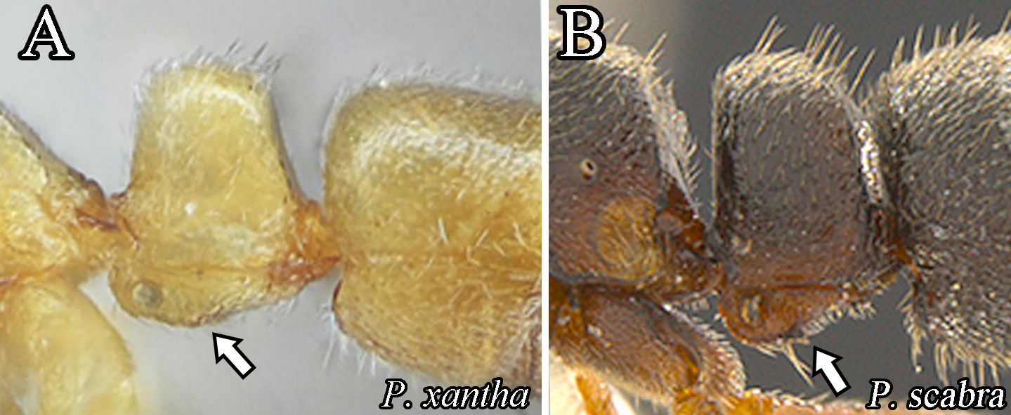
FIGURE 15. Lateral view of petiolar node (the arrow indicates the posterovental teeth in subpetiolar process), A: P. xantha, B: P. scabra.

FIGURE 16. Lateral view of mesosoma (the arrow indicates the metanotal groove), A: P. yuhuang, B: P. nangongshan.
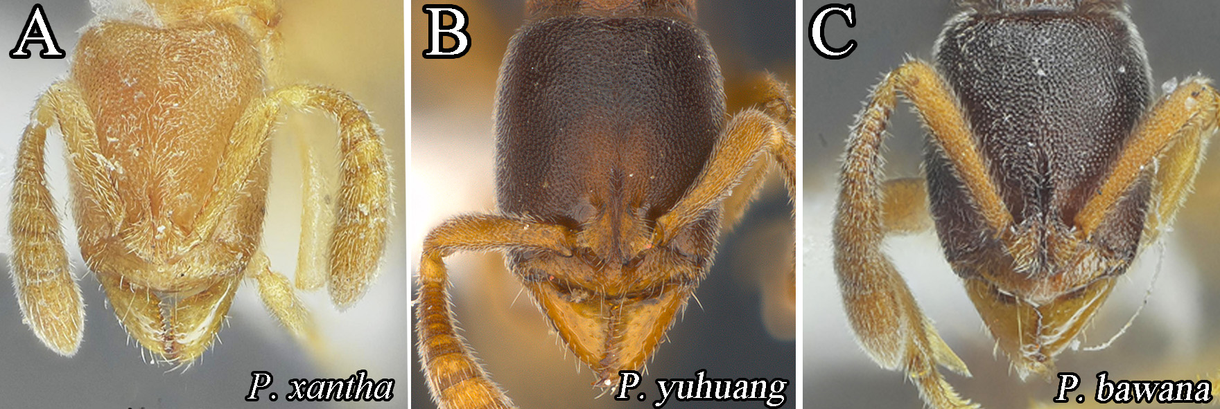
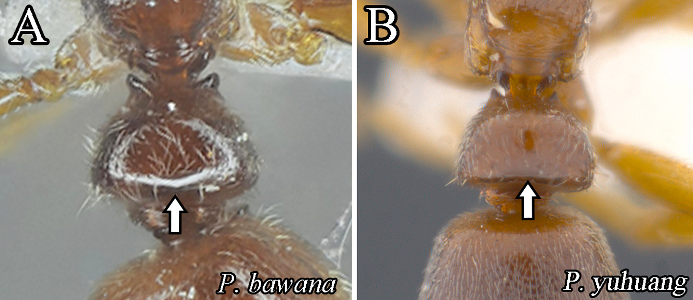
FIGURE 18. Dorsal view of petiolar node (the arrow indicates the posterior margin of petiolar node), A: P. bawana, B: P. yuhuang.
FIGURE 19. Full-face view of head (the arrow indicates the antennal segment), a: P. szaboi (CASENT0907297, Zach Lieberman, AntWeb, 2017), b: P. incerta (CASENT0178448, April Nobile, AntWeb, 2017).
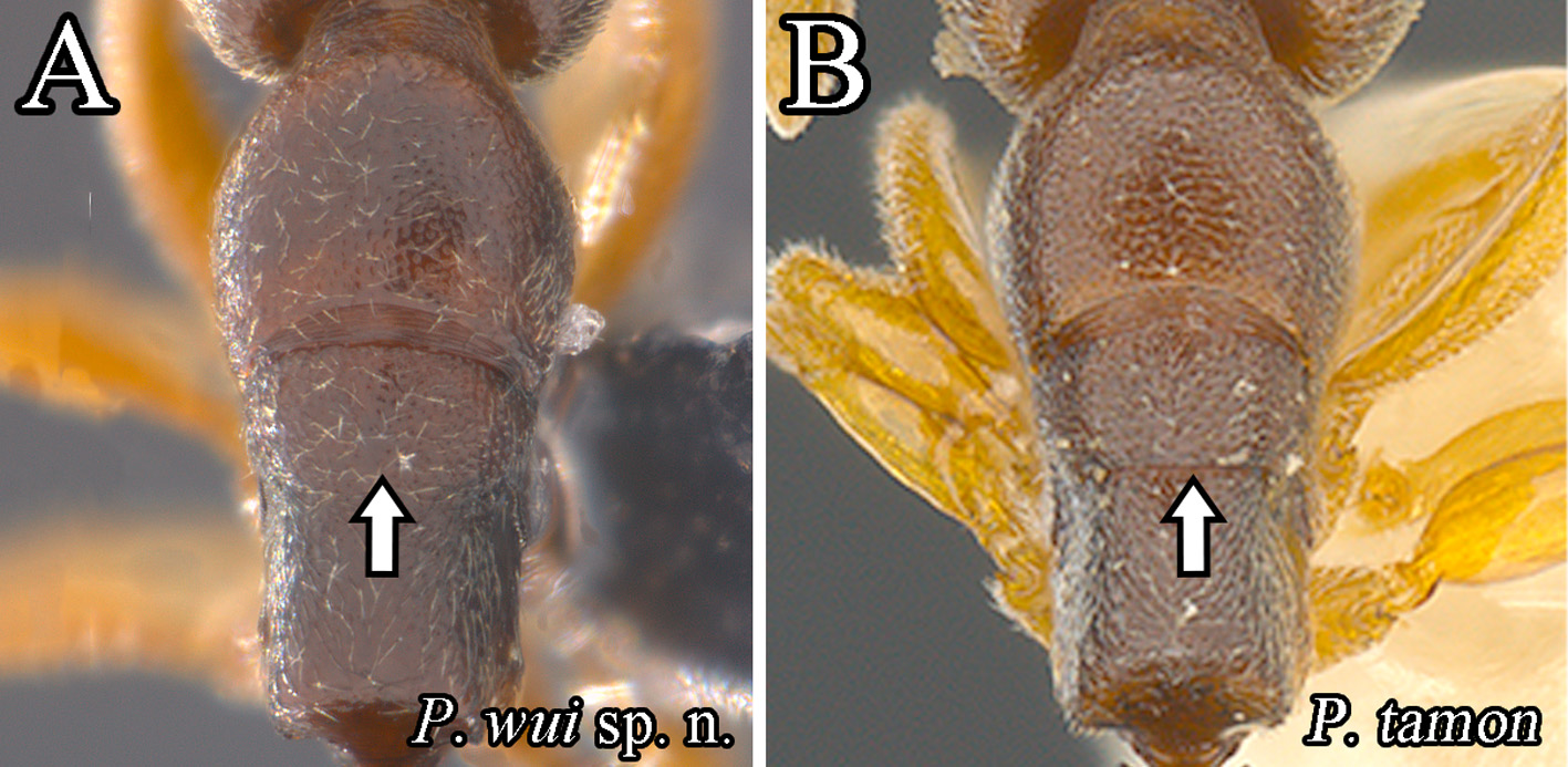
FIGURE 20. Dorsal view of mesosoma (the arrow indicates the metanotal groove), A: P. wui sp. n., B: P. tamon.
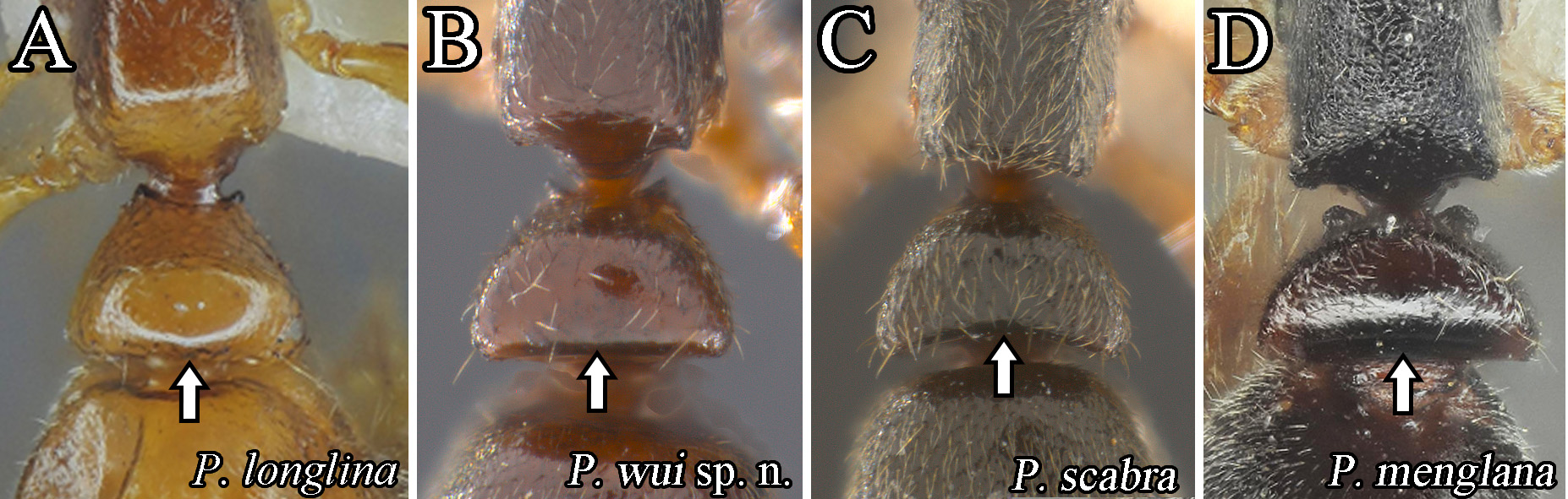
FIGURE 21. Dorsal view of petiolar node (the arrow indicates the posterior margin of petiolar node), A: P. longlina, B: P. wui sp. n., C: P. scabra, D: P. menglana.

FIGURE 22. Lateral view of propodeum (the arrow indicates the contained portion between metapleuron and propodeum), A: P. scabra, B: P. menglana, C: P. paedericera, D: P. wui sp. n.

FIGURE 23. Lateral view of petiole (the arrow indicates the anteriorodorsal corner of petiolar node), A: P. diodonta, B: P. incerta (CASENT0178448, April Nobile, AntWeb, 2017), C: P. oreas, D: P. pianmana.
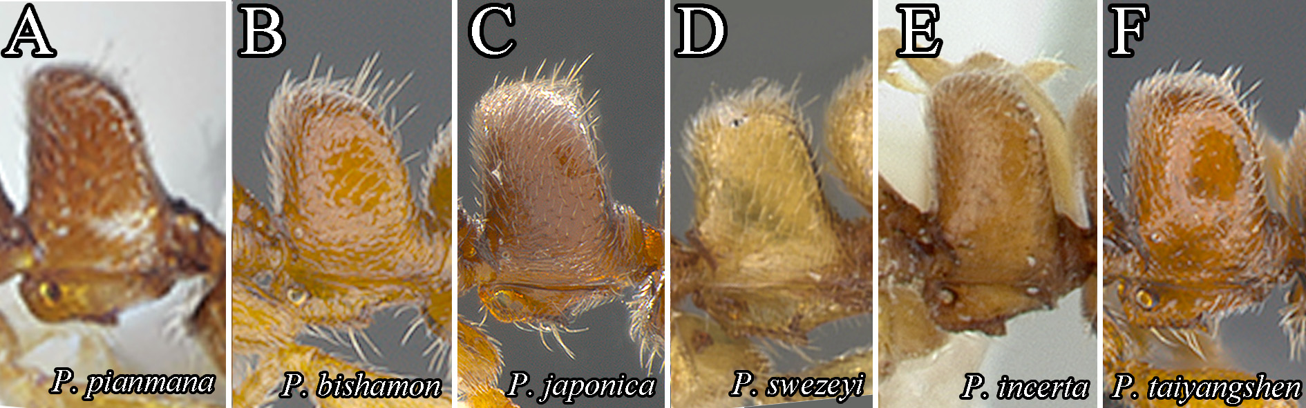
FIGURE 24. Lateral view of petiole, A: P. pianmana, B: P. bishamon, C: P. japonica, D: P. swezeyi (CASENT0900367, Will Ericson, AntWeb, 2017), E: P. incerta (CASENT0178448, April Nobile, AntWeb, 2017), F: P. taiyangshen.
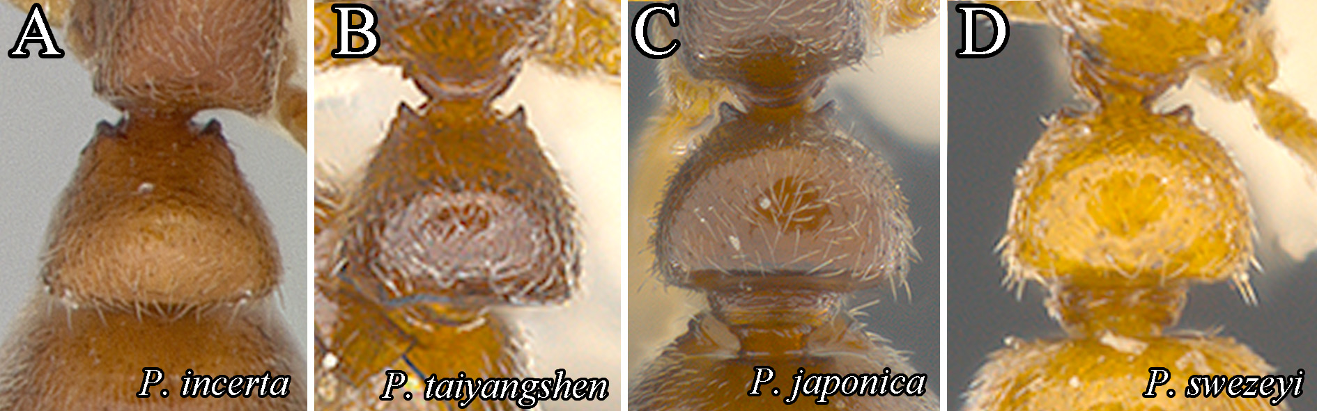
FIGURE 25. Dorsal view of petiole, A: P. incerta (CASENT0178448, April Nobile, AntWeb, 2017), B: P. taiyangshen, C: P. japonica, D: P. swezeyi.

FIGURE 26. Full-face view of head, A: P. incerta (CASENT0178448, April Nobile, AntWeb, 2017), B: P. taiyangshen.
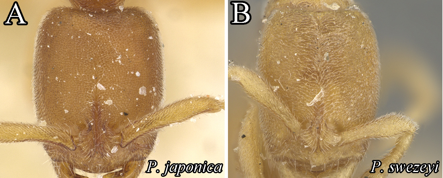
FIGURE 27. Full-face view of head, A: P. japonica (CASENT0915295, Zach Lieberman, AntWeb, 2017), B: P. swezeyi (MCZ-ENT00593416, Michele Esposito, AntWeb, 2017).

FIGURE 28. Lateral view of petiole (the arrow indicates the posterior margin of petiolar node), A: P. sinensis, B: paratype of P. tamon, C: syntype of P. oreas, D: paratype of P. guangxiensis.

FIGURE 29. Dorsal view of petiole (the dotted line indicates the posterior margin of petiolar node), A: P. sinensis, B: P. tamon, C: P. oreas, D: P. guangxiensis.
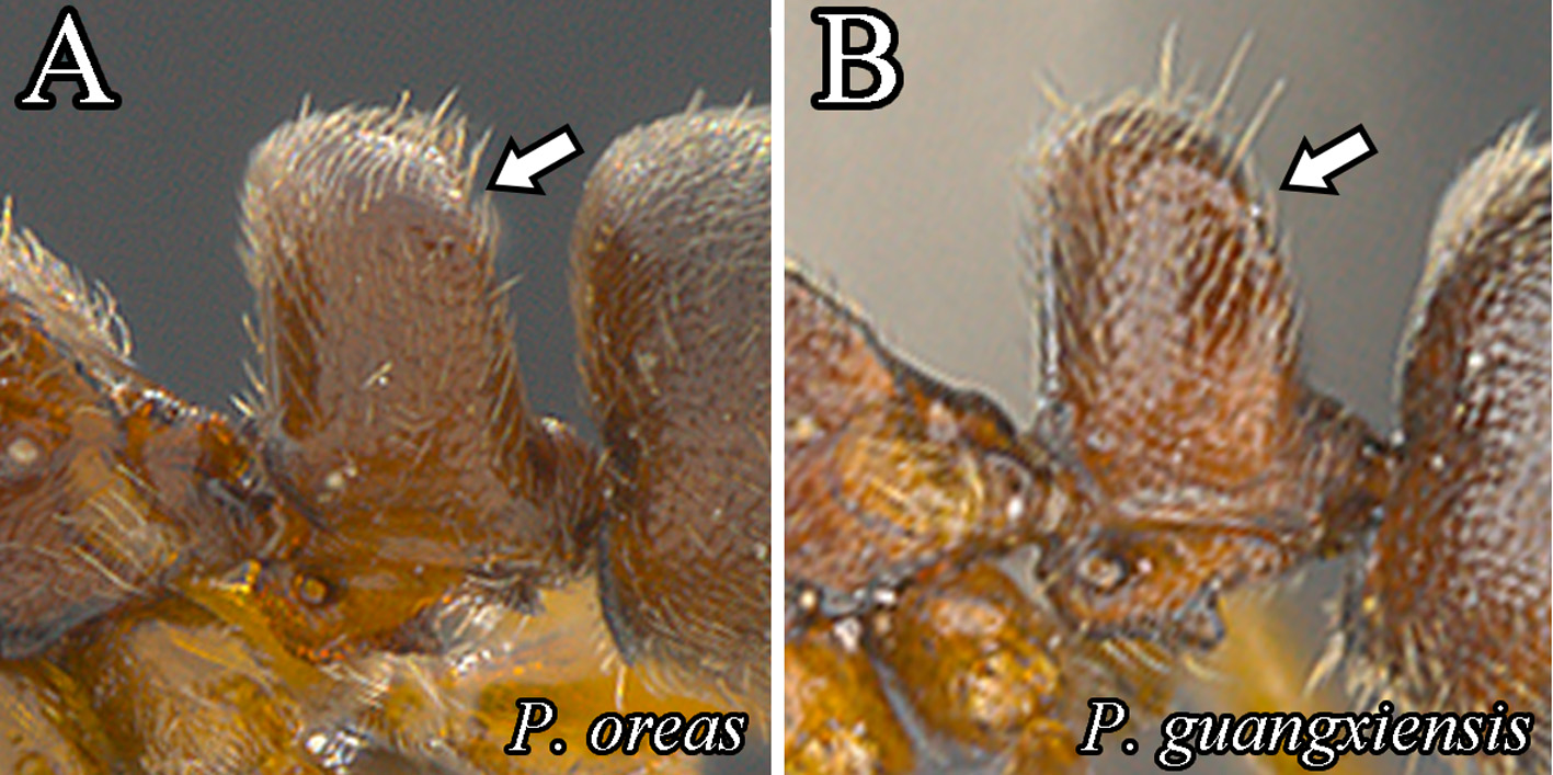
FIGURE 30. Lateral view of petiole, A: P. oreas collected in Sarawak, Malaysia, B: guangxiensis collected in Vietnam.
No known copyright restrictions apply. See Agosti, D., Egloff, W., 2009. Taxonomic information exchange and copyright: the Plazi approach. BMC Research Notes 2009, 2:53 for further explanation.
|
Kingdom |
|
|
Phylum |
|
|
Class |
|
|
Order |
|
|
Family |
1 (by plazi, 2019-05-04 09:51:11)
2 (by ImsDioSync, 2019-05-04 10:03:00)
3 (by plazi, 2019-08-29 11:40:11)
4 (by plazi, 2019-09-10 08:08:50)
5 (by plazi, 2019-09-10 10:31:41)
6 (by ExternalLinkService, 2019-09-25 22:42:36)
7 (by ExternalLinkService, 2021-11-09 13:31:12)
8 (by ExternalLinkService, 2021-11-09 23:24:26)
9 (by plazi, 2023-10-30 14:01:53)