
Plesioaxymyia, Sinclair, Bradley J., 2013
|
publication ID |
https://doi.org/ 10.11646/zootaxa.3682.1.7 |
|
publication LSID |
lsid:zoobank.org:pub:A652476E-EBB7-42EB-B5B6-5E391CA4318F |
|
DOI |
https://doi.org/10.5281/zenodo.6157764 |
|
persistent identifier |
https://treatment.plazi.org/id/5E1AE42D-0C55-FFDF-FF5A-FB72FCB7F8B8 |
|
treatment provided by |
Plazi |
|
scientific name |
Plesioaxymyia |
| status |
gen. nov. |
Plesioaxymyia View in CoL gen. nov.
(Figs 1–5)
Type-species: P. vespertina sp. nov.
Etymology. The genus is named in reference to its plesiomorphic position in comparison to Axymyia . Its gender is feminine.
Diagnosis. This is the only extant axymyiid genus with well developed mouthparts; R2+3 fork V-shaped; R2 distant from apex of R1; distal stem of M1+2 elongate; CuP very short, two-thirds length of cell bm.
Description. Head: Compound eyes broadly separated in female, each eye divided longitudinally into upper and lower hemispheres by fine groove starting at deep, broad, triangular incision at antennal bases, extending for more than 10 facets (Fig. 2); upper hemisphere smaller than lower hemisphere with facets uniform in size in both sections; facets with numerous ommatrichia; ocellar triangle elevated above eye with three prominent ocelli. Antenna with 14 moniliform flagellomeres, scape and pedicel with circlet of setulae. Mouthparts well-developed; clypeus prominent, strongly convex; palpus apparently 4-segmented (segment 1 possibly obscured), second segment broader than other segments; labellum developed, distinct. Thorax: Postpronotum depressed, triangular, separated by prominent prescutum; prescutal suture not prominent, very short, terminating at shallow, shiny rectangular depression. Scutum not distinctly swollen; transverse suture arising at base of wing, distinctly invaginated, terminating before midline of scutum; postsutural area of scutum shallowly concave medially. Scutellum rounded, strongly domed. Mediotergite twice length of scutellum, not as strongly prominent as scutellum. Prosternum large, separated by broad membrane. Anepisternum large, divided by anepisternal cleft into large anterior portion and narrow posterior portion; katepisternum prominent, overhanging mid coxa. Meron large, more than half as long as katepisternum. Legs: Fairly short, lacking spurs and long setae. Fore coxa shorter than katepisternum; mid and hind coxae half as long as fore coxa, entirely sclerotized medially; hind coxa broader than mid coxa. Hind tibia with posteroventral apical comb.
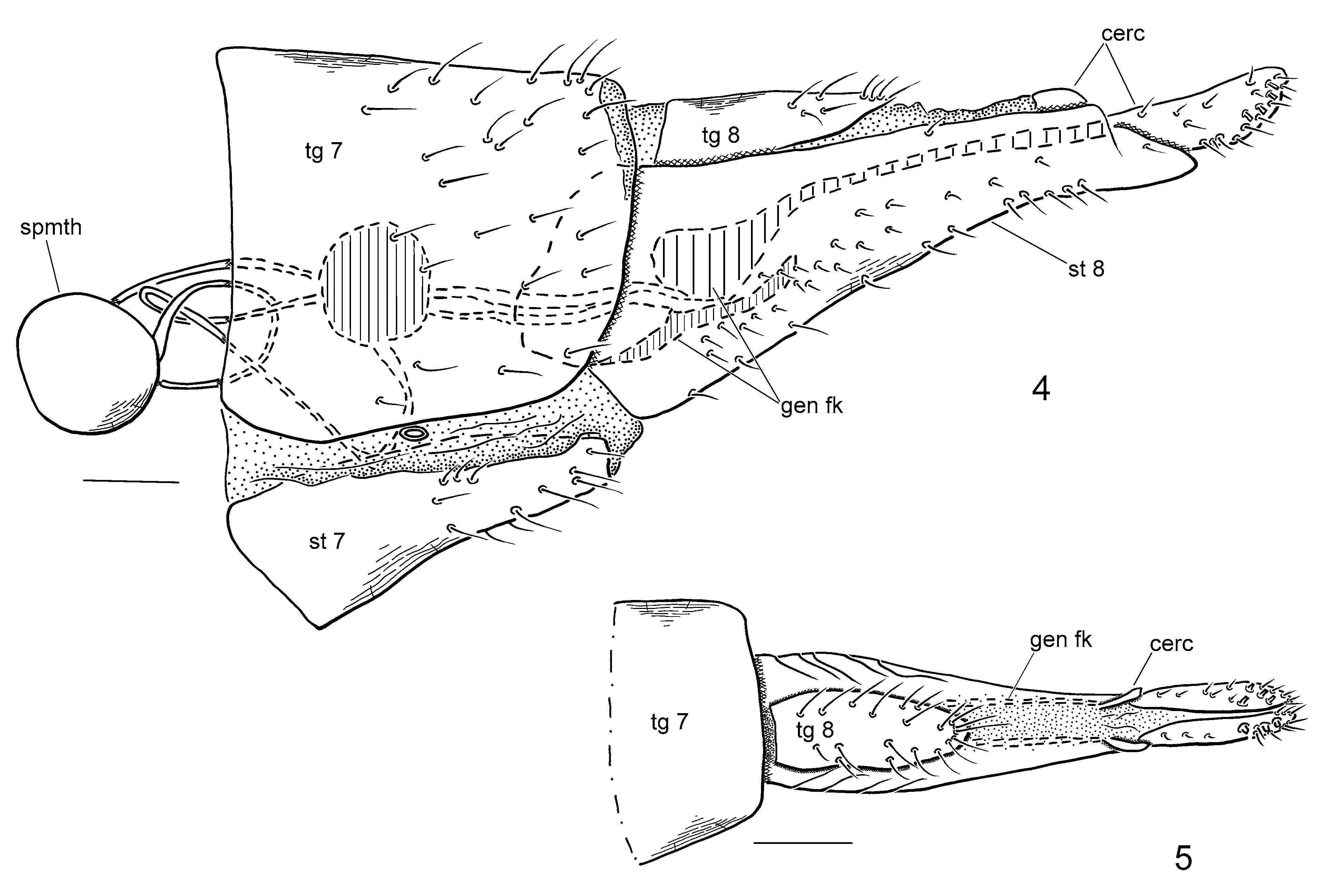
Mediolobus spatulate and elongate, more than half length of tarsomere 5, apex truncate; pulvillus shorter than claw. Wing: Costa greatly reduced slightly beyond apex of R4+5 (Fig. 3). Sc very short, extended to basal quarter of wing, strongly curved to costa proximal to vein Rs; sc-r reduced to faint streak. Rs with kink at mid-length; R2+3 branched slightly beyond apex of R1; R2 reaching costa well-separated from R1; R2+3 fork V-shaped. Crossvein r-m perpendicular to R4+5. Medial veins weaker than radial veins, distinctly pale in comparison; M1+2 with long distal stem, branching proximal to R2+3 fork; M4 straight. Stem of M vein with apex distinct, basal portion reduced to faint crease, indistinctly separating basal cells. CuP very short, extended to half length of cell bm. Alula greatly reduced; anal lobe well-developed. Halter with very long stem, lacking cluster of setae. Abdomen: Sparsely setose. Segments 1–7 each with spiracles in membranous pleural region. Tergites considerably broader than sternites; tergite 8 one-third length of sternite, undivided; sternite 8 greatly lengthened, slender, tapered apically, clothed in short setae. Genital fork (sternite 9) with V-shaped ventral portion, dorsal portion prolonged posteriorly as pair of slender rod-like extensions to apex of sternite 8 ( Fig. 4 View FIGURES 4 – 5 ). Spermathecae spherical, paired, well-sclerotized; spermathecal ducts paired, separated throughout, slightly inflated distally to genital fork; duct broad exiting spermatheca. Cercus two-segmented; basal segment very small reduced, slender, flap-like, surrounding anus; apical segment longer than wide, bearing several rod-like sensilla (Figs 4,5). Male and immatures: Unknown.
Remarks. Plesioaxymyia will key with some difficulty to Protaxymyia in the key to genera in Zhang (2010). The new genus clearly differs from the latter genus in the shape of R2, shortened Sc and CuP, and the very long distal stem of M1+2.
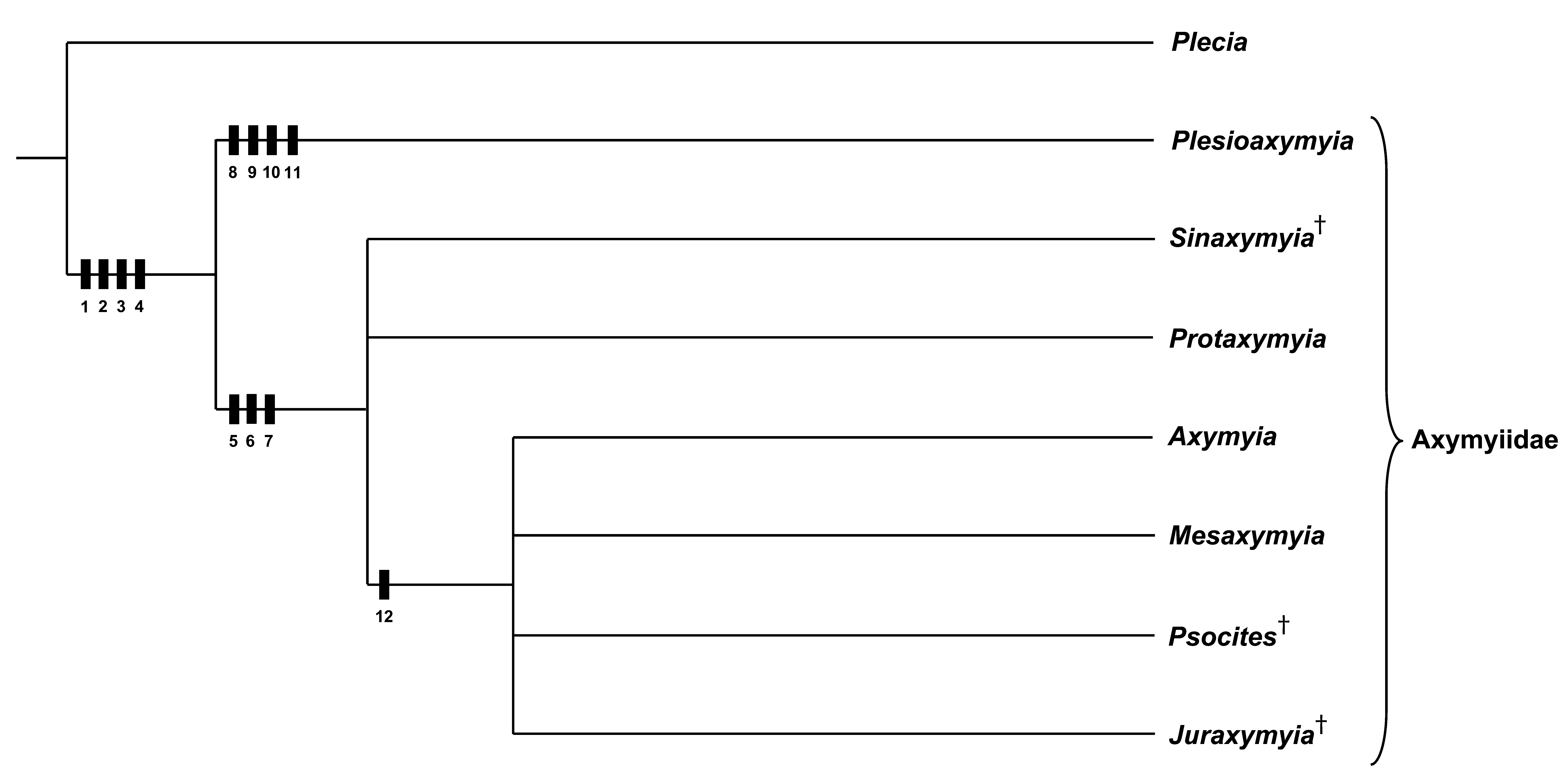
A preliminary phylogenetic assessment of the infrafamilial relationships of Axymyiidae is summarized in Figure 6 View FIGURE 6 and characters listed and described in Table 1. The Bibionidae was used to polarize the characters. The assignment of Plesioaxymyia to the family Axymyiidae is supported on the basis of the four family-level synapomorphies: female cercus with rod-like sensilla (1); vein Rs with kink at mid-length (2); compound eye divided longitudinally (3); and prescutal suture ending at an elongate depression (shiny spot in Axymyia ) (4). All axymyiid genera, exclusive of Plesioaxymyia form a clade, supported by the reduction of the mouthparts (5), presence of a cluster of setae on the stem of the halter (6) (although both characters scored as unknown in the fossil genera) and R2 directed towards R1 (7). The monophyly of Plesioaxymyia is supported on the basis of four synapomorphies: shortened Sc (8), elongate distal stem of M1+2 (longer than M2) (9), greatly weakened M vein (10), and shortened CuP (anal vein), two-thirds length of cell bm (11). Four genera are apparently most closely related ( Fig. 6 View FIGURE 6 ) on the basis of the closely positioned apex of veins R1 and R2 (12). The three fossil genera (Juraxymyia Zhang, Psocites Hong and Sinaxymyia Zhang) clearly belong to the sister group of Plesioaxymyia and provides strong evidence that this genus has its roots in the Jurassic or earlier.
The shape and size of the female ovipositor (e.g., length and shape of sternite 8 and cercus) requires further evaluation. The apical segment of the cercus is longer than wide in Plesioaxymyia and sternite 8 is greatly lengthened in Plesioaxymyia and Protaxymyia japonica (Ishida) .
1. Female cercus: lacking rod-like sensilla (0); rod-like sensilla present (1). 2. Vein R s: straight (0); with kink (1).
3. Compound eye: undivided longitudinally (0); divided longitudinally (1). 4. Prescutal suture: unmodified (0); ending at elongate depression (1). 5. Mouthparts: unmodified (0); shortened and reduced (1).
6. Stem of halter: bare (0); with cluster of setae (1).
7. R2 orientation: directly apically (0); directed towards apex of R1 (1). 8. Sc vein: elongate, extending to beyond Rs (0); short, not reaching Rs (1). 9. Stem of M1+2: short, less than half length of M2 (0); elongate nearly subequal in length to M2 (1). 10. M vein: well defined (0); greatly weakened (1).
11. CuP length: elongate, at least three-quarters length of cell bm (0); short, two-thirds length of cell bm (1). 12. R1 and R2: separated by length of R2 (0); nearly touching (1).
No known copyright restrictions apply. See Agosti, D., Egloff, W., 2009. Taxonomic information exchange and copyright: the Plazi approach. BMC Research Notes 2009, 2:53 for further explanation.