
Liogluta
|
publication ID |
https://doi.org/10.11646/zootaxa.4193.2.5 |
|
publication LSID |
lsid:zoobank.org:pub:87F6E94A-67BB-4A9B-8EED-1C7B20E6E9D3 |
|
DOI |
https://doi.org/10.5281/zenodo.6085382 |
|
persistent identifier |
https://treatment.plazi.org/id/5667352A-E85B-FFD4-609B-FD33FA32FA11 |
|
treatment provided by |
Plazi (2016-11-16 07:38:57, last updated 2024-11-27 02:39:19) |
|
scientific name |
Liogluta |
| status |
|
Key to adults of species of Korean Liogluta View in CoL
1. Body fusiform ( Fig. 1 View FIGURE 1 H)...................................................................... Liogluta ursi View in CoL
- Body subparallel to parallel-sided ( Figs. 1 View FIGURE 1 A–G)............................................................ 2
2. Body yellowish to reddish brown (1B–D, G)............................................................... 3
- Body dark brown to black (1A, E–F)..................................................................... 6
3. Eyes small, shorter than temples.................................................................... L. infans View in CoL
- Eyes not small, about as long as or longer than temples........................................................ 4
4. Body length more than 3.5 mm; antennomere 4 about as long as wide; pronotum with impression in median region................................................................................................. .. L. granigera View in CoL
- Body length less than 3.5 mm; antennomere 4 slightly transverse ( Figs. 4 View FIGURE 4 A, 8A); pronotum without impression in median region.............................................................................................. 5
5. Body length more than 2.5 mm; labium with ligula divided into 2 lobes at base; posterior margin of male tergite VIII with slightly crenate process in median region ( Fig. 4 View FIGURE 4 B).................................................... L. distans View in CoL
- Body length less than 2.5 mm; labium with ligula divided into 2 lobes in basal half; posterior margin of male tergite VIII with denticulate process in median region ( Fig. 8 View FIGURE 8 B)............................................... L. rufescens View in CoL sp. nov.
6. Body relatively broad and more sclerotized ( Fig. 1 View FIGURE 1 E); abdominal tergite III with one pair of anterior macrosetae; posterior mar- gin of male tergite VIII not modified ( Fig. 6 View FIGURE 6 B)................................................. L. jinilli View in CoL sp. nov.
- Body relatively elongate and less sclerotized ( Figs. 1 View FIGURE 1 A–F); abdominal tergite III with two pairs of anterior macrosetae; poste- rior margin of male tergite VIII modified ( Figs. 3 View FIGURE 3 B, 7A)....................................................... 7
7. Body length less than 3.0 mm; antennomere 4–5 slightly transverse ( Fig. 3 View FIGURE 3 A); abdominal tergite III with one pair of posterior macrosetae......................................................................... L. changwhani View in CoL sp. nov.
- Body length more than 3.0 mm; antennomere 4–5 about as long as wide ( Fig. 7 View FIGURE 7 A); abdominal tergite III with three pairs of posterior macrosetae........................................................................ L. pyonganica View in CoL

FIGURE 1. Habitus of Liogluta. A, L. changwhani sp. nov.; B, L. distans; C, L. granigera; D, L. infans. E, L. jinilli sp. nov.; F, L. pyonganica; G, L. rufescens sp. nov.; H, L. ursi.
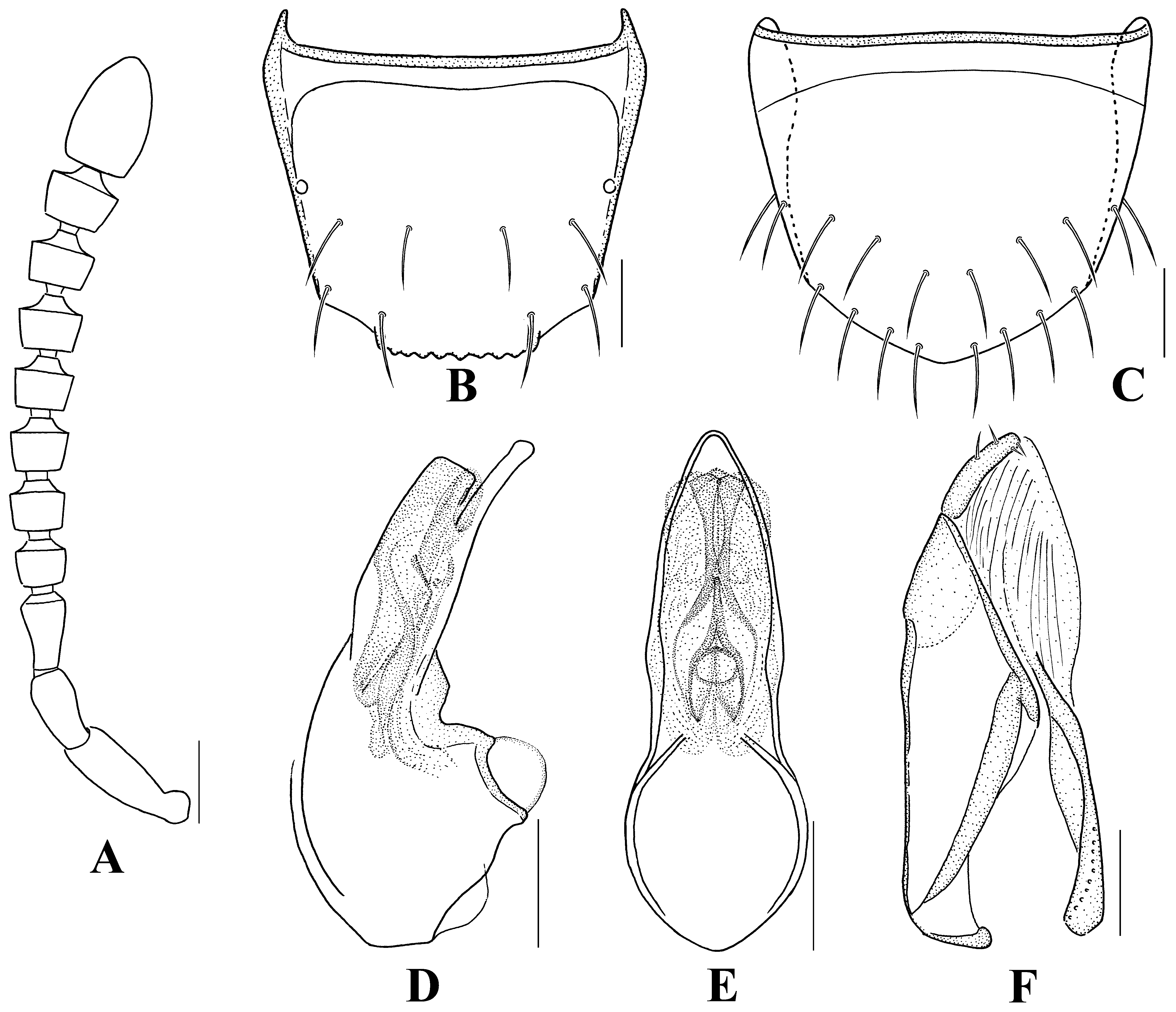
FIGURE 3. Liogluta changwhani sp. nov. A, antenna; B, male abdominal tergite VIII (dorsal aspect); C, male abdominal sternite VIII (ventral aspect); D, female abdominal tergite VIII (dorsal aspect); E, female abdominal sternite VIII (ventral aspect); F, median lobe of aedeagus (lateral aspect); G, median lobe of aedeagus (ventral aspect); H, paramere (lateral aspect); I: spermatheca. Scales = 0.1 mm.
FIGURE 4. Liogluta distans. A, antenna; B, male abdominal tergite VIII (dorsal aspect); C, male abdominal sternite VIII (ventral aspect); D, female abdominal tergite VIII (dorsal aspect); E, female abdominal sternite VIII (ventral aspect); F, median lobe of aedeagus (lateral aspect); G, median lobe of aedeagus (ventral aspect); H, paramere (lateral aspect). Scales = 0.1 mm.
FIGURE 6. Liogluta jinilli sp. nov. A, antenna; B, male abdominal tergite VIII (dorsal aspect); C, male abdominal sternite VIII (ventral aspect); D, female abdominal tergite VIII (dorsal aspect); E, female abdominal sternite VIII (ventral aspect); F, median lobe of aedeagus (lateral aspect); G, median lobe of aedeagus (ventral aspect); H, paramere (lateral aspect); I: spermatheca. Scales = 0.1 mm.
FIGURE 7. Liogluta pyonganica. A, antenna; B, male abdominal tergite VIII (dorsal aspect); C, male abdominal sternite VIII (ventral aspect); D, female abdominal tergite VIII (dorsal aspect); E, female abdominal sternite VIII (ventral aspect); F, median lobe of aedeagus (lateral aspect); G, median lobe of aedeagus (ventral aspect); H, paramere (lateral aspect); I: spermatheca. Scales = 0.1 mm.
No known copyright restrictions apply. See Agosti, D., Egloff, W., 2009. Taxonomic information exchange and copyright: the Plazi approach. BMC Research Notes 2009, 2:53 for further explanation.
|
Kingdom |
|
|
Phylum |
|
|
Class |
|
|
Order |
|
|
Family |
|
|
SubFamily |
Aleocharinae |
1 (by plazi, 2016-11-16 07:38:57)
2 (by ImsDioSync, 2016-11-16 07:42:40)
3 (by ImsDioSync, 2017-01-24 22:32:36)
4 (by ImsDioSync, 2017-02-08 05:42:14)
5 (by ExternalLinkService, 2019-09-26 07:49:30)
6 (by ExternalLinkService, 2022-01-30 00:20:03)
7 (by ExternalLinkService, 2022-02-15 04:10:55)
8 (by plazi, 2023-10-27 08:00:51)