
Cephalosphaera, Enderlein, 1936
|
publication ID |
https://doi.org/10.11646/zootaxa.5178.4.1 |
|
publication LSID |
lsid:zoobank.org:pub:6E706C39-8F42-4050-8792-0423F4267D2B |
|
DOI |
https://doi.org/10.5281/zenodo.7043650 |
|
persistent identifier |
https://treatment.plazi.org/id/41113E13-FF9B-FFC1-FF12-93624BC4B6C9 |
|
treatment provided by |
Plazi (2022-08-29 10:28:58, last updated 2024-11-26 06:12:37) |
|
scientific name |
Cephalosphaera |
| status |
|
Key to males of the Neotropical species of Cephalosphaera View in CoL View at ENA and Neocephalosphaera
This key is a result of the modification of those by Rafael (1992) and Souza & Ale-Rocha (2009). The initial for genera were adapted from Skevington & Yeates (2001) and Rafael & Skevington (2010).
1 Hind tibia with a comb of longer setae apically; male abdominal syntergosternite 8 with the membranous area not reaching epandrium ( Fig. 8 View FIGURES 1–13 )...................................................... Cephalosphaera Enderlein, 1936 View in CoL ... 2
- Hind tibia without comb of longer setae apically; male abdominal syntergosternite 8 with the membranous area reaching epandrium and almost reaching tergite 5 ( Figs 19, 21 View FIGURES 14–26 , 32, 34 View FIGURES 27–39 , 48, 50 View FIGURES 43–55 , 64, 66 View FIGURES 59–71 , 77, 79 View FIGURES 72–84 , 90, 92 View FIGURES 85–97 , 103, 105 View FIGURES 98–110 , 118, 120 View FIGURES 114–125 )................................................................................. Neocephalosphaera De Meyer, 1994 View in CoL . 13
2 All femora predominantly yellow, sometimes with light brown to brown spots apically or dorsally..................... 3
- All femora predominantly brown to black, sometimes with bases and apices yellow................................ 9
3 All femora completely yellow, apex of phallic guide with a small spine laterally [see figure 3F in Souza & Ale-Rocha (2009)] or with a rigid lobe laterally [see figure 3N, presented by Souza & Ale-Rocha (2009)]............................... 4
- All femora yellow, with bases and apices or at least in the middle of the femur with a small spotlight brown to brown dorsoapically; apex of phallic guide with a strong lobe apically [see figure 3H in Souza & Ale-Rocha (2009)] or a row or tuft of strong spines ventroapically [see figures 3M, 4D, in Souza & Ale-Rocha (2009)] or a tuft of fine setae ventrally [ Fig. 12 View FIGURES 1–13 and see figure 3O in Souza & Ale-Rocha (2009)]........................................................................ 5
4 Both surstyli with tips acute inward directed, with inner margins straight and outer margins curved, with short and fine setae marginally [see figure 2F in Souza & Ale-Rocha (2009)]; apex of phallic guide with acute lobe apically downward directed and one short spine laterally (see figure 3F in Souza & Ale-Rocha (2009)]; phallus trifid with two lateral projections apically, giving the false impression of being five ejaculatory ducts [see figure 3F in Souza & Ale-Rocha (2009)]........................................................................................ C. guanacastensis Rafael & Menezes, 1999 View in CoL
- Both surstyli with subrounded tips, slightly downwards directed, with inner and outer margins curved, without marginal setae [see figure 2N in Souza & Ale-Rocha (2009)]; apex of phallic guide with a rigid and acute lobe laterally, giving the false impression of being bifid [see figure 3N in Souza & Ale-Rocha (2009)]; phallus trifid, with one long projection dorsally, forward directed and ejaculatory ducts slightly coiled [see figure 3N in Souza & Ale-Rocha (2009)]... C. panamensis ( Hardy, 1948)
5 Surstyli asymmetrical [see figure 2M in Souza & Ale-Rocha (2009)]; apex of phallic guide with a tuft of strong and short spine apically (see figure 3M in Souza & Ale-Rocha (2009)]............................. C. pacaraima Rafael & Rosa, 1991 View in CoL
- Surstyli subsymmetrical [ Fig. 9 View FIGURES 1–13 and see figures 2H, 2O, 4C in Souza & Ale-Rocha (2009)]; apex of phallic guide with a strong lobe apically [see figure 3H in Souza & Ale-Rocha (2009)] or with a row of strong and long spines ventroapically [see figure 4D in Souza & Ale-Rocha (2009)] or with a tuft of short and fine setae ventroapically ( Fig. 12 View FIGURES 1–13 and see figure 3O in Souza & Ale-Rocha (2009)].................................................................................... 6
6 Wing with membrane hyaline; phallus long (see figures 3O, 4.D in Souza & Ale-Rocha (2009)]....................... 7
- Wing with membrane faintly light brown to brown infuscated; phallus short [ Figs 12 View FIGURES 1–13 , see figure 3H in Souza & Ale-Rocha (2009)]............................................................................................. 8
7 Surstyli about equal in length to epandrium, both surstyli equal in length [see figure 4C in Souza & Ale-Rocha (2009)]; apex of phallic guide with a row of strong and long spines ventroapically (see figure 4D in Souza & Ale-Rocha (2009)]; phallus with ejaculatory ducts stout, with distal 1/3 distinctly striated (see figure 4D in Souza & Ale-Rocha (2009)].......................................................................................... C. aurata Souza & Ale-Rocha, 2009
- Surstyli distinctly shorter than the length of epandrium, left surstylus shorter than right (see figure 2O in Souza & Ale-Rocha (2009)]; apex of phallic guide with a tuft of fine setae ventroapically (see figure 3O in Souza & Ale-Rocha (2009)]; phallus with ejaculatory ducts thin, with tips slightly coiled (see figure 3O in Souza & Ale-Rocha (2009)]................................................................................................. C. procera Rafael & Menezes, 1999 View in CoL
8 Postpronotal lobe brown ( Fig. 1 View FIGURES 1–13 ) both surstyli stout, thickened basally and medially, thin apically, with inner and outer margins sinuous ( Fig. 9 View FIGURES 1–13 ); apex of phallic guide with a tuft of fine setae ventrally ( Fig. 12 View FIGURES 1–13 ); phallus somewhat triangle-shaped in lateral view, with ejaculatory ducts slightly coiled ( Fig. 12 View FIGURES 1–13 ).................................... C. munchiquensis View in CoL sp. nov.
- Postpronotal lobe yellow; both surstyli slender, thickened basally, thin medially and apically, inner and outer margins slightly curved [see figure 2H in Souza & Ale-Rocha (2009)]; apex of phallic guide with strong lobe apically [see figure 3H in Souza & Ale-Rocha (2009)]; phallus stem-shaped, with ejaculatory ducts simple, not coiled, backwards directed [see figure 3H in Souza & Ale-Rocha (2009)]............................................................... C. insularis Rafael, 1996 View in CoL
9 Section between cell dm and vein M 2 equal than vein dm-m ( Fig. 102 View FIGURES 98–110 ); all femora entirely black; both surstyli with apical sinus on outer margins, forming acute tips, right surstylus shorter than left [see figure 2G in Souza & Ale-Rocha (2009)]; phallus thin [see figure 3G in Souza & Ale-Rocha (2009)]......................................... C. incomitata (Hardy, 1965) View in CoL
- Section between cell dm and vein M 2 greater than vein dm-m ( Figs 5 View FIGURES 1–13 , 18 View FIGURES 14–26 , 31 View FIGURES 27–39 , 47 View FIGURES 43–55 , 63 View FIGURES 59–71 , 76 View FIGURES 72–84 , 89 View FIGURES 85–97 ); all femora brown to black, with bases and apices yellow; both surstyli without apical sinus on outer margins, not forming acute tips, both surstyli equal in length [see figures 2E, 2J, 2L, 2Q in Souza & Ale-Rocha (2009)]; phallus thickened [see figures 3E, 3J, 3L, 3R in Souza & Ale-Rocha (2009)]............................................................................................ 10
10 Mesopleuron and mediotergite brown, brown pruinose; surstyli asymmetrical [see figure 2E in Souza & Ale-Rocha (2009)]; apex of phallic guide triangular shaped [see figure 3E in Souza & Ale-Rocha (2009)]; phallus stouter apically, one ejaculatory duct with two small protuberances dorsally [see figure 3E in Souza & Ale-Rocha (2009)]........ C. fairchildi Rafael, 1992 View in CoL
- Mesopleuron and mediotergite dark brown to black, gray pruinose; surstyli subsymmetrical [see figures 2J, 2L, 2Q in Souza & Ale-Rocha (2009)]; apex of phallic guide rectangular or slightly rectangular shaped [see figures 3J, 3L, 3R in Souza & AleRocha (2009)]; phallus not stouter apically, with ejaculatory ducts simple, without protuberances dorsally (see figures 3J, 3L, 3R in Souza & Ale-Rocha (2009)]....................................................................... 11
11 Postpronotal lobe yellow; all tibiae brown to black; both surstyli distinctly narrowing in the pre-apical portion, with short and fine setae in the inner and outer margins [see figure 2Q in Souza & Ale-Rocha (2009)]; apex of phallic guide with tip truncated and one lobe dorsoapically [see figure 3R in Souza & Ale-Rocha (2009)]; phallus with a dorsal lobe [see figure 3R in Souza & Ale-Rocha (2009)]...................................................... C. zumbadoi Rafael & Menezes, 1999 View in CoL
- Postpronotal lobe brown to black; all tibiae yellow; both surstyli without distinctly narrowing in the pre-apical portion, without setae in the inner and outer margins [see figures 2J, 2L in Souza & Ale-Rocha (2009)]; apex of phallic guide with acute tip and without a lobe dorsally apically [see figures 3J, 3L in Souza & Ale-Rocha (2009)]; phallus simple, without lobe [see figures 3J, 3L in Souza & Ale-Rocha (2009)]....................................................................... 12
12 Postpedicel yellow, concolor with scape and pedicel; postpronotal lobe brown, gray pruinose; scutellum brown, grayish pruinose; third section costal longer than the length of fourth ( Figs 31 View FIGURES 27–39 , 76 View FIGURES 72–84 ); vein dm-m slightly curved ( Figs 5 View FIGURES 1–13 , 18 View FIGURES 14–26 , 47 View FIGURES 43–55 , 76 View FIGURES 72–84 , 89 View FIGURES 85–97 , 102 View FIGURES 98–110 , 117 View FIGURES 114–125 ); hind femora with ctenidia not prominent; both surstyli with inner margin sinuous and tips truncated inward directed [see figure 2L in Souza & Ale-Rocha (2009)]; phallus with ejaculatory ducts stout (see figure 3L in Souza & Ale-Rocha (2009)]................................................................................ C. mocaensis ( Hardy, 1948) View in CoL
- Pospedicel yellow, scape, and pedicel brown; postpronotal lobe shining black, brown pruinose; scutellum shining black, brown pruinose; third section costal shorter than the length of fourth ( Figs 5 View FIGURES 1–13 , 18 View FIGURES 14–26 , 47 View FIGURES 43–55 , 63 View FIGURES 59–71 , 89 View FIGURES 85–97 , 102 View FIGURES 98–110 , 117 View FIGURES 114–125 ); vein dm-m straight ( Figs 31 View FIGURES 27–39 , 63 View FIGURES 59–71 ); hind femur with ctenidia very prominent; both surstyli with inner margin slightly curved [see figure 3J in Souza & AleRocha (2009)]; phallus with ejaculatory ducts thin [see figure 3J in Souza & Ale-Rocha (2009)]..................................................................................................... C. macroctenia Rafael, 1992 View in CoL
13 All femora predominantly yellow to dark yellow, sometimes with distal half or spotlight brown to brown dorsally; both surstyli conical shaped, thickened basally, narrowing medially and apically and inner and outer margins sinuous [ Figs 93 View FIGURES 85–97 , 121 View FIGURES 114–125 , and see figures 2K in Souza & Ale-Rocha (2009)]................................................................. 14
- All femora predominantly brownish yellow to black, sometimes with bases and apices yellow to brown or with brown spot or band dorsally; both surstyli not conical shaped, at least one margin straight or slightly curved ( Figs 22 View FIGURES 14–26 , 35 View FIGURES 27–39 , 51 View FIGURES 43–55 , 67 View FIGURES 59–71 , 106 View FIGURES 98–110 , and see figures 2A, 2B, 2C, 2D, 2I, 2P in Souza & Ale-Rocha (2009)]................................................. 17
14 Postpronotal lobe yellow; all coxae black or at least mid coxa black; fore and hind trochanters yellow, mid trochanter black 15
- Postpronotal lobe whitish yellow or light brown; fore and hind coxae whitish yellow, mid coxae brown; fore and hind trochanter whitish yellow, mid trochanter dark yellow to brown........................................................ 16
15 Postpedicel light yellow, discrepant with scape and pedicel yellow; scutellum yellow; vein r-m located before the basal third of the upper section of the cell dm; section between cell dm and vein M 2 greater than vein dm-m; fore and hind coxae yellow, mid coxa black; all femora with spot brown dorsally; left surstylus with two ventral lobes when seen in lateral view [see figures 9–10 in Rafael & Rosa (1991)].............................................. N. semispiralis Rafael & Rosa, 1991 View in CoL
- Postpedicel yellow, concolor with scape and pedicel; scutellum dark brown to black; vein r-m located after the basal third of the upper section of the cell dm [see figures 42.22 in Rafael (1992)]; section between cell dm and vein M 2 equal than vein dm-m [see figure 42.22 in Rafael (1992)]; all coxae black; fore femur completely yellow, mid and hind femora with brown discoloration on distal half; left surstylus with subapical sinus when seen in lateral view [see figure 42.58 in Rafael (1992)]....................................................................................... N. miriamae Rafael, 1992 View in CoL
16 Postpronotal lobe whitish yellow ( Fig. 85 View FIGURES 85–97 ); wing almost hyaline ( Fig. 89 View FIGURES 85–97 ); section between cell dm and vein M 2 greater than vein dm-m ( Fig. 89 View FIGURES 85–97 ); both surstyli with tips outward directed, right surstylus slightly shorter and thinner than left ( Fig. 93 View FIGURES 85–97 ); both surstyli with a small lobe in the proximal third when seen in lateral view ( Figs 94–95 View FIGURES 85–97 )............... N. spinifera View in CoL sp. nov.
- Postpronotal lobe light brown ( Fig. 114 View FIGURES 114–125 ); wing faintly brown infuscated ( Fig. 117 View FIGURES 114–125 ); section between cell dm and vein M 2 smaller than vein dm-m ( Fig. 114 View FIGURES 114–125 ); both surstyli with tips downward directed, right surstylus slightly longer than left ( Fig. 121 View FIGURES 114–125 ); both surstyli with a small lobe in the middle third when seen in lateral view ( Figs 122–123 View FIGURES 114–125 ).......... N. sumapazensis View in CoL sp. nov.
17 Pospronotal lobe yellow; all femora completely brown, only hind femur with base yellow; fore and mid tibiae completely yellow, hind tibia brown, with white to light yellow ring medially; both surstyli very thickened, with apices thickener than bases [see figure 2D in Souza & Ale-Rocha (2009)]............................................. N. cristata Rafael, 1992 View in CoL
- Postpronotal lobe brown to black; all femora brownish yellow to black, sometimes with bases dark brown and band dark brown dorsally or bases and apices yellow to light brown or yellow on proximal half; all tibiae completely yellow or fore and hind tibiae completely yellow and hind tibia with brown spot medially or posteriorly; both surstyli not very thickened as above, with apices thinner than bases [ Figs 22 View FIGURES 14–26 , 35 View FIGURES 27–39 , 51 View FIGURES 43–55 , 67 View FIGURES 59–71 , 80 View FIGURES 72–84 , 106 View FIGURES 98–110 , see figures 2A, 2B, 2C, 2I, 2P in Souza & Ale-Rocha (2009)]..... 18
18 Both surstyli with carinae on apical inner edges [ Figs 22 View FIGURES 14–26 , 51 View FIGURES 43–55 , 67 View FIGURES 59–71 , 106 View FIGURES 98–110 , see Fig. 2.I View FIGURES 1–13 , presented by Souza & Ale-Rocha (2009)]; phallus completely spiralized ( Figs 25 View FIGURES 14–26 , 109 View FIGURES 98–110 ) or only from distal 3/4 [ Figs 54 View FIGURES 43–55 , 70 View FIGURES 59–71 , and see figure 3I in Souza & Ale-Rocha (2009)] not forming a knot at the apex.................................................................... 19
- Both surstyli without carinae on apical inner edges [ Figs 35 View FIGURES 27–39 , 80 View FIGURES 72–84 and see figures 2A, 2B, 2C, 2P in Souza & Ale-Rocha (2009)]; phallus slightly spiralized only in distal 1/4 [see figures 3A, 3B, 3P in Souza & Ale-Rocha (2009)] or forming a knot at the apex [ Fig. 38 View FIGURES 27–39 , 83 View FIGURES 72–84 and see figure 3D in Souza & Ale-Rocha (2009)]................................................. 23
19 Antenna with short acuminate apex [see figure 42.6 in Rafael (1992)]; all trochanters yellow; apex of phallic guide with ventral and dorsal margins slightly straight, without lobes ventrally or dorsally [see figure 3I in Souza & Ale-Rocha (2009)].................................................................................. N. jamaicensis ( Johnson, 1919) View in CoL
- Antenna with long acuminate apex ( Figs 6 View FIGURES 1–13 , 45 View FIGURES 43–55 , 61 View FIGURES 59–71 , 100 View FIGURES 98–110 ); all trochanters brown to dark brown or at least mid trochanter brown; apex of phallic guide with ventral or dorsal margins sinuous, with a lobe ventrally or dorsally ( Figs 25 View FIGURES 14–26 , 54 View FIGURES 43–55 , 70 View FIGURES 59–71 , 109 View FIGURES 98–110 )...... 20
20 Both surstyli with acute apices when seen in lateral view ( Figs 23–24 View FIGURES 14–26 , 68–69 View FIGURES 59–71 ); apex of phallic guide with a rigid lobe dorsomedially ( Figs 25 View FIGURES 14–26 , 70 View FIGURES 59–71 ); ejaculatory apodeme with apex thickened ( Figs 26 View FIGURES 14–26 , 71 View FIGURES 59–71 )...................................... 21
- Both surstyli with subrounded apex when seen in lateral view ( Figs 52–53 View FIGURES 43–55 , 107–108 View FIGURES 98–110 ); apex of phallic guide with a translucent lobe dorsoapically ( Figs 54 View FIGURES 43–55 , 109 View FIGURES 98–110 ); ejaculatory apodeme with apex narrowed ( Figs 55 View FIGURES 43–55 , 110 View FIGURES 98–110 ).......................... 22
21 Frons brown pruinose; vein dm-m slightly curved ( Fig. 18 View FIGURES 14–26 ); syntergosternite 8 without prominent crestlike membranous area ( Figs 15, 19, 21 View FIGURES 14–26 ); both surstyli with carinae, forming a straight line ventrally when they join ( Fig. 22 View FIGURES 14–26 ); phallus with ejaculatory ducts completely spiralized ( Fig. 25 View FIGURES 14–26 )........................................................ N. carinae View in CoL sp. nov.
- Frons gray pruinose; vein dm-m slightly straight ( Fig. 63 View FIGURES 59–71 ); syntergosternite 8 with prominent crestlike membranous area ( Figs 59–60, 64, 66 View FIGURES 59–71 ); both surstyli with carinae, not forming a straight line ventrally when they join ( Fig. 67 View FIGURES 59–71 ); phallus with ejaculatory ducts spiralized, only in distal 3/4 ( Fig. 70 View FIGURES 59–71 )................................................... N. muisca View in CoL sp. nov.
22 Frons yellow pruinose; postcranium brown, gray-brown pruinose dorsally ( Fig. 44 View FIGURES 43–55 ); notopleuron brown, yellow pruinose ( Fig. 43 View FIGURES 43–55 ); section between cell dm and vein M 2 greater than vein dm-m ( Fig. 47 View FIGURES 43–55 ); all femora light brown ( Fig. 43 View FIGURES 43–55 ); syntergosternite 8 with prominent crestlike membranous area ( Figs 43–44, 48, 50 View FIGURES 43–55 ); phallus with ejaculatory ducts spiralized, only in distal 3/4 ( Fig. 54 View FIGURES 43–55 ); ejaculatory apodeme somewhat nail-shaped ( Fig. 55 View FIGURES 43–55 )............................... N. iguaquensis View in CoL sp. nov.
- Frons light brown pruinose; postcranium dark brown to black, brown pruinose dorsally ( Fig. 99 View FIGURES 98–110 ); notopleuron black, gray pruinose; section between cell dm and vein M 2 equal than vein dm-m ( Fig. 102 View FIGURES 98–110 ); all femora dark brown to black ( Fig. 98 View FIGURES 98–110 ); syntergosternite 8 without prominent crestlike membranous area ( Figs 98–99, 103, 105 View FIGURES 98–110 ); phallus with ejaculatory ducts completely spiralized ( Fig. 109 View FIGURES 98–110 ); ejaculatory apodeme somewhat funnel-shaped narrowed ( Fig. 110 View FIGURES 98–110 ).............. N. spiralis View in CoL sp. nov.
23 Postpronotal lobe brown ( Fig. 27 View FIGURES 27–39 ); scutellum brown ( Figs 28, 30 View FIGURES 27–39 , 73, 75 View FIGURES 72–84 ); third section costal longer than the length of fourth ( Figs 31 View FIGURES 27–39 , 76 View FIGURES 72–84 )........................................................................................ 24
- Postpronotal lobe black; scutellum black; third section costal shorter or equal than the length of fourth ( Figs 5 View FIGURES 1–13 , 18 View FIGURES 14–26 , 47 View FIGURES 43–55 , 63 View FIGURES 59–71 , 89 View FIGURES 85–97 , 102 View FIGURES 98–110 , 117 View FIGURES 114–125 )........................................................................................... 25
24 Frons yellow pruinose; vein dm-m straight ( Fig. 31 View FIGURES 27–39 ); all trochanters light brown; all femora brownish yellow, with dark brown bases and dark brown band dorsally ( Figs 27–28 View FIGURES 27–39 ); both surstyli with apices forming a distinctive rounded lobe, tips downward directed, never converging ( Fig. 35 View FIGURES 27–39 ); apex of phallic guide without lobes dorsally and ventrally, tip upward directed ( Fig. 38 View FIGURES 27–39 ); ejaculatory apodeme fan-shaped ( Fig. 39 View FIGURES 27–39 ); phallus with ejaculatory ducts spiralized, only in distal 3/4 ( Fig. 38 View FIGURES 27–39 ).................................................................................................. N. grisea View in CoL sp. nov.
- Frons gray pruinose; vein dm-m slightly curved ( Fig. 76 View FIGURES 72–84 ); all trochanters yellow, all femora brown, yellow on proximal half ( Figs 72–73 View FIGURES 72–84 ); both surstyli with apices forming a distinctive acute lobe, tips inward directed that converging apically ( Fig. 80 View FIGURES 72–84 ); apex of phallic guide with distinct translucent lobes dorsally and ventrally, tip downward directed ( Fig. 83 View FIGURES 72–84 ); ejaculatory apodeme funnel-shaped ( Fig. 84 View FIGURES 72–84 ); phallus with ejaculatory ducts spiralized, only in distal 1/4 ( Fig. 83 View FIGURES 72–84 )... N. paolae View in CoL sp. nov.
25 Section between cell dm and vein M 2 equal than vein dm-m [ Figs 102 View FIGURES 98–110 , and see figures 42.14, 42.25 in Rafael (1992)]; all trochanters brown to black; both surstyli with apices acute or slightly acute [see figures 2A, 2P in Souza & Ale-Rocha (2009)]25
- Section between cell dm and vein M 2 smaller or greater than vein dm-m ( Figs 5 View FIGURES 1–13 , 18 View FIGURES 14–26 , 31 View FIGURES 27–39 , 47 View FIGURES 43–55 , 63 View FIGURES 59–71 , 76 View FIGURES 72–84 , 89 View FIGURES 85–97 ); all trochanters yellow; both surstyli with apex subrounded [see figures 2B, 2C in Souza & Ale-Rocha (2009)]............................. 26
25 Vein r-m located before the basal third of the upper section of the cell dm [see figure 42.14 in Rafael (1992)]; all trochanters black; all femora black with bases and apices yellow; all tibiae yellow; both surstyli equal in length [see figure 2A in Souza & Ale–Rocha (2009)]; apex of phallic guide with a rigid lobe and a tuft of short setae ventrally [see figure 3A in Souza & Ale–Rocha (2009)]................................................................. N. aequatorialis ( Becker, 1919) View in CoL
- Vein r-m located after the basal third of the upper section of the cell dm [see figure 42.25 in Rafael (1992)]; all trochanters brown; all femora completely black; fore and mid tibiae yellow, hind tibia with a distinctly black spot medially; left surstylus slightly thinner and longer than right [see figure 2P in Souza & Ale-Rocha (2009)]; apex of phallic guide without a rigid lobe and with a tuft of long setae ventrally [see figure 3A in Souza & Ale-Rocha (2009)]........ N. santiagonensis Rafael, 1992
26 Postpedicel black concolor with scape and pedicel, with short acuminate apex [see figure 42.2 in Rafael (1992)]; third section costal shorter than the length of fourth [see figure 42.15 in Rafael (1992)]; section between cell dm and vein M 2 smaller than vein dm-m [see figure 42.15 in Rafael (1992)]; vein dm-m slightly curved [see figure 42.15 in Rafael (1992)]; all coxae black; all femora black with bases and apices yellow; hind tibia with erect setae medially; left surstylus slightly longer than right [see figure 3B in Souza & Ale-Rocha (2009)]; phallus with ejaculatory ducts slightly coiled at apex, not forming knot [see figure 3B in Souza & Ale-Rocha (2009)]........................................................ N. arnaudi Rafael, 1992 View in CoL
- Postpedicel yellow, discrepant with scape and pedicel brown, with long acuminate apex [see figure 42.3 in Rafael (1992)]; third section costal equal in length to fourth [see figure 42.16 in Rafael (1992)]; section between cell dm and vein M 2 greater than vein dm-m [see figure 42.16 in Rafael (1992)]; vein dm-m straight [see figure 42.16 in Rafael (1992)]; all coxae brown; all femora brown with bases and apices yellow; hind tibiae without erect setae medially; both susrtyli equal in length [see figure 3C in Souza & Ale-Rocha (2009)]; phallus with ejaculatory ducts forming a knot at the apex [see figure 3D in Souza & Ale-Rocha (2009)]....................................................................... N. boutropis ( Hardy, 1965a) View in CoL
Becker, T. (1919) Dipteres Brachyceres. Mission du Service Geographique de l´Armee. Mesure d´un Arc de Meridien Equatorial en Amerique du Sud, Paris, 10, 163 - 216.
De Meyer, M. (1994) Phylogenetic relationship within the Cephalopsini (Diptera, Pipunculidae). Bulletin et Annales de la Societe Royale Belge dEntomologie, 130, 7 - 18.
Enderlein, G. (1936) Ordnung Zweiflugler, Diptera. In: Brohmer, P., Ehrmann, P. & Ulmer, G. (Eds.), Die Tierwelt Mitteleuropas 6. Insekten, Leipzig, pp. 3 - 16.
Hardy, D. E. (1948) Neotropical Dorilaidae (Pipunculidae) Studies, Part I (Diptera). Psyche, 55, 1 - 15.
Hardy, D. E. (1965 a) The Pipunculidae of Argentina. Acta Zoologica Lilloana, 19, 187 - 241.
Johnson, C. W. (1919) A revised list of the Diptera of Jamaica. Bulletin of the American Museum of Natural History, 41, 421 - 449.
Rafael, J. A. & Rosa, M. S. S. (1991) Pipunculidae (Diptera) da estacao ecologica de Maraca e da localidade de Pacaraima, Roraima, Brasil. Acta Amazonica, 21, 337 - 350. https: // doi. org / 10.1590 / 1809 - 43921991211350
Rafael, J. A. (1996) Pipunculidae (Insecta: Diptera) of the Dominican Republic: New records and Description of new species. Annals of Carnegie Museum, 65, 363 - 381. https: // doi. org / 10.5962 / p. 215136
Rafael, J. A. & Menezes, M. D. S. (1999) Taxonomic review of Costa Rican Pipunculidae (Insecta: Diptera). Revista de Biologia Tropical, 47, 513 - 534. https: // doi. org / 10.15517 / rbt. v 47 i 3.19190
Rafael, J. A. & Skevington, J. (2010) Pipunculidae. In: Brown, B. V., Borkent, A., Cumming, J. M., Wood, D. M. Woodley, N. E. & Zumbado, M. A. (Eds.), Manual of Central American Diptera, 2010, pp. 793 - 803.
Skevington, J. H. & Yeates, D. K. (2001) Phylogenetic classification of Eudorylini (Diptera: Pipunculidae). Systematic Entomology, 26, 421 - 452. https: // doi. org / 10.1046 / j. 0307 - 6970.2001.00160. x
Souza, B. B. & Ale-Rocha, R. (2009) Descricao de uma especie nova de Cephalosphaera Enderlein, 1936 da Amazonia (Diptera, Pipunculidae). Acta Amazonica, 39, 987 - 996. https: // doi. org / 10.1590 / S 0044 - 59672009000400028

FIGURES 1–13. Cephalosphaera munchiquensis sp. nov. (IAvH–M4969). Male. 1, Habitus, left lateral view; 2, Habitus, dorsal view; 3, Antenna; 4, Thorax, dorsal view; 5, Wing; 6, Abdomen, dorsal view; 7, Tergites and sternites 6 and 7, ventral view; 8, Terminalia, dorsal view; 9, Surstyli, dorsal view; 10, Left surstylus, lateral view; 11, Right surstylus, lateral view; 12, Phallic guide and phallus, left lateral view; 13, Ejaculatory apodeme.

FIGURES 14–26. Neocephalosphaera carinae sp. nov. (IAvH–M752). Male. 14, Habitus, left lateral view; 15, Habitus, dorsal view; 16, Antenna; 17, Thorax, dorsal view; 18, Wing; 19, Abdomen, dorsal view; 20, Tergites and sternites 6 and 7, ventral view; 21, Terminalia, dorsal view; 22, Surstyli, dorsal view; 23, Left surstylus, lateral view; 24, Right surstylus, lateral view; 25, Phallic guide and phallus, left lateral view; 26, Ejaculatory apodeme.

FIGURES 27–39. Neocephalosphaera grisea sp. nov. (IAvH–M3537). Male. 27, Habitus, left lateral view; 28, Habitus, dorsal view; 29, Antenna; 30, Thorax, dorsal view; 31, Wing; 32, Abdomen, dorsal view; 33, Tergites and sternites 6 and 7, ventral view; 34, Terminalia, dorsal view; 35, Surstyli, dorsal view; 36, Left surstylus, lateral view; 37, Right surstylus, lateral view; 38, Phallic guide and phallus, left lateral view; 39, Ejaculatory apodeme.

FIGURES 43–55. Neocephalosphaera iguaquensis sp. nov. (IAvH–M3068). Male. 43, Habitus, left lateral view; 44, Habitus, dorsal view; 45, Antenna; 46, Thorax, dorsal view; 47, Wing; 48, Abdomen, dorsal view; 49, Tergites and sternites 6 and 7, ventral view; 50, Terminalia, dorsal view; 51, Surstyli, dorsal view; 52, Left surstylus, lateral view; 53, Right surstylus, lateral view; 54, Phallic guide and phallus, left lateral view; 55, Ejaculatory apodeme.

FIGURES 59–71. Neocephalosphaera muisca sp. nov. (IAvH–M). Male. 59, Habitus, left lateral view; 60, Habitus, dorsal view; 61, Antenna; 62, Thorax, dorsal view; 63, Wing; 64, Abdomen, dorsal view; 65, Tergites and sternites 6 and 7, ventral view; 66, Terminalia, dorsal view; 67, Surstyli, dorsal view; 68, Left surstylus, lateral view; 69, Right surstylus, lateral view; 70, Phallic guide and phallus, left lateral view; 71, Ejaculatory apodeme.
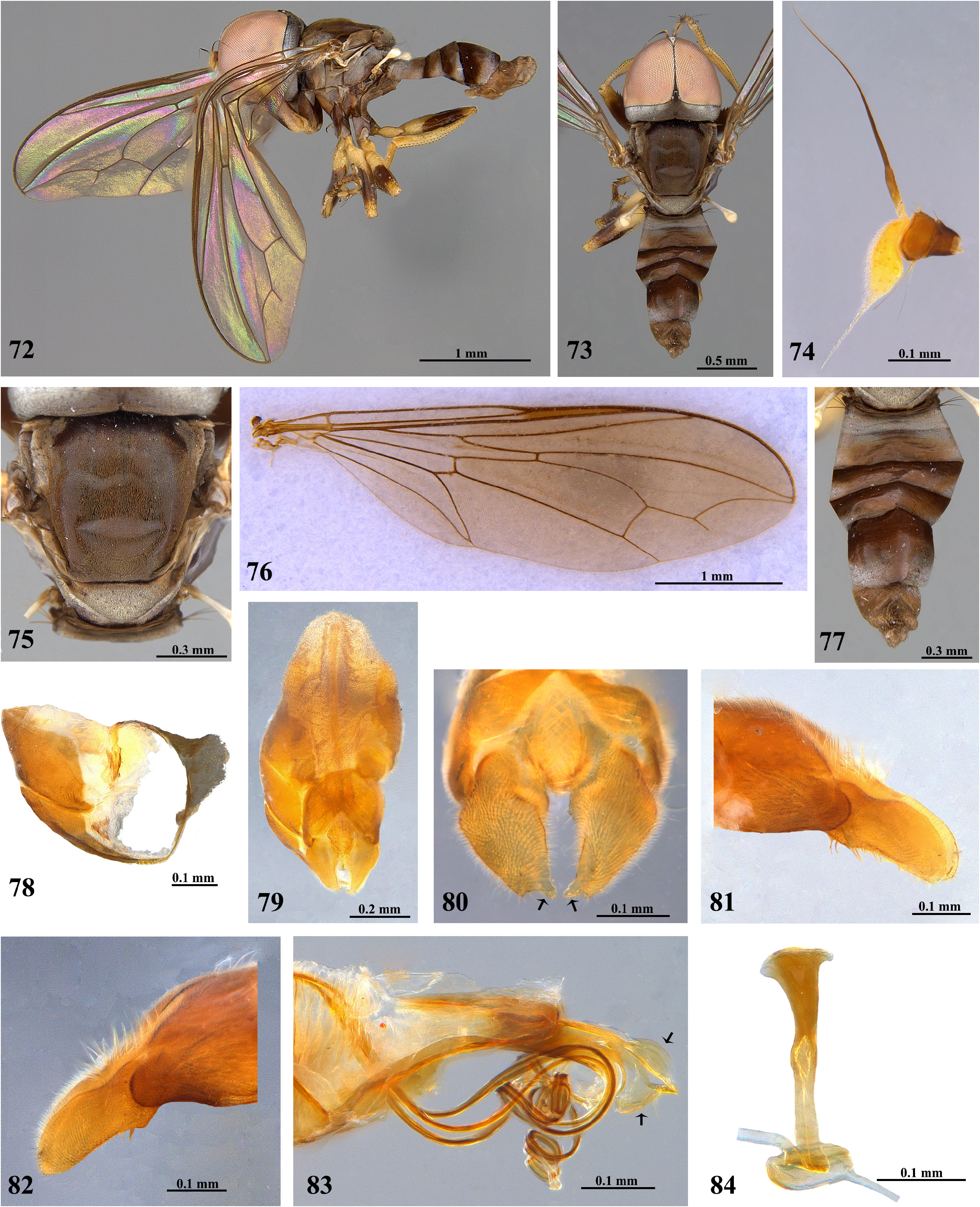
FIGURES 72–84. Neocephalosphaera paolae sp. nov. (IAvH–M03). Male. 72, Habitus, left lateral view; 73, Habitus, dorsal view; 74, Antenna; 75, Thorax, dorsal view; 76, Wing; 77, Abdomen, dorsal view; 78, Tergites and sternites 6 and 7, ventral view; 79, Terminalia, dorsal view; 80, Surstyli, dorsal view; 81, Left surstylus, lateral view; 82, Right surstylus, lateral view; 83, Phallic guide and phallus, left lateral view; 84, Ejaculatory apodeme.
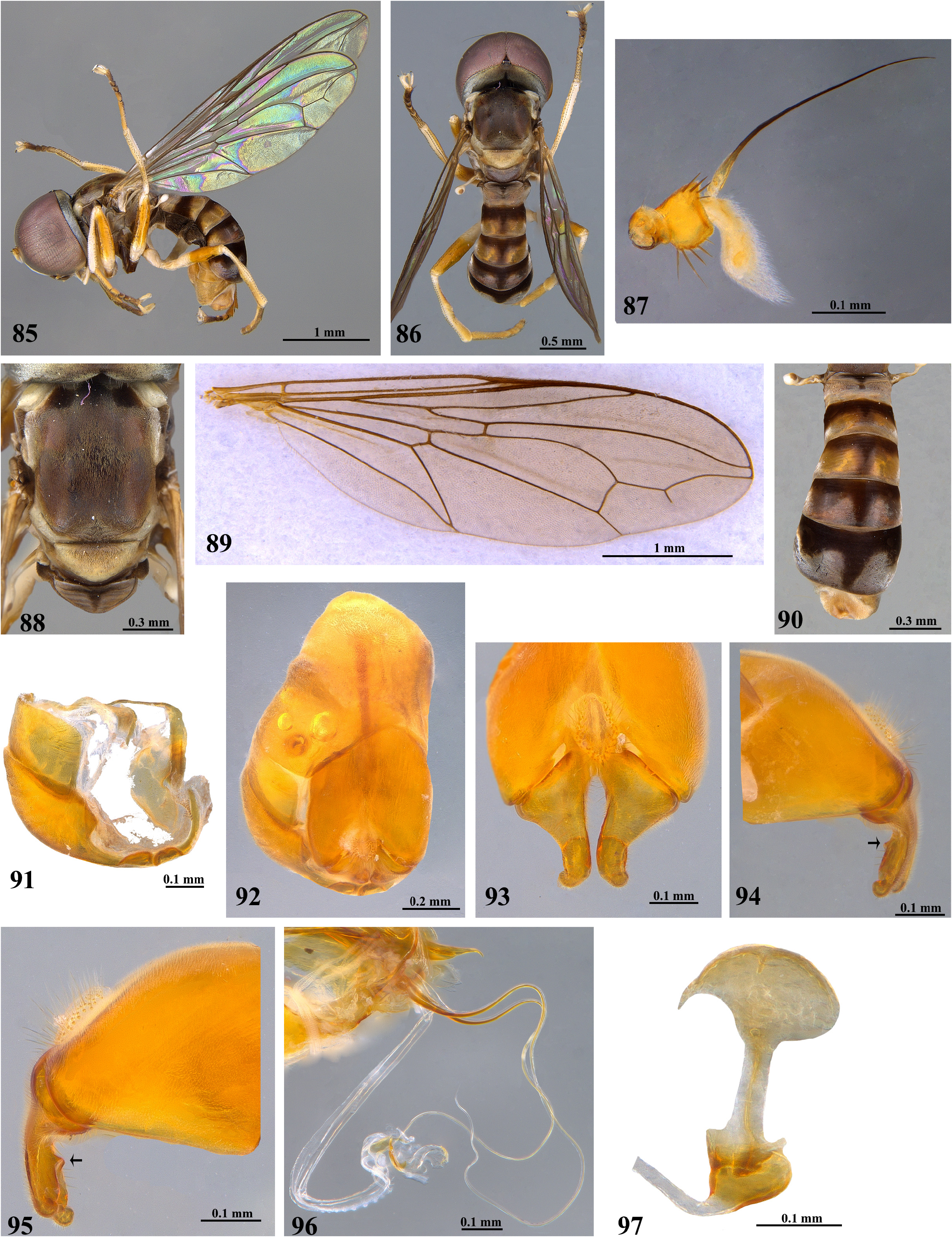
FIGURES 85–97. Neocephalosphaera spinifera sp. nov. (IAvH–M2628). Male. 85, Habitus, left lateral view; 86, Habitus, dorsal view; 87, Antenna; 88, Thorax, dorsal view; 89, Wing; 90, Abdomen, dorsal view; 91, Tergites and sternites 6 and 7, ventral view; 92, Terminalia, dorsal view; 93, Surstyli, dorsal view; 94, Left surstylus, lateral view; 95, Right surstylus, lateral view; 96, Phallic guide and phallus, left lateral view; 97, Ejaculatory apodeme.
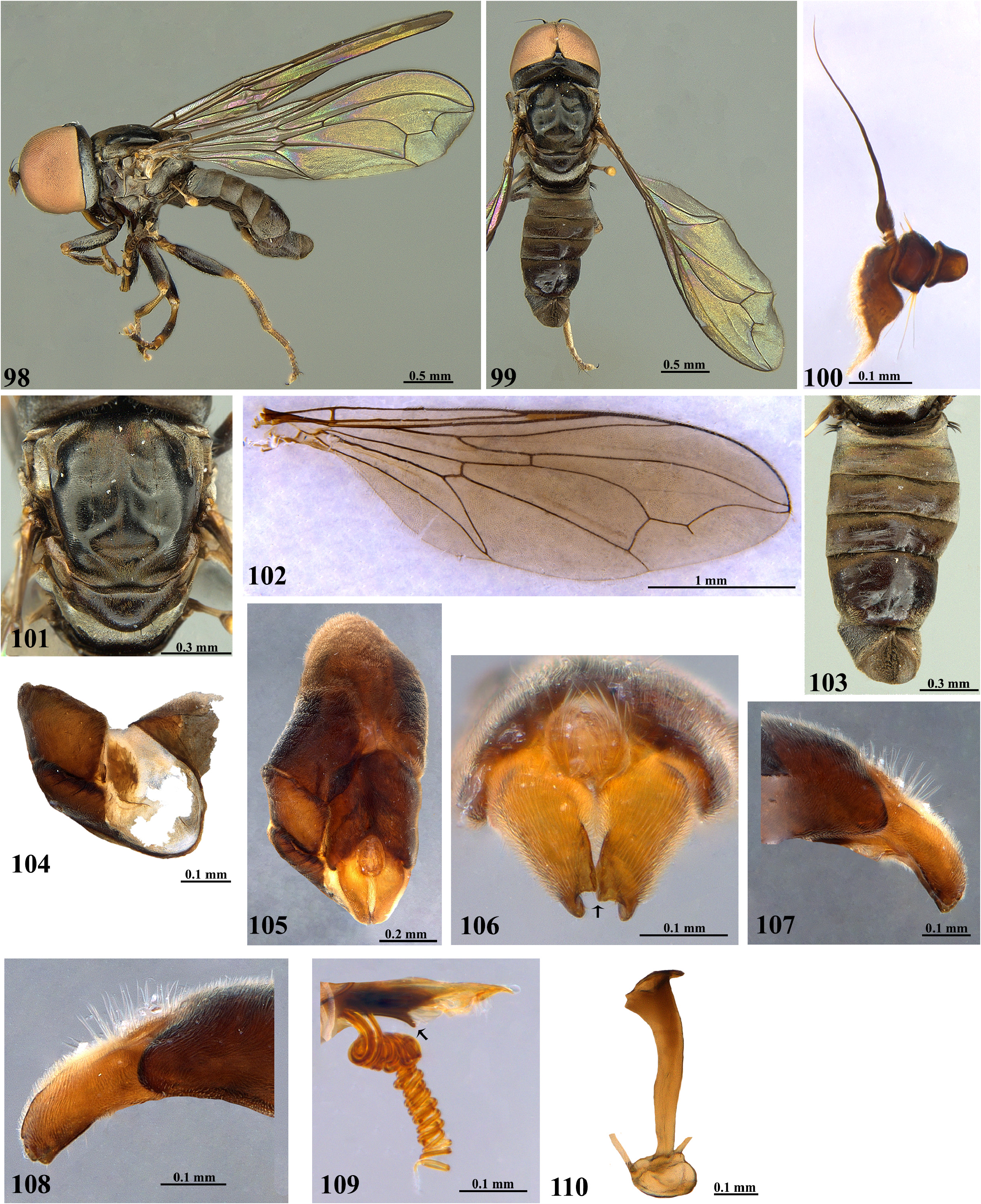
FIGURES 98–110. Neocephalosphaera spiralis sp. nov. (IAvH–M1358). Male. 98, Habitus, left lateral view; 99, Habitus, dorsal view; 100, Antenna; 101, Thorax, dorsal view; 102, Wing; 103, Abdomen, dorsal view; 104, Tergites and sternites 6 and 7, ventral view; 105, Terminalia, dorsal view; 106, Surstyli, dorsal view; 107, Left surstylus, lateral view; 108, Right surstylus, lateral view; 109, Phallic guide and phallus, left lateral view; 110, Ejaculatory apodeme.
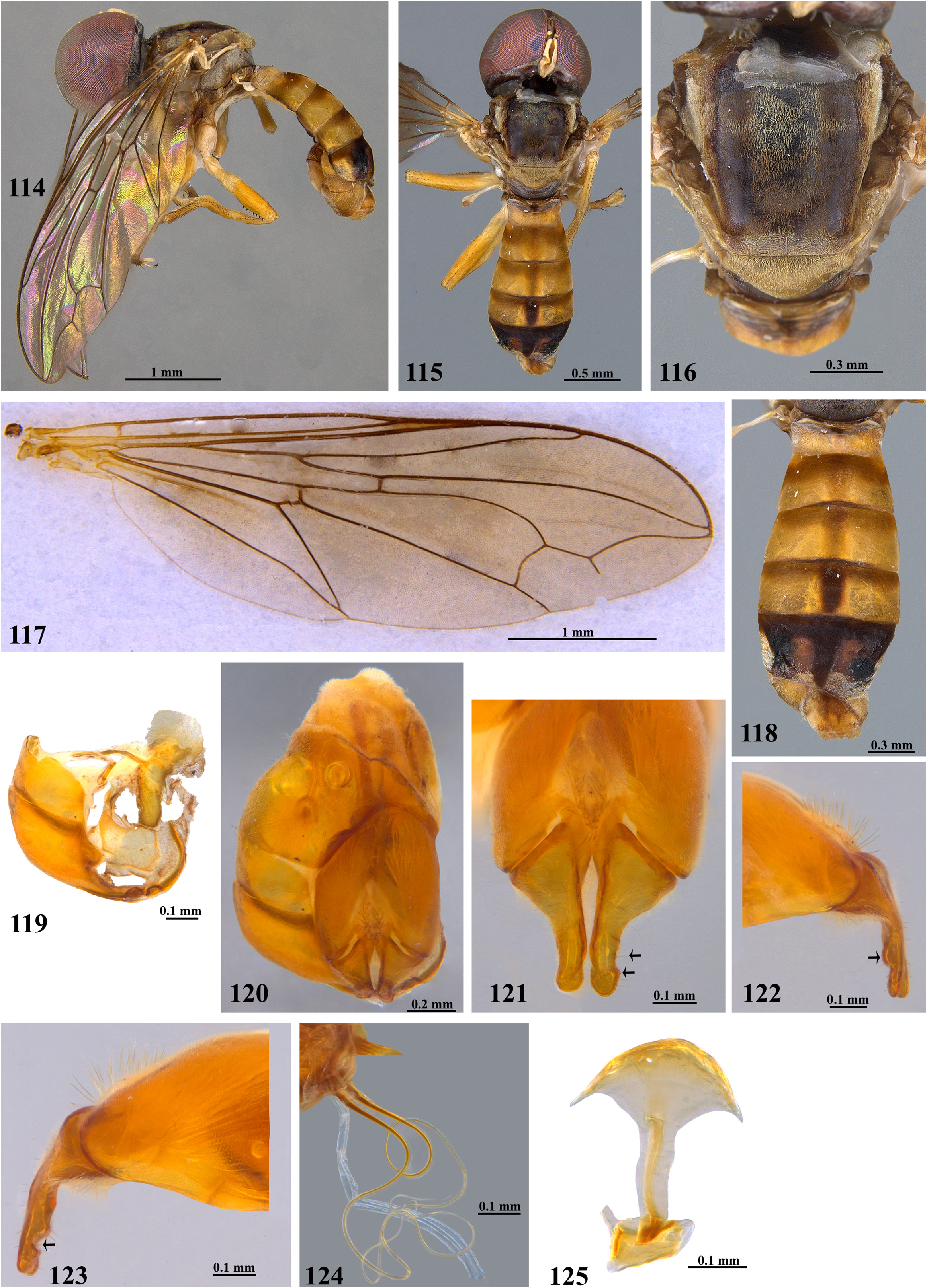
FIGURES 114–125. Neocephalosphaera sumapazensis sp. nov. (IAvH–M4343). Male. 114, Habitus, left lateral view; 115, Habitus, dorsal view; 116, Thorax, dorsal view; 117, Wing; 118, Abdomen, dorsal view; 119, Tergites and sternites 6 and 7, ventral view; 120, Terminalia, dorsal view; 121, Surstyli, dorsal view; 122, Left surstylus, lateral view; 123, Right surstylus, lateral view; 124, Phallic guide and phallus, left lateral view; 125, Ejaculatory apodeme.
No known copyright restrictions apply. See Agosti, D., Egloff, W., 2009. Taxonomic information exchange and copyright: the Plazi approach. BMC Research Notes 2009, 2:53 for further explanation.
|
Kingdom |
|
|
Phylum |
|
|
Class |
|
|
Order |
|
|
Family |
1 (by plazi, 2022-08-29 10:28:58)
2 (by ExternalLinkService, 2022-08-30 21:33:07)
3 (by ExternalLinkService, 2022-08-30 23:02:13)
4 (by ExternalLinkService, 2022-08-31 06:41:43)
5 (by ExternalLinkService, 2022-09-02 12:32:40)
6 (by plazi, 2023-11-07 11:07:27)
7 (by ExternalLinkService, 2023-11-07 16:24:05)