
Rhizorhagium antarcticum ( Hickson & Gravely, 1907 )
|
publication ID |
https://doi.org/10.11646/zootaxa.3972.3.4 |
|
publication LSID |
lsid:zoobank.org:pub:C1698260-269E-4D15-A562-D8FF46751F7F |
|
DOI |
https://doi.org/10.5281/zenodo.5671794 |
|
persistent identifier |
https://treatment.plazi.org/id/3D10878A-FF92-B442-1090-5FB1FEE43BE5 |
|
treatment provided by |
Plazi (2016-04-20 21:45:15, last updated 2024-11-27 06:55:04) |
|
scientific name |
Rhizorhagium antarcticum ( Hickson & Gravely, 1907 ) |
| status |
|
Rhizorhagium antarcticum ( Hickson & Gravely, 1907) View in CoL
( Figs 1 View FIGURE 1 C, D, 2E, F, 3F)
Perigonimus antarcticus Hickson & Gravely, 1907: 4 View in CoL –6, pl. 1 figs 1–3, pl. 4 fig. 32; Rees, 1938: 7; 1956: 338, 347; Naumov & Stepanjants, 1972: 33, 35; Stepanjants, 1972: 56–57, fig. 1; 1979: 10, pl. 1 fig. 1.
Atractylis antarctica — Vanhöffen, 1910: 283, fig. 8;
Atractylis antarcticus — Ritchie, 1913: 10, 11.
Gravelya antarctica — Totton, 1930: 139, fig. 1a, b.
? Gravelya antarcticum — Schuchert, 1996: 41 –42, fig. 21a–d.
Rhizorhagium antarcticum View in CoL — Stechow, 1919: 21; Rees, 1956: 338, 347; Rees & Thursfield, 1965: 55; Millard, 1971: 401, fig. 3; Branch & Williams, 1993: 8, fig.; Peña Cantero, 2004: 768; Bouillon et al., 2006: 137, fig. 79I, J; Peña Cantero et al., 2013: 735 –736, figs 2c–d, 3a–b.
Material examined. Discovery Antarctic Expedition 1901-04: Type (NHM 1907.8.20.1), McMurdo Bay (Ross Sea), 1–20 fms, several stems, with gonophores, on Hydrodendron arboreum .
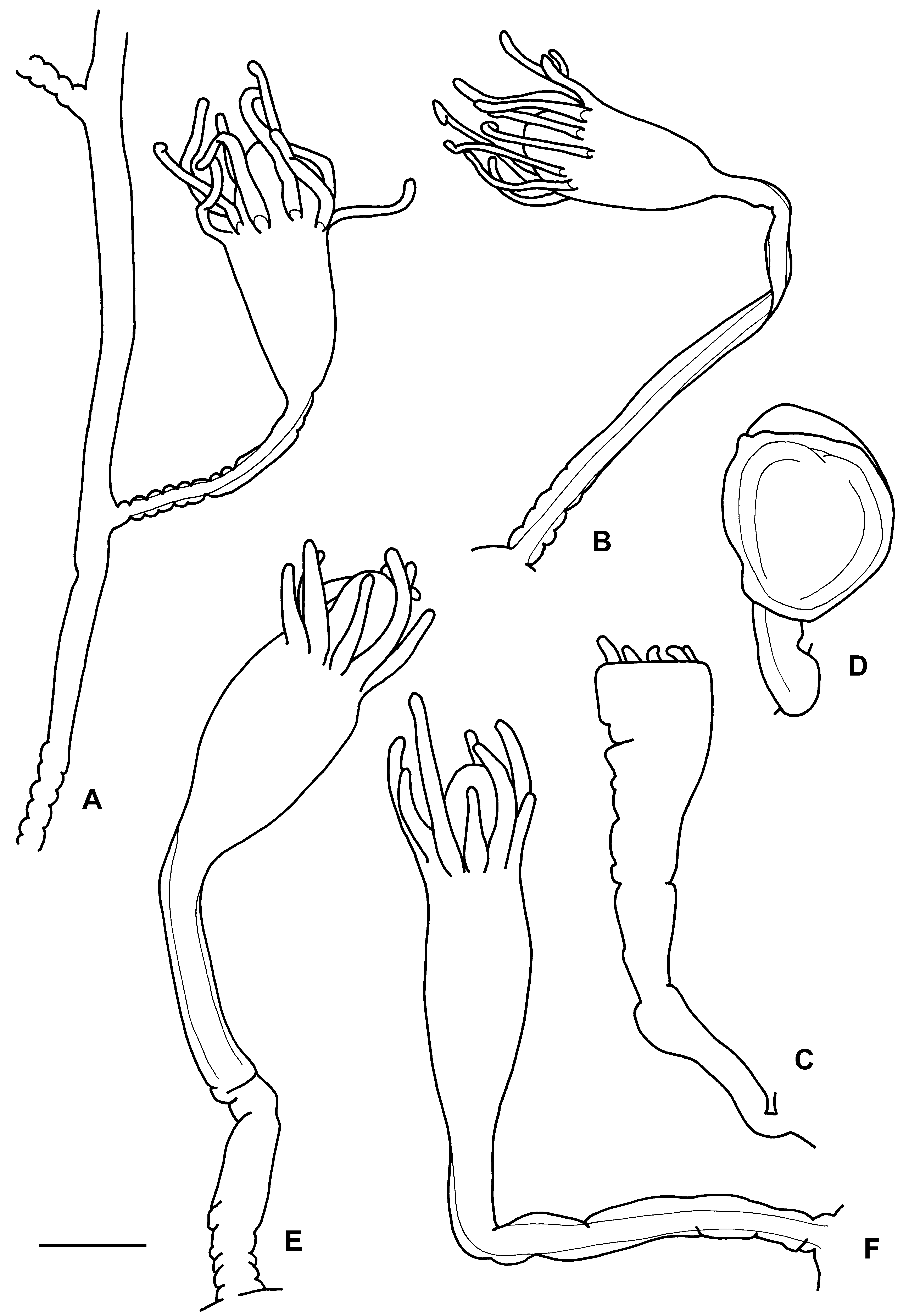
Description. Usually stolonal ( Fig. 1 View FIGURE 1 C, D), but stems with up to three polyps present. Stems covered with perisarc ( Figs 1 View FIGURE 1 C, D, 2E, F), smoothly disappearing on hydranth body ( Fig. 2 View FIGURE 2 E, F). No distinct pseudohydrotheca present. Polyps 800–900 µm high and 250–300 µm in maximum diameter, with conical hypostome and a distal crown of nine to 12 filiform tentacles ( Fig. 2 View FIGURE 2 E, F).
Gonophores c. 950 µm high and c. 680 µm in maximum diameter, on short pedicels, either on hydrorhizal stolon or at basal part of largest stems ( Figs 1 View FIGURE 1 C, D, 3F). “The colonies appear to be invariably dioecious. In both sexes the gonophore is a degenerate medusa ... protected by a thin layer of perisarc. It is larger in the female than in the male (1.1 mm. x 0.9 mm. in the female, and 0.9 mm. x 0.7 mm. in the male). In the female gonophore there is a large sub-umbrella cavity (fig. 3, su.c), the manubrium is well developed and has a well-marked endoderm cavity. In the young gonophore there is a distinct endodermal layer of cells and mesoglea in the umbrella, but in the adult gonophore (fig. 32) these are reduced to a non-cellular mesoglea except at the margin, where a cord of cells represents the ring canal. There are no radial canals in the adult gonophore. In the male gonophore the subumbrella cavity is completely filled with sperm cells (figs. 2 and 32, sp.)” ( Hickson & Gravely, 1907: 5).
Cnidome consisting of two categories of nematocysts, Type I (microbasic euryteles?) and desmonemes.
Measurements (in µm). Cnidome: Type I [range 7.0–7.5 x 4.0–4.5, mean 7.3±0.2 x 4.1±0.2 (n=10); ratio, range 1.6–1.9, mean 1.8±0.1 (n=10)], desmonemes [range 5.5–6.0 x 3.0–3.5].
Remarks. This is a well-characterized species, the only important character missing from the original description was information related to the cnidome. The examination of the type material has allowed me to complete this partially. The heteronemes could not be identified because they could not be seen discharged. However, it is worth mentioning that Peña Cantero et al. (2013), in material that perfectly agrees with this species, identified them as microbasic euryteles. Millard (1971) also indicated the presence of microbasic euryteles.
Hickson & Gravely (1907) gave an excellent description of the gonophores (see above).
Problems with this species were related with its genus allocation. Originally described as Perigonimus antarcticus , it was later considered belonging to Atractylis by Vanhöffen (1910). Totton (1930) even erected the new genus Gravelya to allocate it. Rees (1938) re-erected the genus Rhizorhagium M. Sars, 1874 to include all unbranched colonial bougainvilliids with pseudohydrotheca not enveloping the tentacles, with one crown of filiform tentacles and with fixed sporosacs. Calder (1988) separated members with nipple-shaped hypostomes from this group, including the type species of Rhizorhagium ( R. roseum ). Consequently, Schuchert (1996) saw it necessary to move those species that lack nipple-shaped hypostomes into other genera, in particular to Aselomaris Berrill, 1948 and Gravelya Totton, 1930 , the latter harbouring the present species. More recently, however, Calder’s division based on the shape of the hypostome was not supported (cf. Bouillon et al. 2006: 126) and the species is again considered belonging to Rhizorhagium .
Schuchert’s (1996) material seems to correspond to a different species. The perisarc and the pseudohydrotheca are contaminated with detritus and the size of polyps is smaller. Female gonophores are also distinctly smaller (0.5 x 0.3 mm) than those studied by Hickson & Gravely (1907). He also indicated that there are up to 20 eggs per gonophore; Millard (1971) and Branch & Williams (1993) found 30– 40 eggs.
According to the diagnosis of the genus provided by Bouillon et al. (2006), Rhizorhagium should have ‘perisarc firm, continued over polyp base as a pseudohydrotheca, but never investing tentacle bases’. According to Hickson & Gravely, (1907), ‘the perisarc is continued as an exceedingly thin film over the hydranth as far as the base of the tentacles’. Naumov & Stepanjants (1972: 35) also indicated that ‘the polyp’s body is covered by a fine sheet of perisarc reaching the base of tentacles’. In the material examined the perisarc is not firm and it does not form a distinct pseudohydrotheca; the perisarc sheet covering the polyp body is totally inconspicuous.
Ecology and distribution. Rhizorhagium antarcticum has been found at depths from the tidal level ( Hickson & Gravely 1907) to 450 m ( Totton 1930), epibiotic on hydroids ( Hartlaub 1904; Ritchie 1913; Totton 1930; Naumov & Stepanjants 1972; Stepanjants 1972, 1979; Peña Cantero et al. 2013) and tube of polychaete ( Peña Cantero et al. 2013) and epilithic on stones ( Millard 1971). It is used in turn as substrate by other species of hydroids ( Peña Cantero et al. 2013). Gonophores have been found in colonies collected in January ( Totton 1930; Stepanjants 1979; Peña Cantero et al. 2013), February ( Totton 1930; Stepanjants 1979), from May to July ( Stepanjants 1979), August ( Ritchie 1913; Stepanjants 1979) and in December ( Stepanjants 1979).
Probably Antarctic-Kerguelen distribution, although a careful re-examination of previous records is needed. In Antarctic waters, recorded from the Ross Sea ( Hickson & Gravely 1907; Ritchie 1913; Totton 1930; Peña Cantero et al. 2013), Adélie Land ( Naumov & Stepanjants 1972), Davis Sea ( Vanhöffen 1910; Stepanjants 1972, 1979), and Sodruzestva Sea (Emery glacier) ( Stepanjants 1979), in East Antarctica , and from the Bellingshausen Sea ( Hartlaub 1904), in West Antarctica . In sub-Antarctic waters, it is known from off Marion and Prince Edwards Islands ( Millard 1971; Branch & Williams 1993) and Crozet ( Stepanjants 1979). Schuchert’s (1996) record is from New Zealand.
Pena Cantero, A. L., Boero, F. & Piraino, S. (2013) Shallow-water benthic hydroids from Tethys Bay (Terra Nova Bay, Ross Sea, Antarctica). Polar Biology, 36, 731 - 753. http: // dx. doi. org / 10.1007 / s 00300 - 013 - 1299 - 3
Bouillon, J., Gravili, C., Pages, F., Gili, J. M. & Boero, F. (2006) An introduction to Hydrozoa. Memoires du Museum national d'Histoire naturelle, 194, 1 - 591.
Hickson, S. J. & Gravely, F. H. (1907) Coelenterata. II. Hydroid zoophytes. National Antarctic Expedition (S. S. Discovery) 1901 - 1904, Natural History, 3, 1 - 34, pls 1 - 4.
Branch, M. L. & Williams, G. C. (1993) The Hydrozoa, Octocorallia and Scleractinia of subantarctic Marion and Prince Edward Islands: illustrated keys to the species and results of the 1982 - 1989 University of Cape Town surveys. South African Journal of Antarctic Research, 23 (1 - 2), 3 - 24.
Calder, D. R. (1988) Shallow-water hydroids of Bermuda. The Athecatae. Royal Ontario Museum Life Sciences Contributions, 148, 1 - 107.
Hartlaub, C. (1904) Hydroiden. In: Expedition antarctique Belge. Resultats du voyage du S. Y. Belgica en 1897 - 1899. Rapports scientifiques, Zoologie, 1 - 19.
Millard, N. A. H. (1971) Hydrozoa. In: Van Zinderen Bakker, E. M. Winterbottom, Sr. J. M. & Dyer, R. A. (Eds.), Marion and Prince Edward Islands. A. A. Balkema, Cape Town, pp. 396 - 408, figs. 1 - 7.
Naumov, D. V. & Stepanjants, S. D. (1972) Marine invertebrates from Adelie Land collected by the XIIth and XVth French Antarctic Expeditions. 3. Hydroida. Tethys Supplement, 4, 25 - 60.
Pena Cantero, A. L. (2004) How rich is the deep-sea Antarctic benthic hydroid fauna? Polar Biology, 27, 767 - 774. http: // dx. doi. org / 10.1007 / s 00300 - 004 - 0654 - 9
Rees, W. J. (1938) Observations on British and Norwegian hydroids and their medusae. Journal of the Marine Biological Association of the United Kingdom, 23, 1 - 42. http: // dx. doi. org / 10.1017 / S 0025315400053923
Rees, W. J. (1956) On three northern species of Hydractinia. Bulletin of the British Museum (Natural History), 3, 351 - 362.
Rees, W. J. & Thursfield, S. (1965) The hydroid collection of James Ritchie. Proceedings of the Royal Society of Edinburgh, Section B, 69 (1 - 2), 34 - 220. http: // dx. doi. org / 10.1017 / s 0080455 x 00010122
Ritchie, J. (1913) The hydroid zoophytes collected by the British Antarctic Expedition of Sir Ernest Shackleton, 1908. Proceedings of the Royal Society of Edinburgh, 33 (1), 9 - 34.
Schuchert, P. (1996) The marine fauna of New Zealand: athecate hydroids and their medusa (Cnidaria: Hydrozoa). New Zealand Oceanographic Institute Memoir, 106, 1 - 159.
Stechow, E. (1919) Zur Kenntnis der Hydroidenfauna des Mittelmeeres, Amerikas und anderer Gebiete, nebst Angaben uber einige Kirchenpauer'sche Typen von Plumulariden. Zoologische Jahrbucher, Abteilung fur Systematik, 42 (1), 1 - 172.
Stepanjants, S. D. (1979) Hydroids of the antarctic and subantarctic waters. In: Biological results of the Soviet Antarctic Expedition, 6. Issledovaniya Fauny Morei, 20 (30), 1 - 200, pls 1 - 25. [in Russian]
Totton, A. K. (1930) Coelenterata. Part V. Hydroida. Natural History Report, British Antarctic ' Terra Nova' Expedition, 1910, Zoology, 5 (5), 131 - 252, pls 1 - 3.
Vanhoffen, E. (1910) Die Hydroiden der Deutschen Sudpolar-Expedition 1901 - 1903. Deutsche Sudpolar-Expedition 1901 - 1903, 11 (Zoology 3), 269 - 340.
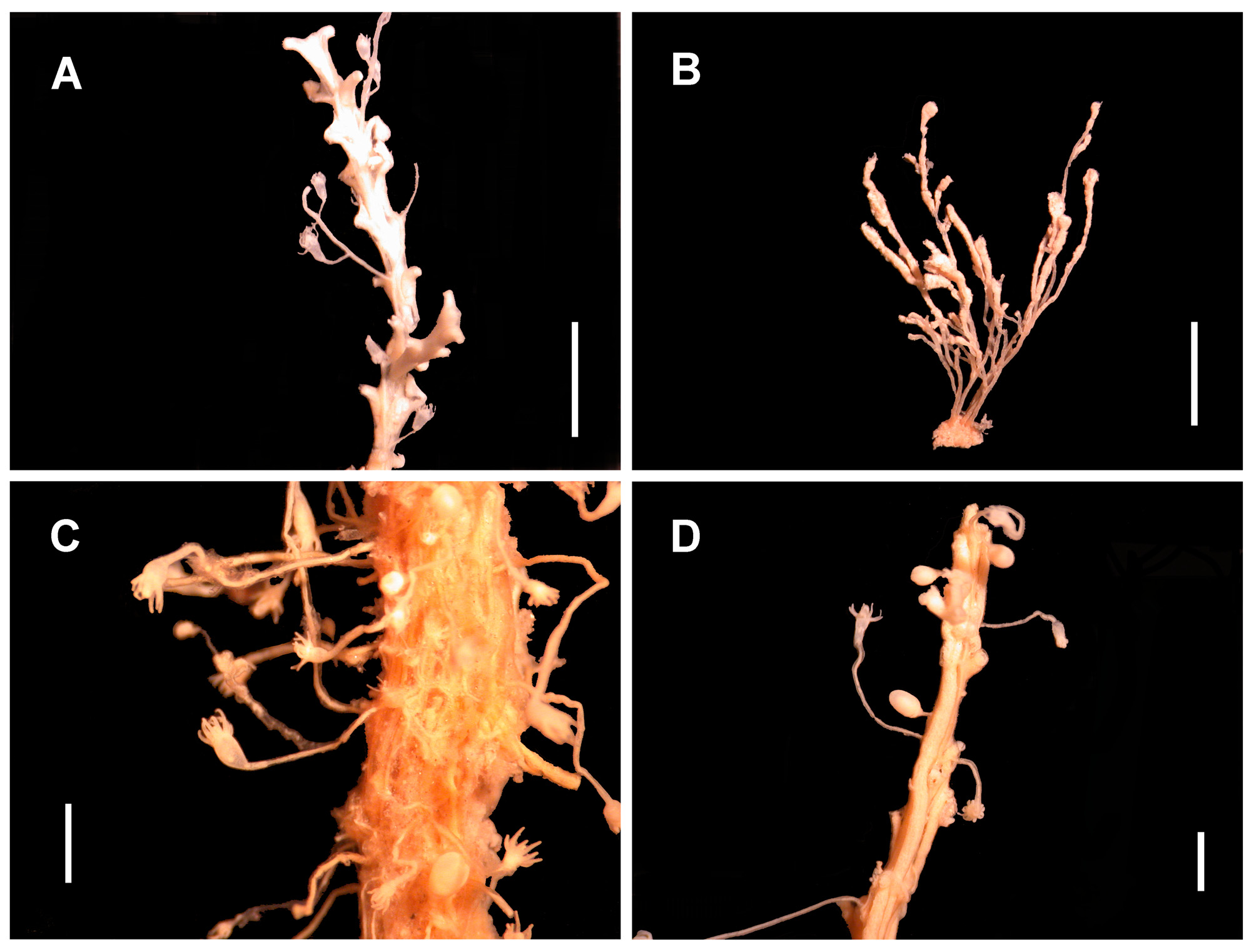
FIGURE 1. Bimeria corynopsis Vanhöffen, 1910: A, a few stems growing on Staurotheca.? Koellikerina belgicae (Vanhöffen, 1910): B, colony with several stems. Rhizorhagium antarcticum (Hickson & Gravely, 1907): C – D, several stems and gonophores. Scale bar: 2 mm (A – B), 1 mm (C – D).
No known copyright restrictions apply. See Agosti, D., Egloff, W., 2009. Taxonomic information exchange and copyright: the Plazi approach. BMC Research Notes 2009, 2:53 for further explanation.
|
Kingdom |
|
|
Phylum |
|
|
Class |
|
|
Order |
|
|
Family |
|
|
Genus |
Rhizorhagium antarcticum ( Hickson & Gravely, 1907 )
| Peña Cantero, Álvaro L. 2015 |
Gravelya antarcticum
| Schuchert 1996: 41 |
Gravelya antarctica
| Totton 1930: 139 |
Rhizorhagium antarcticum
| Pena 2013: 735 |
| Bouillon 2006: 137 |
| Pena 2004: 768 |
| Branch 1993: 8 |
| Millard 1971: 401 |
| Rees 1965: 55 |
| Rees 1956: 338 |
| Stechow 1919: 21 |
Atractylis antarcticus
| Ritchie 1913: 10 |
Atractylis antarctica
| Vanhoffen 1910: 283 |
Perigonimus antarcticus
| Naumov 1972: 33 |
| Rees 1938: 7 |
| Hickson 1907: 4 |
1 (by plazi, 2016-04-20 21:45:15)
2 (by ImsDioSync, 2017-01-10 03:58:00)
3 (by ImsDioSync, 2017-01-10 03:58:56)
4 (by ExternalLinkService, 2019-09-26 11:23:45)
5 (by ExternalLinkService, 2019-10-09 07:43:11)
6 (by ExternalLinkService, 2019-10-18 23:29:35)
7 (by ExternalLinkService, 2019-10-19 19:12:01)
8 (by ExternalLinkService, 2021-11-09 13:54:14)
9 (by ExternalLinkService, 2021-11-10 00:46:39)
10 (by ExternalLinkService, 2021-11-11 00:00:12)
11 (by plazi, 2023-10-29 17:25:24)