
Delarthrum anomalans, Golovatch & Aswathy & Bhagirathan & Sudhikumar, 2021
|
publication ID |
https://doi.org/ 10.11646/zootaxa.5068.4.2 |
|
publication LSID |
lsid:zoobank.org:pub:F769B986-8F7B-4ABF-A7EF-58A813718760 |
|
DOI |
https://doi.org/10.5281/zenodo.5709520 |
|
persistent identifier |
https://treatment.plazi.org/id/3A1C87D9-970D-FFCC-FF1F-2A8CBDD8A0CE |
|
treatment provided by |
Plazi |
|
scientific name |
Delarthrum anomalans |
| status |
sp. nov. |
Delarthrum anomalans sp. nov.
Figs 69–87 View FIGURES 69–73 View FIGURES 74–80 View FIGURES 81–87 .
Material examined. Male holotype (CATE-61604A), 2 male (CATE-61604B and CATE-61604C) and 2 female paratypes (CATE-61604D and CATE-61604E), India, Kerala state, Thrissur district , 10°21’19’’N, 76°12’48’’E, 29 m a.s.l. GoogleMaps , in June 2021 and 1 male (CATE-61604J) and 2 female paratypes (CATE-61604K and CATE-61604L all in 5302B–CATE-5302I) from the sacred groves of Valliyur kavu, Manathavady, Wayanad district , 11°48’08’’N, 76°01’55’’E, 716 m a.s.l., M.D. Aswathy leg. GoogleMaps
Name. To emphasize the anomalous absence of adenostyles from both male legs 1 and 2.
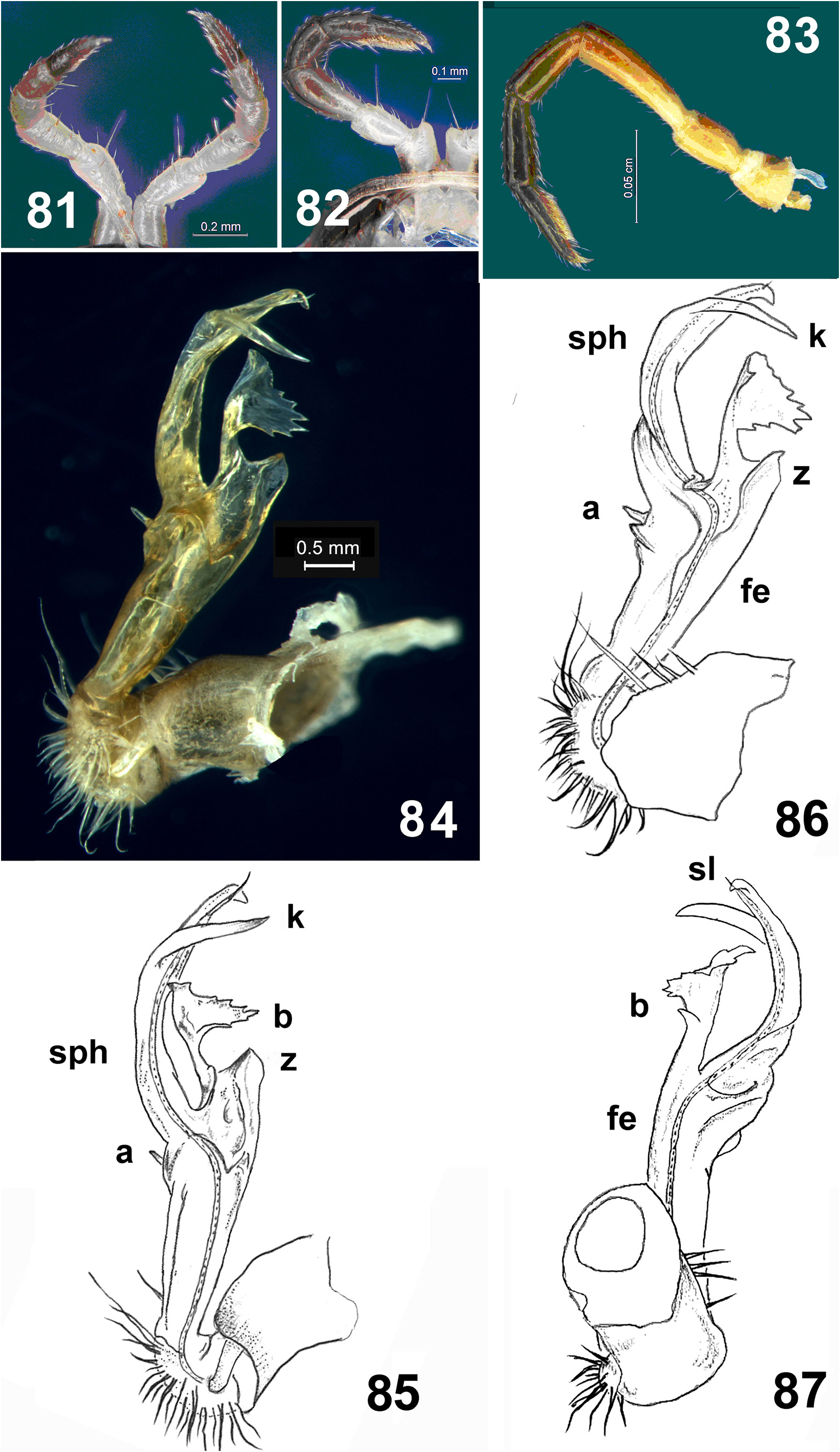
Diagnosis. Differs from congeners by the absence of adenostyles from both male legs 1 and 2, coupled with the sexually dimorphic colour pattern, the transverse tergal sulci starting with the collum, and the peculiar gonopodal conformation, primarily the presence of a small, thumb-shaped, distoventral projection (a) basal to the division of the postfemoral part into (1) the main, dorsal piece of the solenophore (sph), which is long, obliquely truncate dorsad at the base, bifid in the distal third and slightly curved mesad, and (2) a dorsal process that is split immediately at the base into a longer, stalked, leaf-shaped outgrowth (b) with peculiar, ragged, saw-like distal margins, and a much shorter, simple, dorsobasal peg (z) ( Figs 84–87 View FIGURES 81–87 ).
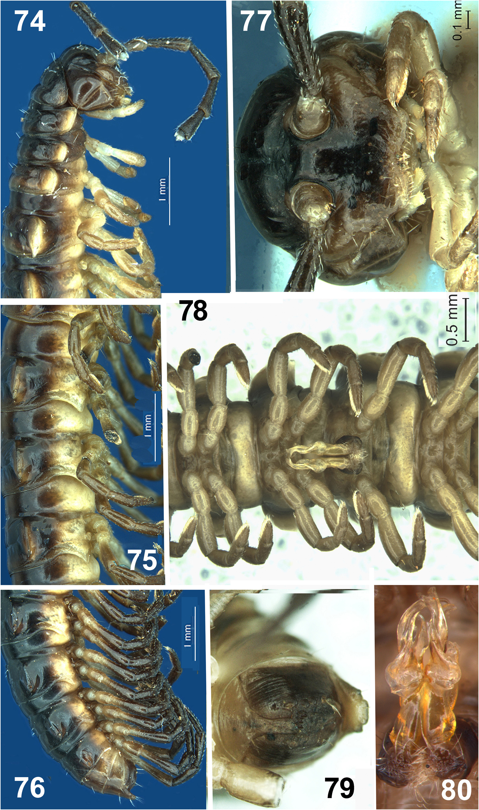
Description. Length of holotype, 17.9 mm, of paratypes, 17.0–17.6 mm (male) or 14.0–14.8 mm (female). Width of midbody rings, 2.6 mm (holotype), 1.9–2.1 mm (male paratypes) or 2.0–2.3 mm (female paratypes). Coloration sex-dimorphic: in vivo, dorsum dark brown to black, venter chestnut brown (male) ( Figs 69, 70 View FIGURES 69–73 ) to yellowish white (female) ( Fig. 71 View FIGURES 69–73 ); paraterga mostly contrasting pale. Legs light brown (male) or whitish (female). Coloration after one month of preservation in 96% ethanol somewhat darkened, but pattern evident ( Figs 69–76 View FIGURES 69–73 View FIGURES 74–80 ).
Body with 20 rings in both sexes. In width, collum <head <ring 2<3<4<5<6<7<8<9=10, body gradually tapering thereafter ( Figs 72, 73 View FIGURES 69–73 ). Vertex glossy and bare; stipes, cardo and clypeus moderately setose, epicranial sulcus distinct and ending before the level of antennal sockets. Clypeal region with ca 5+5 setiferous pits. Antennae moderately long, only slightly clavate, extending back to ring 4 when stretched caudolaterally ( Figs 72 View FIGURES 69–73 , 74 View FIGURES 74–80 ). Relative antennomere lengths: 1<6<4<2<5<3. Antennomere 6 broadest apically, post-antennal groove shallow, diameter of antennal socket and isthmus between sockets subequal, 0.24 and 0.23 mm, respectively ( Fig. 77 View FIGURES 74–80 ). Antennomeres 5–7 each with a distinct dorso-apical group of basiconic sensilla (both male and female), these being especially conspicuous on antennomere 5 ( Figs 72 View FIGURES 69–73 , 74 View FIGURES 74–80 ).
Paraterga mostly well developed, keel-shaped, set at about upper third of body height ( Figs 69–76 View FIGURES 69–73 View FIGURES 74–80 ). Collum ovoid, paraterga small, subtriangular and broadly rounded. Anterior margin of collum with a single transverse row of fragile short setae ( Fig. 72 View FIGURES 69–73 ). Anterior and posterior margins of following rings rather straight and parallel. Anterior margin with a transverse row of 2+2 short setae similar to those on collum. Paraterga 2 as usual, set clearly lower than 3 rd, squarish in shape, anterior corner/lobe drawn anteriad. Following paraterga 3–17 regularly rounded anterolaterally and increasingly drawn caudolaterad into acute triangles, but still lying within rear tergal margins ( Figs 72–76 View FIGURES 69–73 View FIGURES 74–80 ), vs. small, spiniform and slightly drawn past rear tergal margin on rings 18 and 19 ( Figs 73 View FIGURES 69–73 , 76 View FIGURES 74–80 ). Lateral calluses of paraterga thin, only slightly thicker on pore-bearing rings than on poreless ones ( Figs 69, 70 View FIGURES 69–73 ), smooth, delimited by distinct sulci both dorsally and, to a lesser degree, ventrally; usually with a few short setae retained at lateral margin.
Tegument generally smooth and shining, prozonae very delicately shagreened, metazonae mostly striate/striolate at bases of paraterga and near transverse sulci, surface being slightly undulate ( Figs 72, 73 View FIGURES 69–73 ). Tergal setae mostly short and inconspicuous, present both on paraterga and in anterior half of metaterga. Two deep transverse sulci present on collum ( Fig. 72 View FIGURES 69–73 ), followed by one prominent sulcus on metazonae 2–18, smooth at bottom and reaching the base of paraterga ( Figs 72, 73 View FIGURES 69–73 ). A faint axial line traceable on most metazonae until 18 th ( Figs 72, 73 View FIGURES 69–73 ). Strictures between pro- and metazonae narrow, nearly smooth, at most faintly striolate ( Figs 72–76 View FIGURES 69–73 View FIGURES 74–80 ).
Pore formula normal (5, 7, 9, 10, 12, 13, 15–19), ozopores small, circular, opening at ca 1/3 off caudolateral corner of pore-bearing calluses ( Figs 74–76 View FIGURES 74–80 ). Pleurosternal carinae decreasing in size from 2 to 4. Epiproct conical and flattened dorsoventrally, extending well beyond anal valves; tip truncate, subapical lateral incisions evident; hypoproct subtriangular, with 1+1 setae on minute knobs ( Fig. 79 View FIGURES 74–80 ).
Legs moderately long and slender, clearly longer and thickened in male compared to female ( Figs 70, 71 View FIGURES 69–73 ), rather densely setose ventrally and with short claws; male legs with tarsal brushes absent only from last two leg-pairs ( Figs 74–76, 78 View FIGURES 74–80 , 83 View FIGURES 81–87 ). Prefemora and coxae each bearing a moderately long and stiff distoventral seta. Podomere length ratios: coxa <prefemur <tibia = postfemur <tarsus <femur ( Fig. 83 View FIGURES 81–87 ). Neither male leg-pair 1 nor 2 with any adenostyles, only coxae 2 as usual, with gonopores on small knobs ( Figs 81, 82 View FIGURES 81–87 ). Sternal lobe between male coxae 4 small, conical and densely setose.
Gonopodal aperture transverse and suboval in shape, about 2/3 as wide as prozonite 7, its rim being not elevated, but thickened laterally ( Fig. 78 View FIGURES 74–80 ). Gonopods high, complex, in situ held parallel to each other ( Figs 78, 80 View FIGURES 74–80 ). Coxite darkened, about as long as femorite, subcylindrical, setose distoventrally ( Figs 84–87 View FIGURES 81–87 ); cannula a short curved tube, as usual. Prefemoral part short, globose, densely setose as usual, with a particularly long seta distoventrally. Femorite (fe) relatively stout and short, somewhat enlarged distad, subclavate, almost as long as acropodite (= postfemoral part), subcylindrical, without evidence of torsion; a small, thumb-shaped, distoventral projection (a) basal to acropodite bifurcation into a longer, larger, more slender, subacuminate, ventral, solenophore branch (sph), rather slightly curved dorsad and bearing both a strong spine (k) in distal third and a free solenomere (sl), and a shorter, more elaborate, dorsal process divided immediately at base into a longer, stalked, leaf-shaped outgrowth (b) with peculiar, ragged, saw-like distal margins, and a much shorter, simple, dorsobasal peg (z). Seminal groove first running all along mesal side of femorite, then turning slowly towards bifurcation point before moving onto a free, flagelliform sl tightly attached to mesal side of sph, with only a short tip of sl remaining exposed over sph. Division of fe and acropodite through a transverse sulcus, or of sph into laminae, wanting ( Figs 84–87 View FIGURES 81–87 ).

No known copyright restrictions apply. See Agosti, D., Egloff, W., 2009. Taxonomic information exchange and copyright: the Plazi approach. BMC Research Notes 2009, 2:53 for further explanation.
|
Kingdom |
|
|
Phylum |
|
|
Class |
|
|
Order |
|
|
Family |
|
|
Genus |