
Cybaeus septatus, Chamberlin & Ivie, 1942
|
publication ID |
https://doi.org/10.11646/zootaxa.5100.2.2 |
|
publication LSID |
lsid:zoobank.org:pub:1D783E41-8DF8-4D3C-9853-38C41CCEBB30 |
|
DOI |
https://doi.org/10.5281/zenodo.6313355 |
|
persistent identifier |
https://treatment.plazi.org/id/39264D0A-FFE5-AD0E-FF6F-658BFD2DFBE3 |
|
treatment provided by |
Plazi (2022-02-18 08:07:17, last updated 2024-11-29 13:12:45) |
|
scientific name |
Cybaeus septatus |
| status |
|
The septatus View in CoL species group
Diagnosis. Unique among the members of the Californian clade of Cybaeus , the species group affiliation of the males of the septatus group is relatively easily to determine. The males of the septatus group are diagnosed by a combination of the prolaterally to slightly anteriorly directed patellar apophysis which is as long as or longer than the width of the palpal patella and bears four to 12 peg setae one of which is in a proximal medial position isolated from, or slightly to noticeably larger than, the remainder of the peg setae ( Figs 1–2 View FIGURES 1–4 , 5–7 View FIGURES 5–9 , 22 View FIGURES 22–24 , 25 View FIGURES 25–26 , 34 View FIGURES 33–36 , 44 View FIGURES 43–46 , 47 View FIGURES 47–48 ). To our knowledge, no males of other Californian clade Cybaeus species groups ( adenes , aspenicolens , consocius , devius , and tardatus ) or unplaced Californian species ( C. cribelloides Chamberlin & Ivie , C. gidneyi Bennett , C. irreverens Bennett spec. nov.) show this combination of characters.
The females of the septatus group are diagnosed by a combination of features of the epigynum, atrium, and copulatory ducts. The epigynum is marked anteriorly by one or more “wrinkles” in the integument ( Figs 10, 13 View FIGURES 10–15 , 27 View FIGURES 27–32 ) and the atrium features paired longitudinal atrial openings medially ( Figs 10, 13 View FIGURES 10–15 , 16 View FIGURES 16–21 , 27 View FIGURES 27–32 , 37, 40 View FIGURES 37–42 ). The copulatory ducts ( Figs 11–12, 14–15 View FIGURES 10–15 , 18–19 View FIGURES 16–21 , 28–29 View FIGURES 27–32 , 38, 41 View FIGURES 37–42 ) are short and lead from the atrial openings medially to the midline of the vulva where they are contiguous prior to diverging and leading laterally to the spermathecal heads. As with the septatus group males, no females of other Californian clade Cybaeus species groups or unplaced Californian species are known which possess this combination of characters.
Description. As in diagnosis. Medium-sized spiders: carapace lengths averaging 2.15–2.5 mm (females). Males subequal. Abdomen usually patterned. Legs unbanded (femora rarely lightly banded). Two or occasionally three complete pairs of ventral tibia I macrosetae (distal pair present, absent, or incomplete).
Male: Retrolateral tibial apophysis ( Figs 1 View FIGURES 1–4 , 33 View FIGURES 33–36 , 43 View FIGURES 43–46 ) carinate, nearly as long as tibia. Embolus relatively short and thin, describing a compound ( Figs 3 View FIGURES 1–4 , 23 View FIGURES 22–24 , 45 View FIGURES 43–46 ) or simple ( Fig. 35 View FIGURES 33–36 ) curve. Tegular apophysis ( Figs 3 View FIGURES 1–4 , 23 View FIGURES 22–24 , 35 View FIGURES 33–36 , 45 View FIGURES 43–46 ) with distal arm short, unmodified, with rounded or angular terminus; proximal arm prolaterally directed, slightly swollen basally, and tip slightly twisted and swollen ( Figs 3–4 View FIGURES 1–4 , 8–9 View FIGURES 5–9 ), bluntly acuminate ( Figs 23–24 View FIGURES 22–24 , 26 View FIGURES 25–26 ), or sharply acuminate ( Figs 35 View FIGURES 33–36 , 45 View FIGURES 43–46 , 48 View FIGURES 47–48 ).
Female: Atrial openings ( Figs 10, 13 View FIGURES 10–15 , 16 View FIGURES 16–21 , 27 View FIGURES 27–32 , 37, 40 View FIGURES 37–42 ) conspicuous or not. Spermathecal heads ( Figs 12, 15 View FIGURES 10–15 , 20–21 View FIGURES 16–21 , 29–30 View FIGURES 27–32 , 38–39, 41–42 View FIGURES 37–42 ) small dorsolateral lobes; spermathecal stalks short ( Figs 12, 14 View FIGURES 10–15 , 20 View FIGURES 16–21 , 38 View FIGURES 37–42 ) or relatively long ( Figs 29–30 View FIGURES 27–32 , 42 View FIGURES 37–42 ); Bennett’s glands near junction with spermathecal bases ( Figs 12, 14 View FIGURES 10–15 , 20–21 View FIGURES 16–21 , 39 View FIGURES 37–42 , 32 View FIGURES 27–32 , 39, 42 View FIGURES 37–42 ); bases large, rounded; fertilization ducts exit bases posteriorly.
Composition and distribution. Cybaeus chauliodous Bennett , C. lockeae Bennett spec. nov., C. septatus Chamberlin & Ivie , C. somesbar Bennett , and C. viator Bennett spec. nov. Cybaeus septatus and C. chauliodous are relatively common within their known distributions; the other species are uncommon. All have restricted ranges within an area encompassing southwestern Oregon (Jackson and Josephine Counties) and northern California south to Alameda and Sierra Counties ( Figs 49–50 View FIGURE 49 View FIGURE 50 ).
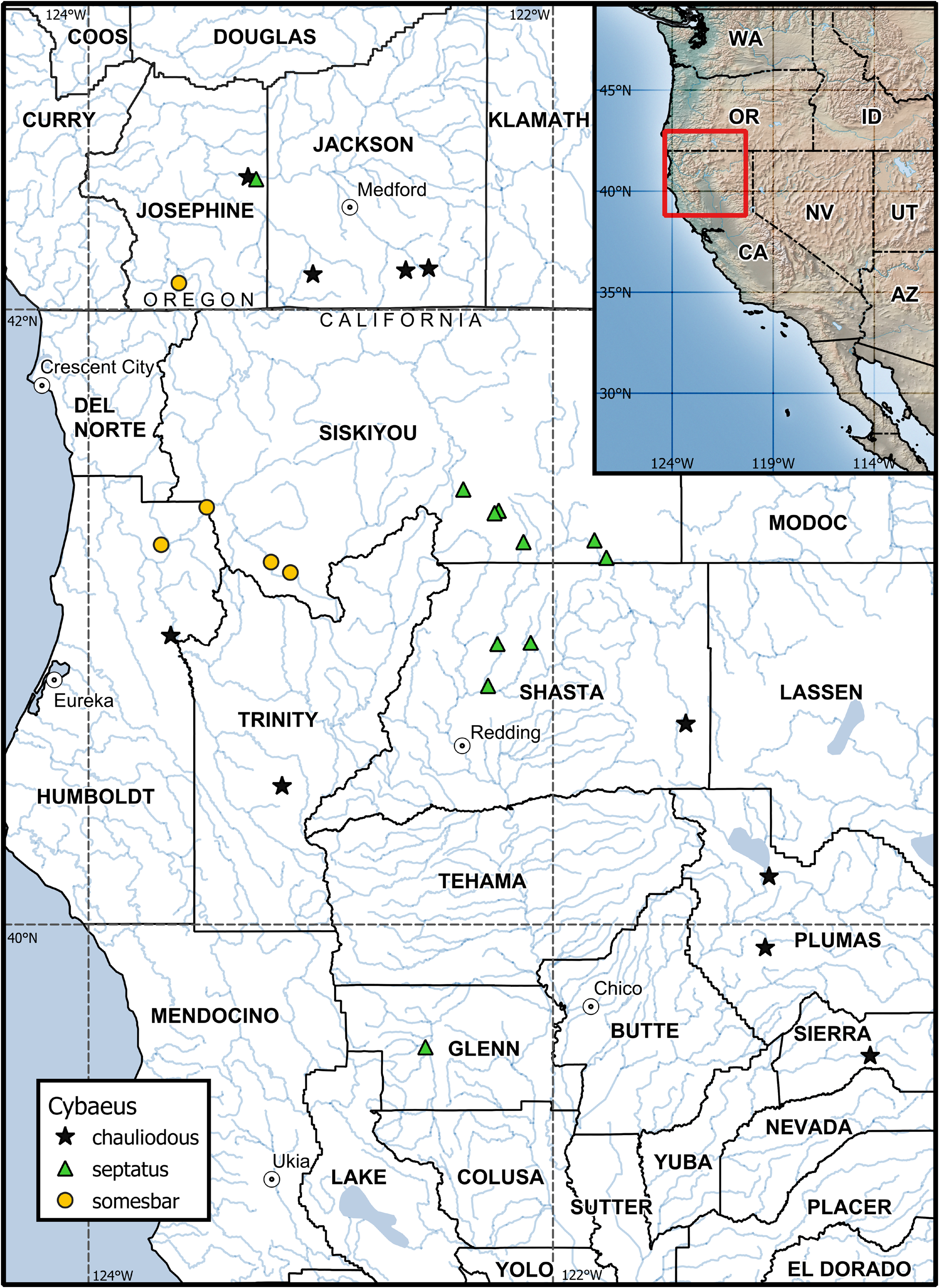
FIGURE 49. Distributions of Cybaeus chauliodous, C. septatus, and C. somesbar. AZ—Arizona, CA—California, ID—Idaho, OR—Oregon, NV—Nevada, UT—Utah, WA—Washington. California and Oregon county names capitalized.
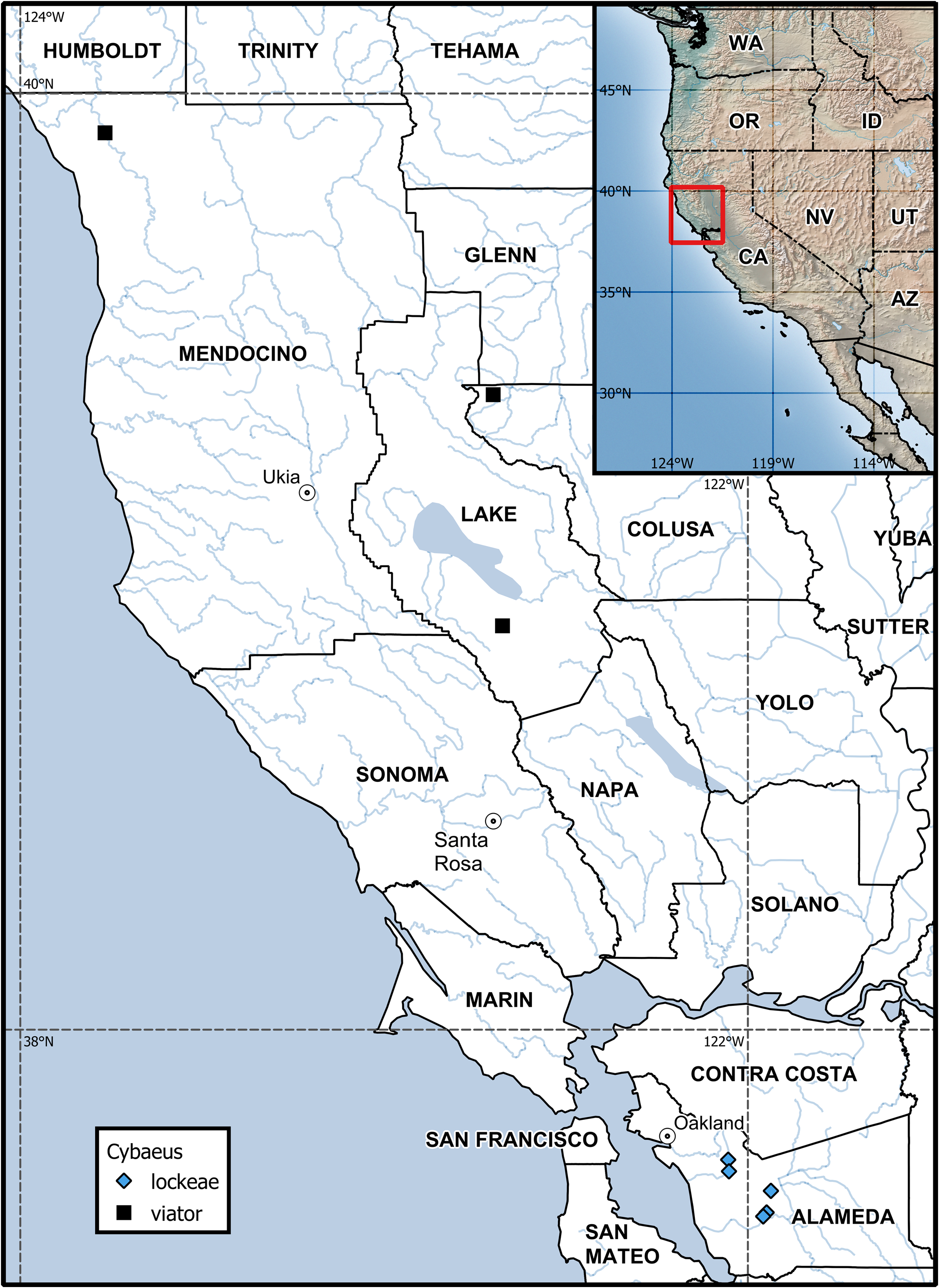
FIGURE 50. Distributions of Cybaeus lockeae spec. nov. and C. viator spec. nov. AZ—Arizona, CA—California, ID—Idaho, OR—Oregon, NV—Nevada, UT—Utah, WA—Washington. California county names capitalized.
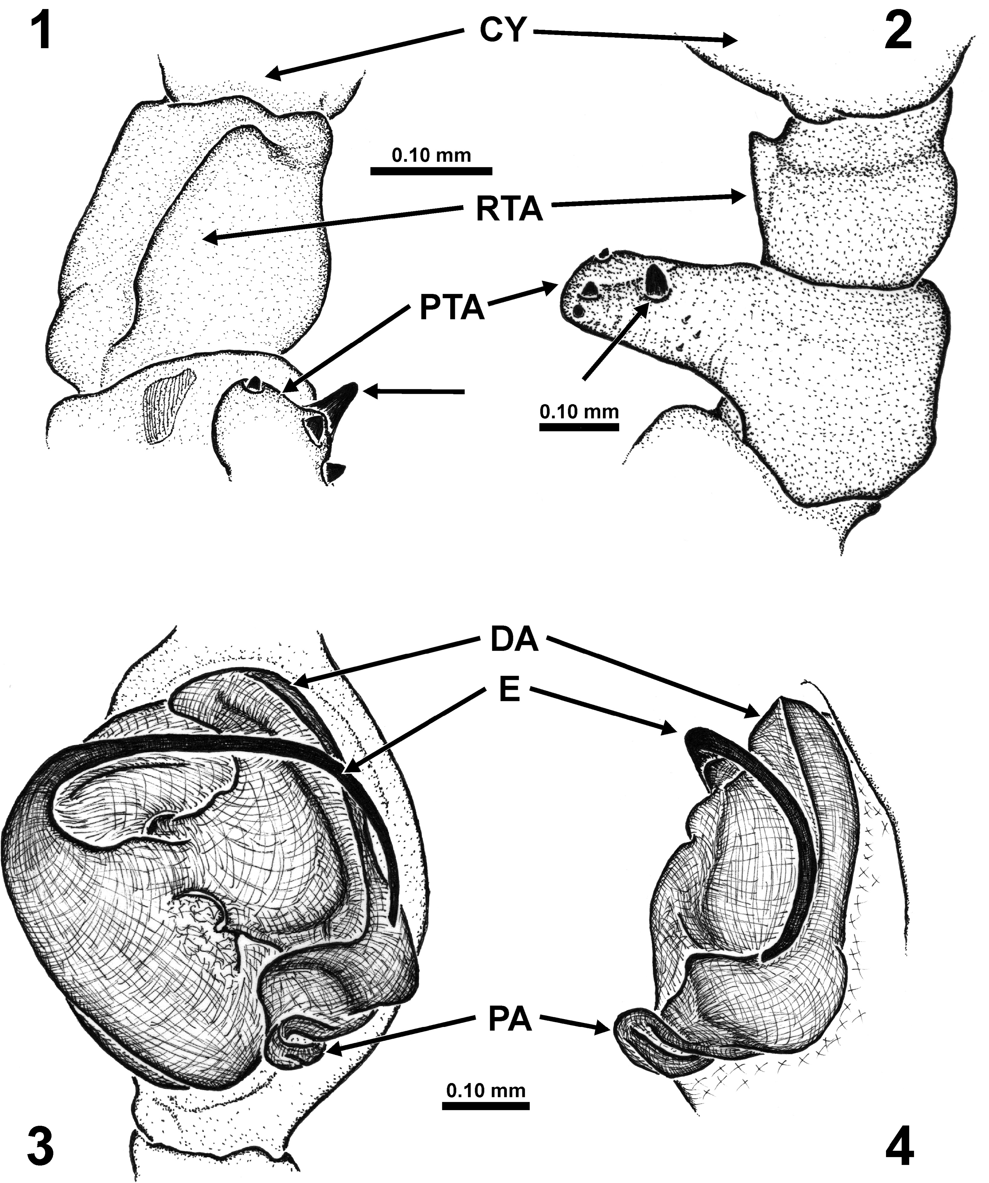
FIGURES 1–4. Cybaeus chauliodous, males (1, 3–4 from Ashland Summit, Oregon; 2 from Lake Almanor, California), left palpus. 1–2 Patella and tibia (1 retrolateral, 2 dorsal). 3–4 Genital bulb (3 ventral, 4 retrolateral). CY—cymbium, DA—distal arm of tegular apophysis, E—embolus, PA—proximal arm of tegular apophysis, PTA—patellar apophysis, RTA—retrolateral tibial apophysis. Unlabelled arrows indicate single enlarged peg seta on patellar apophysis.
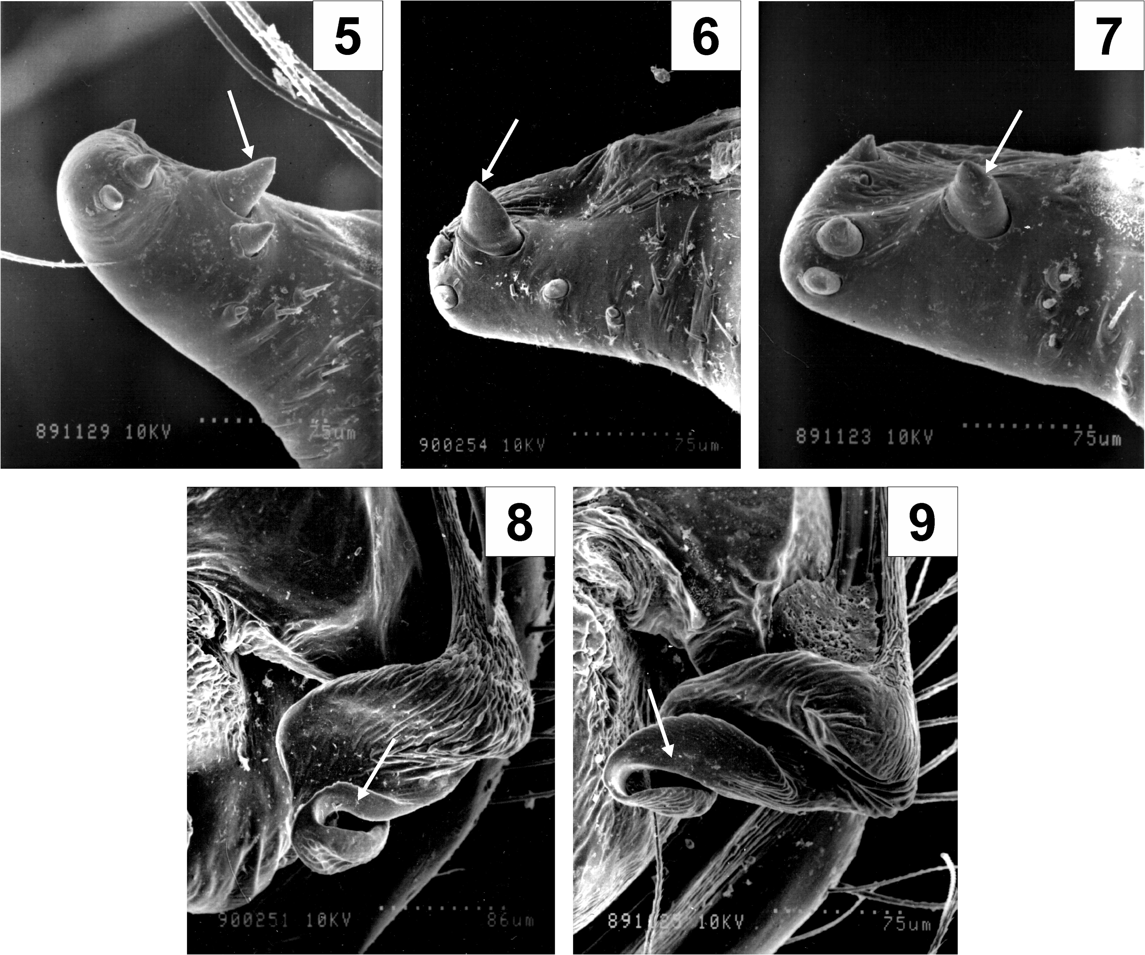
FIGURES 5–9. Cybaeus chauliodous, males (5, 7, 9 from Lake Almanor, California; 6, 8 from Ashland Summit, Oregon), left palpus. 5–7 Patellar apophysis, dorsal. 8–9 Proximal arm of tegular apophysis, ventral. Unlabelled arrows indicate diagnostic characters (5–7 single enlarged peg seta on patellar apophysis, 8–9 swollen lip-like tip of proximal arm of tegular apophysis).
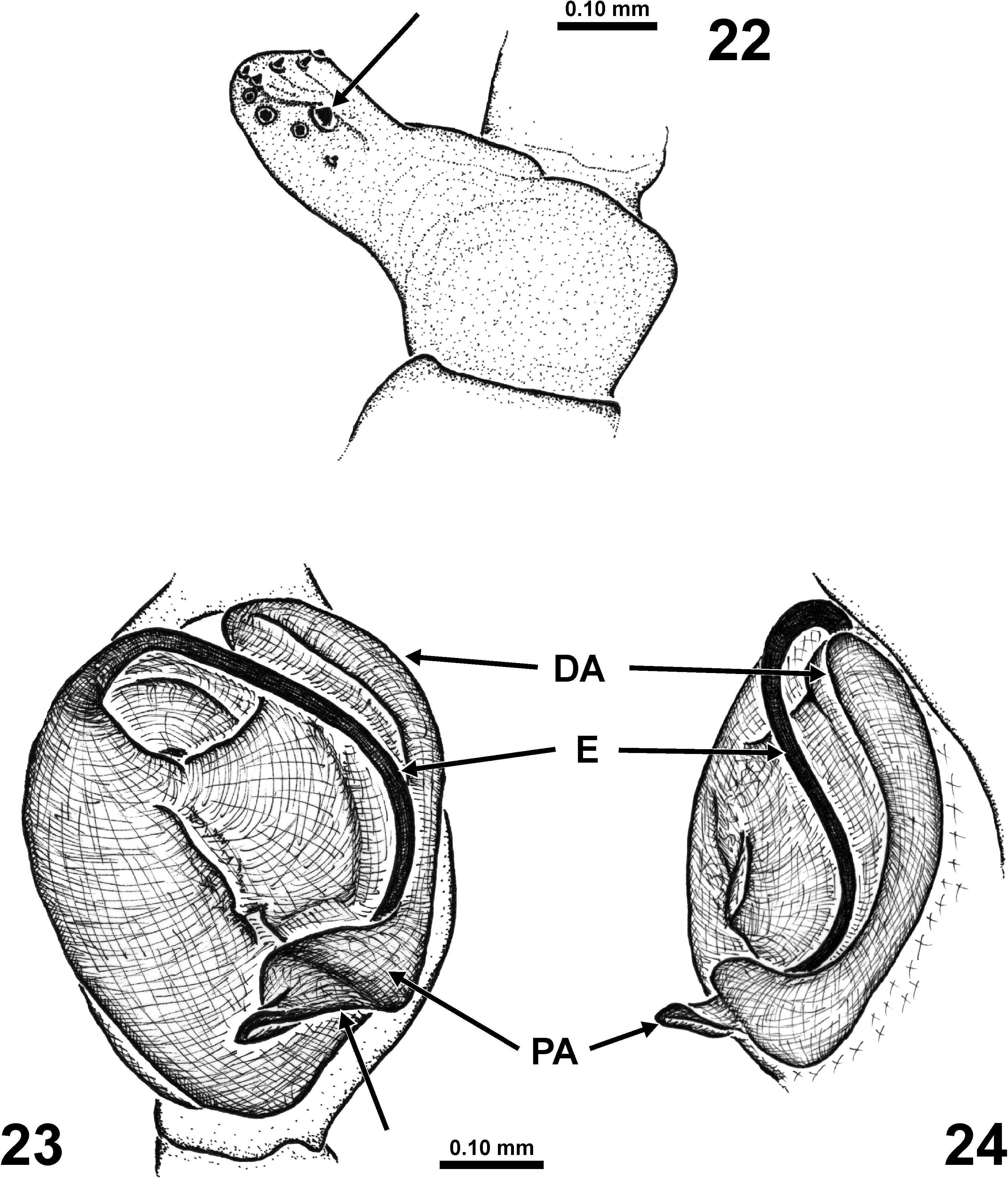
FIGURES 22–24. Cybaeus septatus, male from Mt. Shasta, California, left palpus. 22 Patella, dorsal. 23–24 Genital bulb (23 ventral, 24 retrolateral). DA—distal arm of tegular apophysis, E—embolus, PA—proximal arm of tegular apophysis. Unlabelled arrows indicate diagnostic characters (22 single slightly enlarged peg seta on patellar apophysis, 23 weakly concave posterior margin of PA).
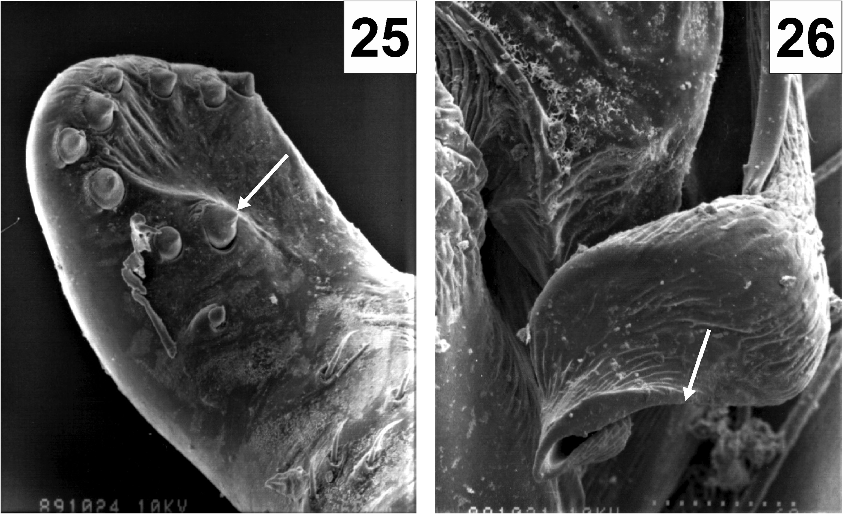
FIGURES 25–26. Cybaeus septatus, male from Mt. Shasta, California, left palpus. 25 Patellar apophysis, dorsal. 26 Proximal arm of tegular apophysis, ventral. Unlabelled arrows indicate diagnostic characters (25 single slightly enlarged peg seta on patellar apophysis; 26 weakly concave posterior margin of proximal arm of tegular apophysis).
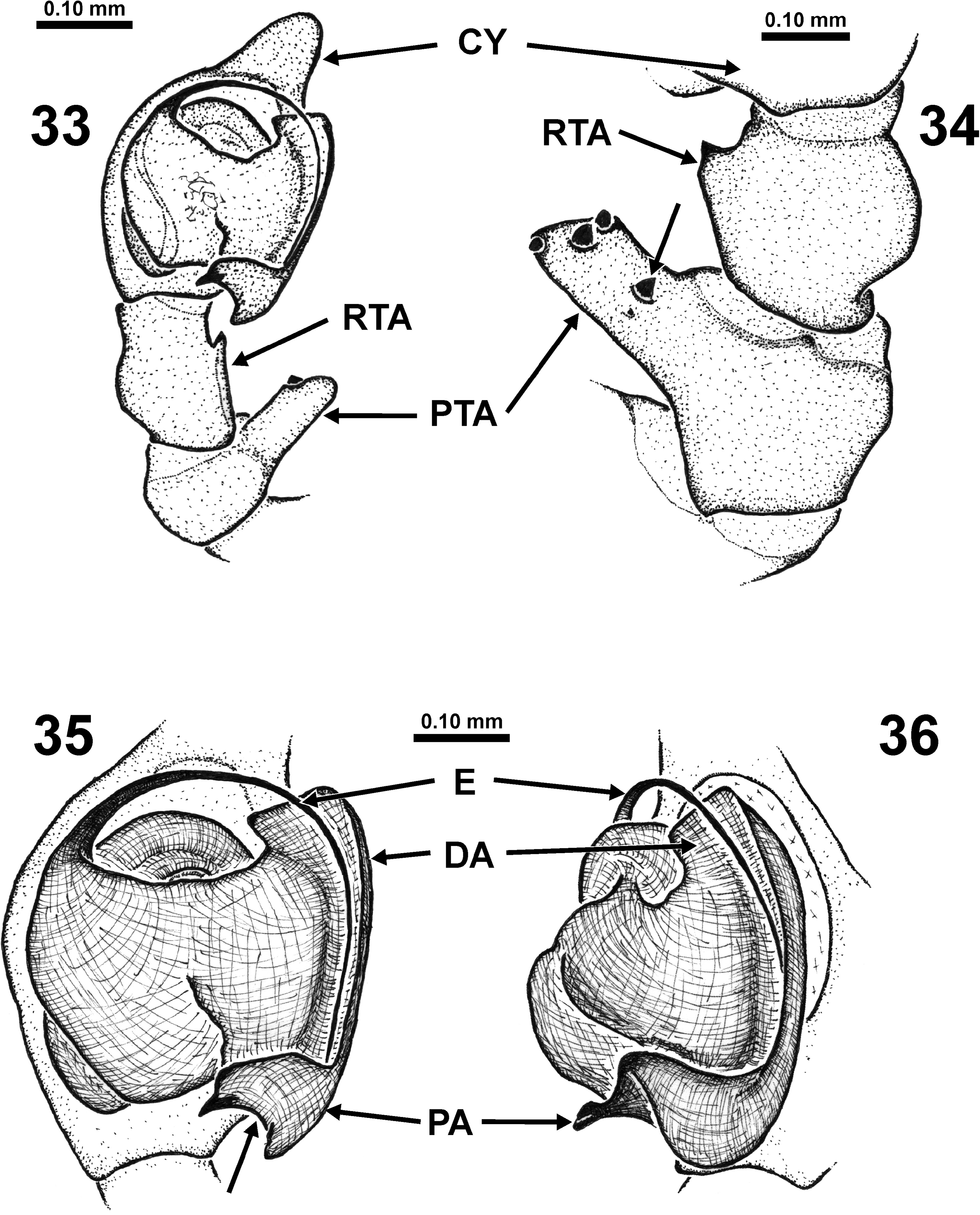
FIGURES 33–36. Cybaeus somesbar, male from Slide Creek, California, left palpus. 33 Patella, tibia, and genital bulb, ventral. 34 Patella and tibia, dorsal. 35–36 Genital bulb (35 ventral, 36 retrolateral). CY—cymbium, DA—distal arm of tegular apophysis, E—embolus, PA—proximal arm of tegular apophysis, PTA—patellar apophysis, RTA—retrolateral tibial apophysis. Unlabelled arrows indicate diagnostic characters (34 single isolated peg seta medially on PTA, 35 strongly concave posterior margin of PA).
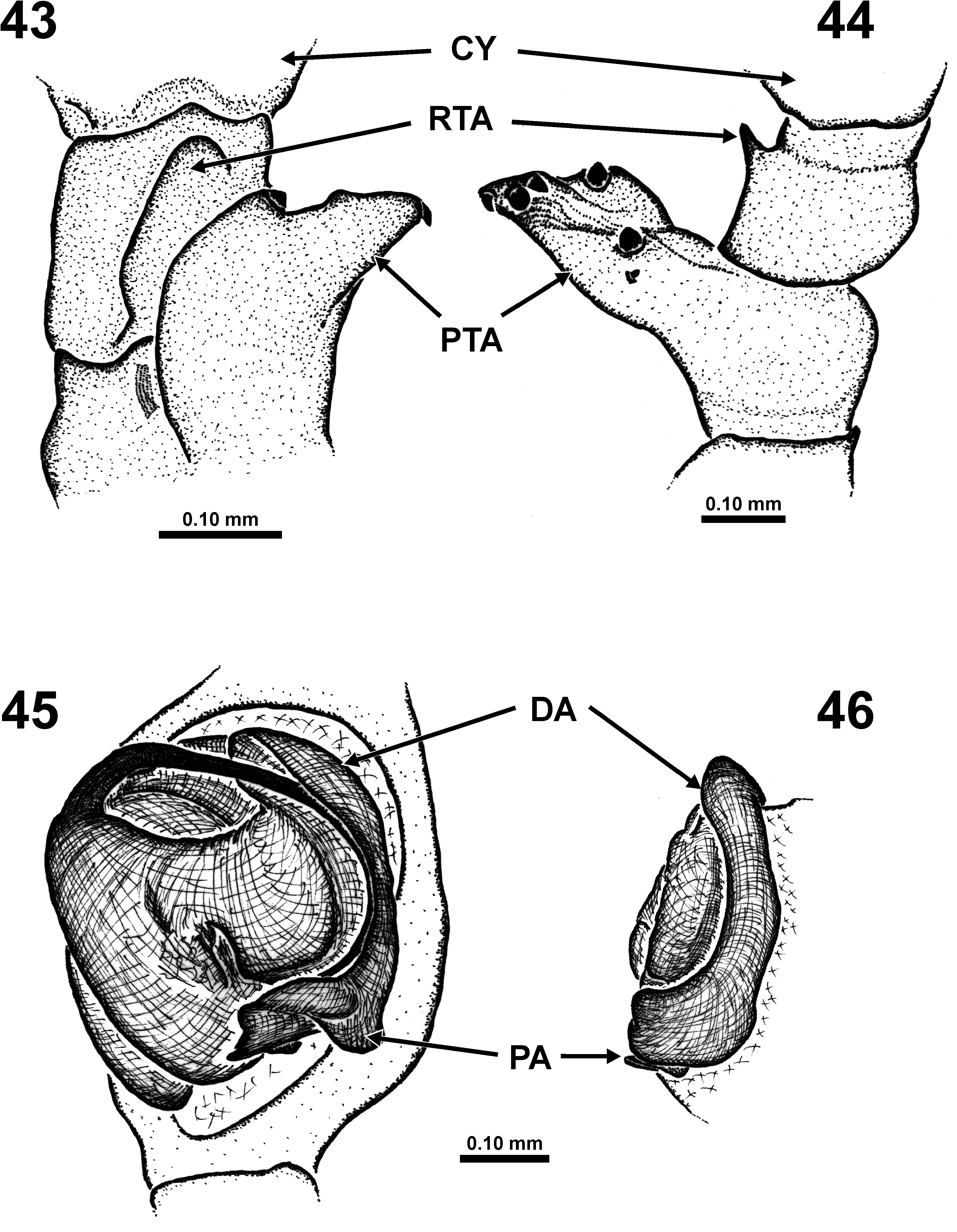
FIGURES 43–46. Cybaeus viator spec. nov., male from Fouts Springs, California, left palpus. 43–44 Patella and tibia (43 retrolateral, 44 dorsal). 45–46 Genital bulb (45 ventral, 46 retrolateral). CY—cymbium, DA—distal arm of tegular apophysis, PA—proximal arm of tegular apophysis, PTA—patellar apophysis, RTA—retrolateral tibial apophysis.
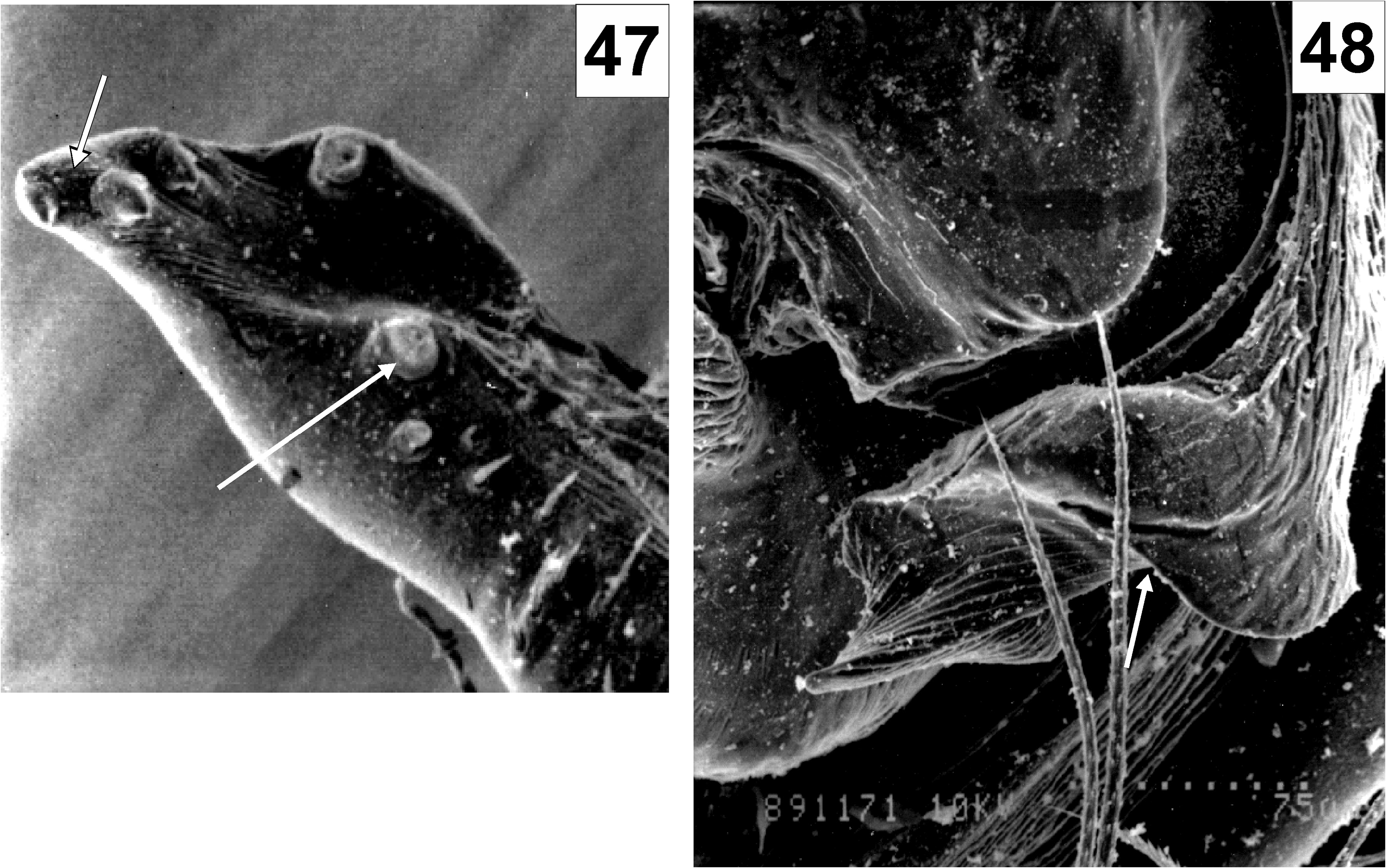
FIGURES 47–48. Cybaeus viator spec. nov., male from Fouts Springs, California, left palpus. 47 Patellar apophysis, dorsal. 48 Proximal arm of tegular apophysis, ventral. Unlabelled arrows indicate diagnostic characters (47 pointed angular tip of patellar apophysis with three peg setae, single isolated peg seta medially; 48 angular posterior margin of proximal arm of tegular apophysis).
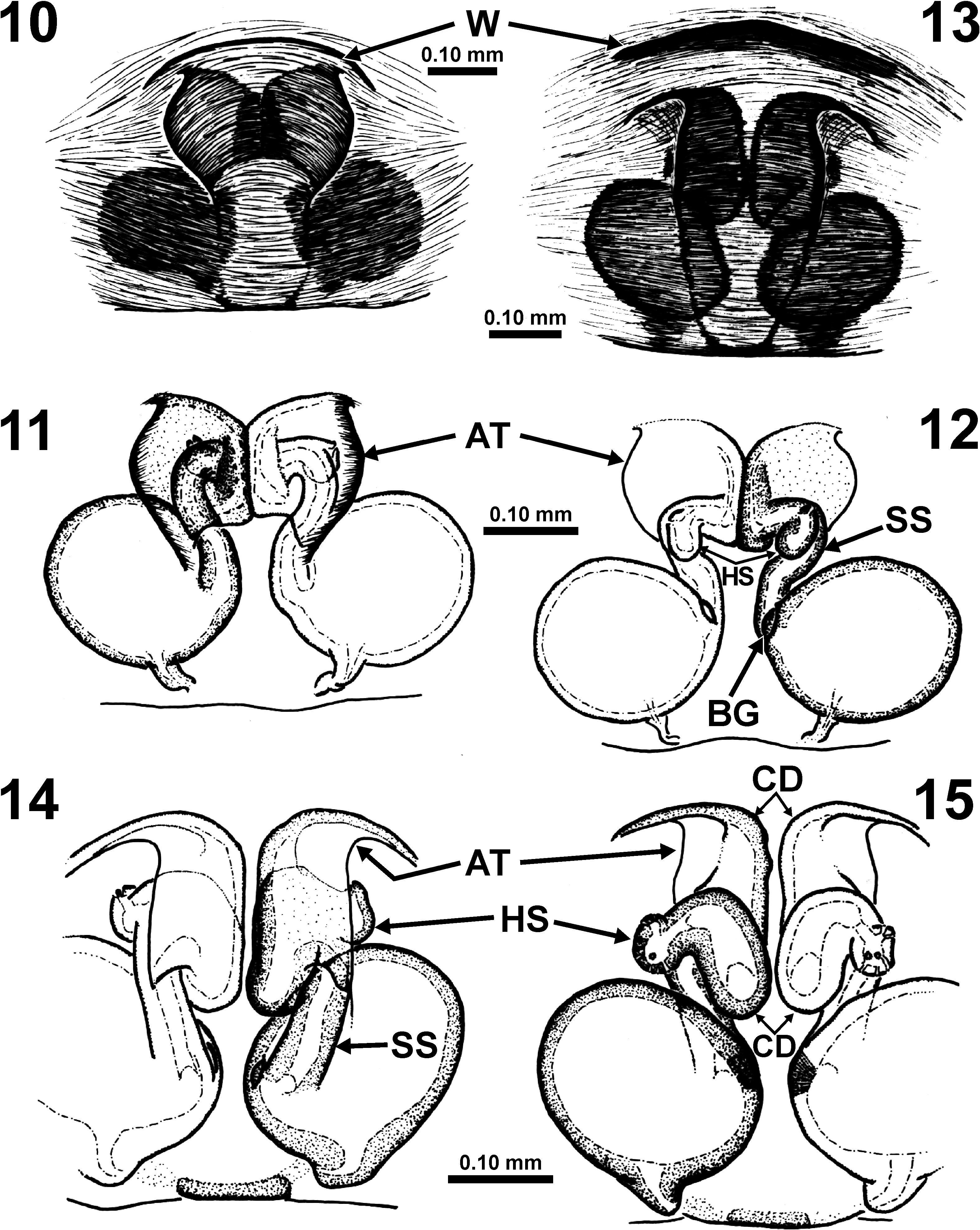
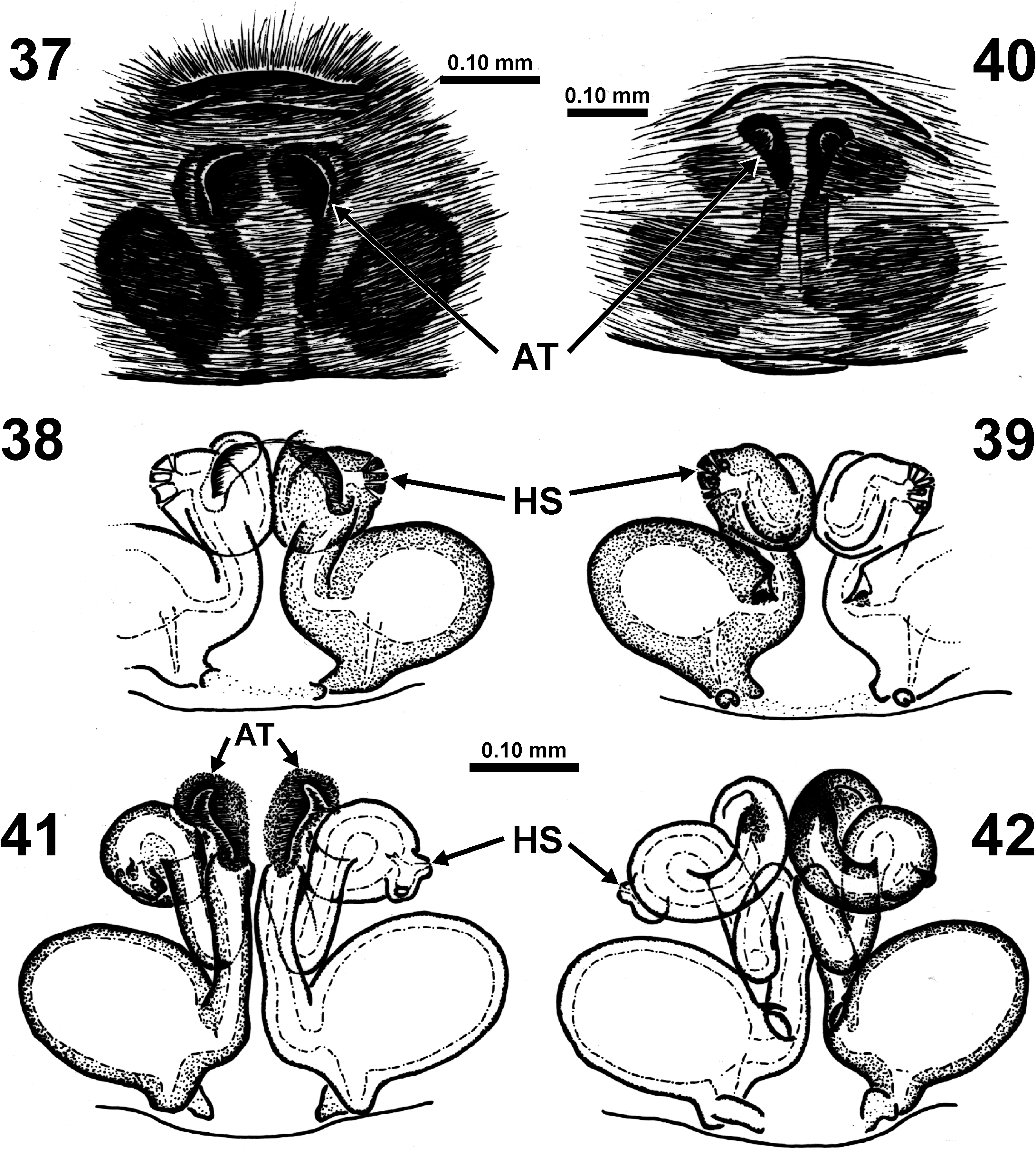
FIGURES 10–15. Cybaeus chauliodous, females (10–12 from Lake Almanor, California; 13–15 from Applegate River, Oregon), copulatory organ. 10, 13 Epigynum, ventral. 11–12, 14–15 Vulva (11, 14 ventral, 12, 15 dorsal). AT—Atrial opening, BG—Bennett’s gland, CD—copulatory duct, HS—head of spermatheca, SS—stalk of spermatheca, W—epigynal “wrinkle”.
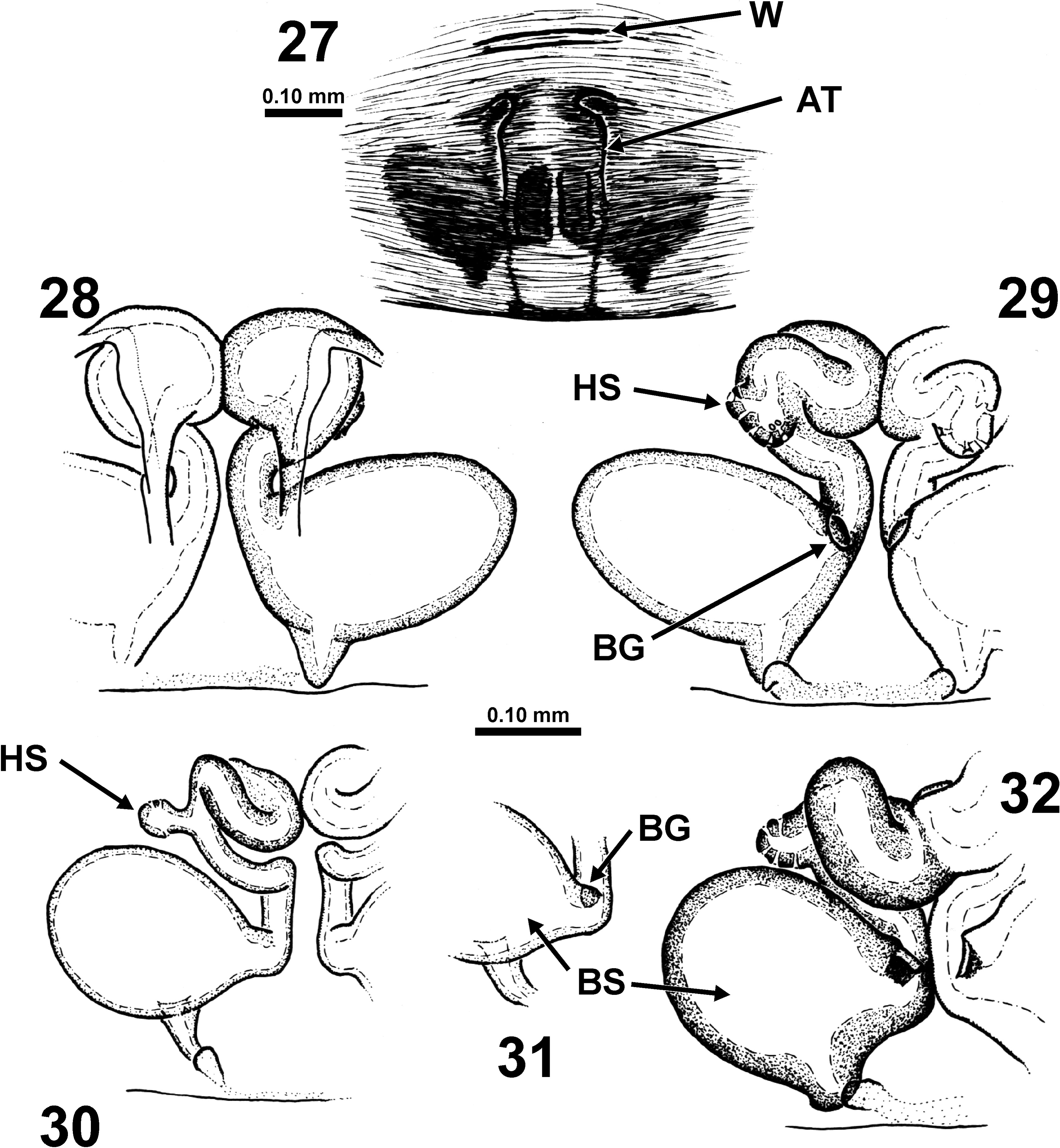
FIGURES 27–32. Cybaeus septatus, females from California (27 from Mt. Shasta, 28–29 holotype, 30–32 from McCloud), copulatory organ. 27 Epigynum, ventral. 28–29, 32 Vulva (28, 32 ventral, 29 dorsal). 30 Subadult vulva, dorsal. 31 Portion of teneral vulva (Bennett’s gland and base of spermatheca), dorsal. AT—Atrial opening, BG—Bennett’s gland, BS—base of spermatheca, HS—head of spermatheca, W—epigynal “wrinkle”.
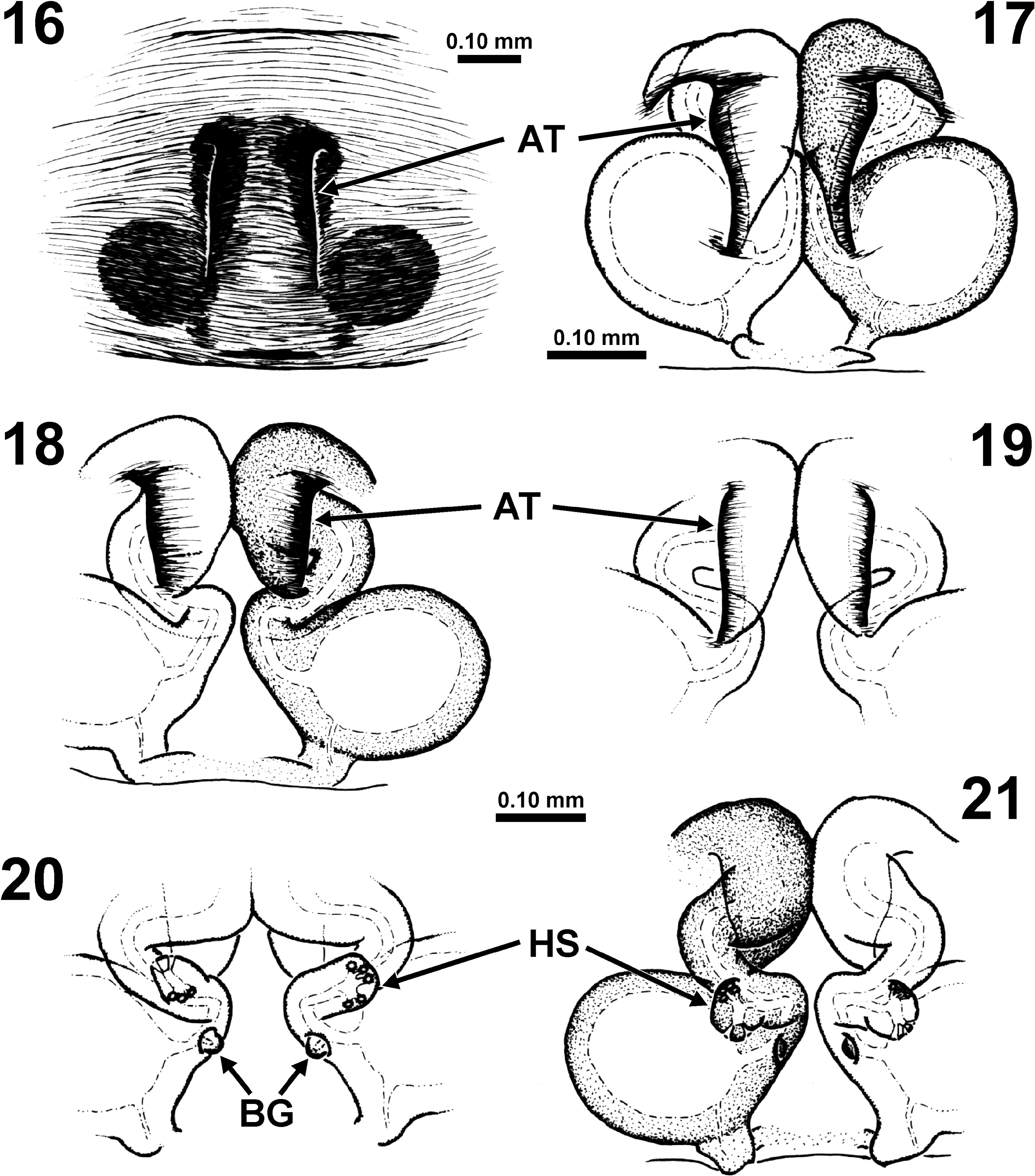
FIGURES 16–21. Cybaeus lockeae spec. nov., females from Castro Valley, California, copulatory organ. 16 Epigynum, ventral. 17–21 Vulva (17–19 ventral, 20–21 dorsal). AT—Atrial opening, BG—Bennett’s gland, HS—head of spermatheca.
No known copyright restrictions apply. See Agosti, D., Egloff, W., 2009. Taxonomic information exchange and copyright: the Plazi approach. BMC Research Notes 2009, 2:53 for further explanation.
1 (by plazi, 2022-02-18 08:07:17)
2 (by ExternalLinkService, 2022-02-18 13:43:16)
3 (by ExternalLinkService, 2022-02-18 15:08:43)
4 (by ExternalLinkService, 2022-02-18 18:41:24)
5 (by ExternalLinkService, 2022-02-28 22:09:30)
6 (by plazi, 2023-11-06 19:15:01)