
Galeripora galeriformis, González-Miguéns & Soler-Zamora & Villar-Depablo & Todorov & Lara, 2022
|
publication ID |
https://doi.org/10.1093/zoolinnean/zlab074 |
|
publication LSID |
lsid:zoobank.org:pub:53637D76-285D-4AB8-9E52-6CDB6F6738D3 |
|
DOI |
https://doi.org/10.5281/zenodo.6461298 |
|
persistent identifier |
https://treatment.plazi.org/id/1C22923F-2949-0977-FC09-4853FD69FB1B |
|
treatment provided by |
Plazi (2022-04-13 07:07:28, last updated 2024-11-25 17:34:24) |
|
scientific name |
Galeripora galeriformis |
| status |
SP. NOV. |
GALERIPORA GALERIFORMIS GONZÁLEZ- MIGUÉNS, SOLER- ZAMORA, VILLAR- DEPABLO, TODOROV & LARA, SP. NOV.
( FIG. 4 View Figure 4 )
Z o o b a n k r e g i s t r a t i o n: u r n: l s i d: z o o b a n k. org:act: 09628A92-07D4-4DA7-BF68-A803B3A9D5D5.
Holotype: MA-Algae11253 .
Specific diagnosis: Test diameter: 71.65–74.95 µm, average 73.20 µm (N = 4); aperture 11.15 to 12.10 µm average 11.50 µm (N = 4). Colour ranges from transparent to yellow-orange. General test shape is rounded and flattened.
The aboral side of the test has a small elevation at the top that gives the test a helmet shape; the surface does not have pores and presents a granular pattern of irregular shape. Large ridges cross the aboral side of the test. The oral side of the test is smooth, covered with an organic matrix that prevents the observation of test building units, with a central aperture. Pores are localized on the brim of the oral side and around the aperture, following a circular pattern. The aperture is invaginated outwards forming a short ring or lip.
Intraspecific variability: The shape and number of ridges on the aboral side of the test can be variable. The number of pores around the aperture is variable. There may be certain deformations in the test that prevent it from having a perfectly circular morphology.
Diagnosis with closely related species: Galeripora galeriformis can be diagnosed by its specific sequences of the mtDNA markers and by its phylogenetic placement (see Molecular phylogeny; Fig. 1 View Figure 1 ). Differs morphologically from other species closely related to Galeripora arenaria by presenting (1) morphometric differences (see Morphometrics and morphology; Fig. 2 View Figure 2 ), with a smaller test and aperture diameters than all species presented here; (2) pores along the edge of the base test; (3) presence of a dome in the top part of the test; and (4) irregular granulations of the top surface of the test.
Habitat: Moss on dry gypsum; terrestrial.
Type locality: Spain, Madrid, Rivas-Vaciamadrid, near Laguna del Campillo (40°19’N 3°30’W).
Etymology: The name is derived from the Latin galerus, helmet, and forma, shape. We propose this name because a Roman helmet is round and wide with a flat brim, similar to the test of this species.
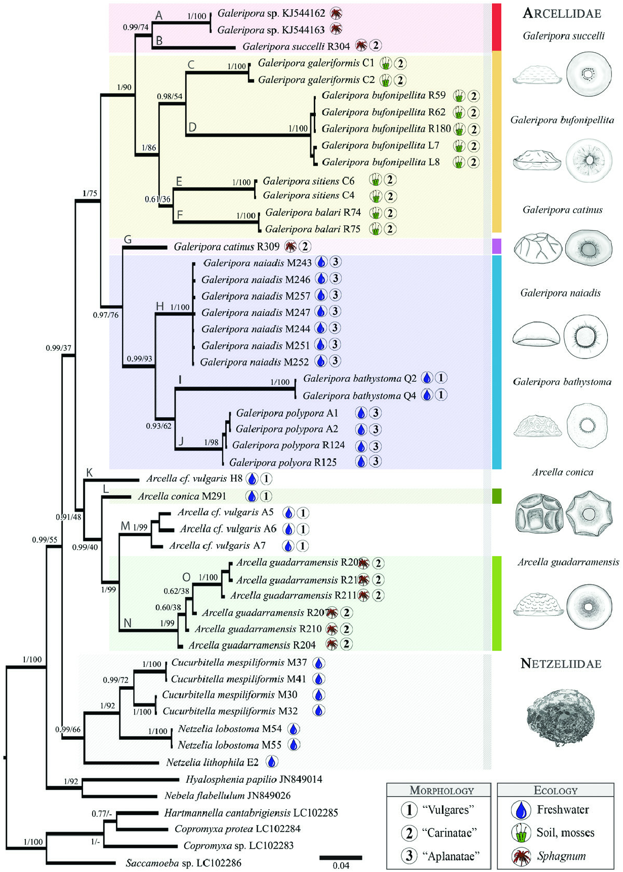
Figure 1. Bayesian phylogenetic tree based on 52 partial sequences COI mtDNA data, 618-nucleotide position alignment. The posterior probability values (Bayesian analysis) and bootstrap values (maximum-likelihood) are represented at each node,with aletterrepresenting the differentmitochondrialclades along thebranches.The coloursrepresent themitochondrial clades that compose the different figures.Next to each species name is the original habitat (freshwater/Sphagnum/terrestrial mosses) and the section according to Deflandre (1928). The drawings show the tests of illustrative species in lateral and oral side views. Drawings by CSZ.
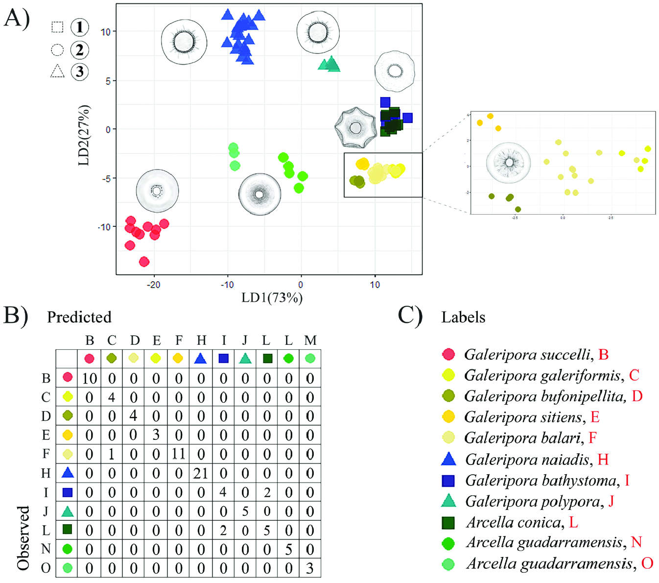
Figure 2. A, scatterplot of the scores of linear discriminants with x-axis representing discriminant function 1 (LD1) and y-axis representing discriminant function 2 (LD2). Colours represent the different mitochondrial clades and symbols refer to the different sections after Deflandre (1928): squares are for Section 1 ‘Vulgares’, circles for Section 2 ‘Carinatae’ and triangles for Section 3 ‘Aplanatae’. The drawings represent the different morphotypes. B, the table represents the results of a linear discriminant analysis which determines the relationship between predicted and observed specimens cells for each mitochondrial clade.
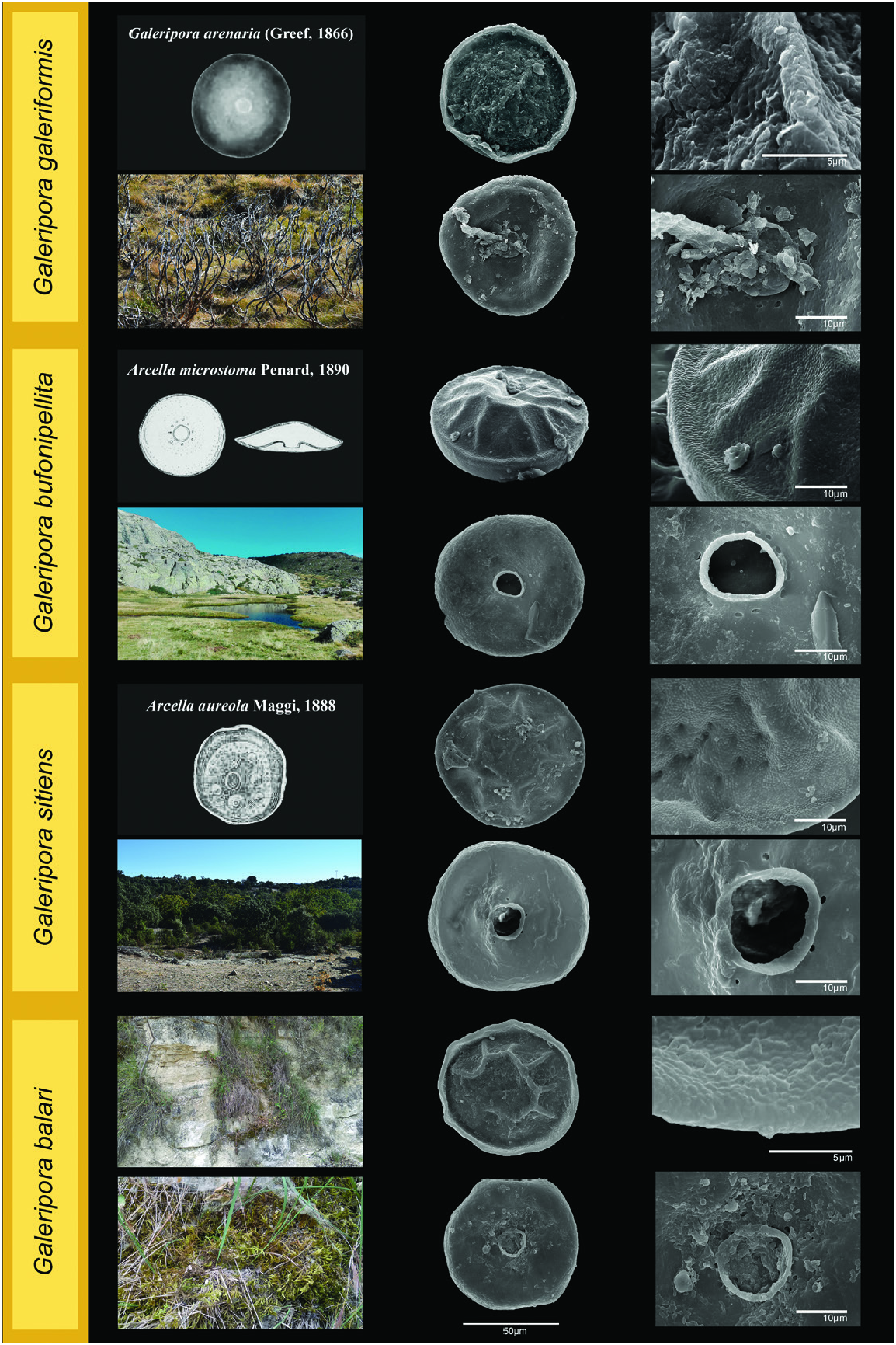
Figure 4. Galeripora galeriformis, Galeripora bufonipellita, Galeripora sitiens and Galeripora balari: scanning electron micrographs of the aboral and oral sides of the test. The images on the right represent detail of the test and the structure of the aperture. On the left, a photograph of a typical habitat for each species, original drawing of the closest resembling species Galeripora arenaria (Greef, 1866), and original drawings of the synonymized species Arcella microstoma Penard, 1890 and Arcella aureola Maggi, 1888.
No known copyright restrictions apply. See Agosti, D., Egloff, W., 2009. Taxonomic information exchange and copyright: the Plazi approach. BMC Research Notes 2009, 2:53 for further explanation.
|
Kingdom |
|
|
Phylum |
|
|
Class |
|
|
Order |
|
|
SubOrder |
Glutinoconcha |
|
InfraOrder |
Sphaerothecina |
|
Family |
|
|
Genus |