
Hebridosimulium, Grenier & Rageau, 1961
|
publication ID |
https://doi.org/10.11646/zootaxa.1380.1.1 |
|
publication LSID |
lsid:zoobank.org:pub:ADA6B48B-CF5D-43A2-8E66-CA946A79A8F8 |
|
persistent identifier |
https://treatment.plazi.org/id/1C1B2B5D-FFB5-FFE5-8748-FB034C1FFB44 |
|
treatment provided by |
Felipe (2021-07-05 19:19:28, last updated by Plazi 2023-11-03 05:45:21) |
|
scientific name |
Hebridosimulium |
| status |
|
Key to later instar larvae of Hebridosimulium
Larvae of S. lucyae and S. anatolicum are unknown.
1. Posterior abdomen steatopygous (expanded posterolaterally and posteroventrally) ( Figs. 11h View FIGURE 11 , 12b View FIGURE 12 ), circlet of hooks directed posteriorly, more than 5,000 hooks ( Fig. 22b View FIGURE 22 ) ........................................................................................................................ 2
- Posterior abdomen callipygous (not markedly expanded) ( Figs. 11a View FIGURE 11 , 12a View FIGURE 12 ), circlet of hooks directed more ventrally, less than 4,000 hooks ( Fig. 22a View FIGURE 22 ) ....................... 3
2 (1). Head darkly pigmented … ( adsonense View in CoL subgroup. Figs. 11m, n View FIGURE 11 ; 14e, f View FIGURE 14 ) ............... 7
- Head pattern with anterior apotome pale or lightly pigmented ( Fig. 13 View FIGURE 13 ) ............... 8
3 (1). Abdomen with dorsolateral tubercles ( tuberculum View in CoL subgroup. Figs. 11b–e View FIGURE 11 ) .......... 4
- Abdomen lacking dorsolateral tubercles ( Fig. 11a View FIGURE 11 ) ........................... S. callipygium View in CoL
4 (3). Head lightly pigmented overall, posteromedial head spots not marked ( Fig. 13b View FIGURE 13 ). .................................................................................................................... S. banksi View in CoL
- Head pale anteriorly, pigmented posteriorly, head spots more distinct ................. 5
5 (4). Posteromedial head spots in distinct Eiffel Tower configuration, lateral head spots distinct ( Fig. 15a View FIGURE 15 . Tanna), or less so (Erromango) .............................. S. johnfrumi View in CoL
- Head spot not as above, posterior apotome pigmented, posterolateral head spots pale ( Figs. 13 View FIGURE 13 , g, h) ................................................................................................. 6
6 (5). Abdomen markedly pigmented and banded. Malekula ( Fig. 11d View FIGURE 11 ). S. pankumuense View in CoL
- Abdomen paler. Efate ( Fig. 11e View FIGURE 11 )......................................................... S. tuberculum View in CoL
7 (2). Ecdysial lines markedly pale and straight, posterior inflexion sharper (Santo) ( Fig. 14e View FIGURE 14 ) ...................................................................................................... S. adsonense View in CoL
- Ecdysial lines markedly pale but curved, posterior inflexion more rounded (Malekula) ( Fig. 14f View FIGURE 14 )............................................................................................. S. spiroi View in CoL
8 (2). Head pale with well-developed posteromedial head spots in Eiffel Tower configuration ....................................................................................................................... 9
- Head pale without distinct head spots, or more evenly, heavily pigmented......... 12
9 (8). Lacking posterolateral pigmentation on apotome ( Fig. 13f View FIGURE 13 ) ......................... S. jolyi View in CoL
- With posterolateral pigmentation on apotome ( Figs. 14a, b View FIGURE 14 )................................ 10
10 (9). Head evenly light brown, posterolateral pigmentation not markedly developed ( Fig. 14a View FIGURE 14 ) ......................................................................................... S. steatopygium View in CoL
- Head markedly pale with posterolateral pigmentation distinct ( Figs. 14b, d View FIGURE 14 )...... 11
11 (10). Posteromedial head spots in Eiffel Tower configuration, posterior edge of apotome markedly emarginate ( Fig. 14d View FIGURE 14 ) ................................................. S. undulatum View in CoL
- Lacking Eiffel Tower configuration, posterior edge of apotome not emarginate ( Fig. 14b View FIGURE 14 ) ...................................................................................... S. subparadisium View in CoL
12 (8). Head evenly pigmented, but anterior apotome pale, muscle scars prominent...... 13
- Head evenly pale, muscle scars not markedly developed ( Fig. 14c View FIGURE 14 ). Distinct dark line and spot above lateral stemmata ( Fig. 15c View FIGURE 15 ) .............................. S. supercilium View in CoL .
13 (12). Anterior genae pale ( Figs. 13g View FIGURE 13 , 15b View FIGURE 15 ; Fiji only) ................................... S. laciniatum View in CoL
- Anterior genae heavily pigmented ( Fig. 13h View FIGURE 13 ) .................................... S. paradisium View in CoL
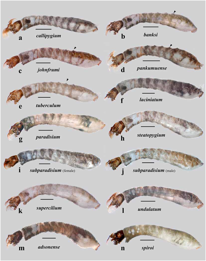
FIGURE 11. Left lateral views of Hebridosimulium species larvae. Last-instar larvae, except l & n, which are penultimate. Arrowheads indicate dorsal tubercles. Scale bar = 1.0 mm.


FIGURE 13. Dorsal views of larval heads of Hebridosimulium species. a–e. callipygium species group, f–h. part of steatopygium species group. Last instars, except f & h, which are penultimate instars. Arrows (b & e) indicate anterior palatal bar. Scale bar = 0.2 mm.

FIGURE 14. Dorsal views of larval heads of Hebridosimulium species. steatopygium species group. Last instars, except d & f, which are penultimate instars. Scale bar = 0.2 mm.
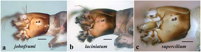
FIGURE 15. Left lateral views of larval heads of Hebridosimulium species. Left labral fan removed for clarity. Last instars except c, which is penultimate. Scale bar = 0.2 mm.

No known copyright restrictions apply. See Agosti, D., Egloff, W., 2009. Taxonomic information exchange and copyright: the Plazi approach. BMC Research Notes 2009, 2:53 for further explanation.
|
Kingdom |
|
|
Phylum |
|
|
Class |
|
|
Order |
|
|
Family |
|
|
Genus |