
Acanthocyrtus, Handschin, 1925
|
publication ID |
https://doi.org/10.11646/zootaxa.5124.3.4 |
|
publication LSID |
lsid:zoobank.org:pub:83E79ECE-70BB-4B88-BBBE-E8DC6565ED49 |
|
DOI |
https://doi.org/10.5281/zenodo.6412886 |
|
persistent identifier |
https://treatment.plazi.org/id/0A09D803-FF8F-242C-FF62-1B11FA139851 |
|
treatment provided by |
Plazi (2022-04-04 06:40:43, last updated 2024-11-27 17:35:25) |
|
scientific name |
Acanthocyrtus |
| status |
|
Key to species of Acanthocyrtus View in CoL
1 Tenent hair chavate ( Fig. 8E View FIGURE 8 ); Australian species............................................................ 2
- Tenent hair pointed; African species................................................ A. marginalis Salmon, 1956 View in CoL
2 Prelabral chaetae ciliate................................................................................ 3
- Prelabral chaetae smooth............................................................................... 4
3 Head with 7 sutural mac; Th II–III respectively with 9 and 7 mac in p1–3 complex; Abd I with 4 mac; Abd IV with 19 central mac.......................................................................... A. lineatus Womersley, 1934 View in CoL
- Head with 5 sutural mac; Th II–III respectively with 4 and 5 mac in p1–3 complex; Abd I with 2 mac; Abd IV with 15 central mac............................................................................ A. spinosus ( Schött, 1917) View in CoL
4 Labral papillae present ( Figs 3E View FIGURE 3 , 9C View FIGURE 9 )..................................................................... 5
- Labral papillae absent.................................................................................. 6
5 Head and Abd III–IV with lateral spots ( Fig. 2 View FIGURE 2 ); labral inner papillae conical, outer papillae reduced to a small rounded projection ( Fig. 3E View FIGURE 3 ); labial papilla E l.p. surpass the base of a.a. ( Fig. 4A View FIGURE 4 ); manubrium ventro-distally with 3 inner chaetae ( Fig. 7B View FIGURE 7 ) A. necropolitanus View in CoL sp. nov.
- Head and trunk depigmented ( Fig. 8 View FIGURE 8 ); labral inner papillae with 3 projections and outer papillae with 4 projections ( Fig. 9C View FIGURE 9 ); labial papilla E l.p. not reach the base of a.a. ( Fig. 9E View FIGURE 9 ); manubrium ventro-distal with 2 inner chaetae ( Fig. 11F View FIGURE 11 )................................................................................................ A. pallidus View in CoL sp. nov.
6 Ant IV apical bulb bilobed; Th II with 24 mac in p1–3 complex; Abd III with 5 lateral mac; Abd IV with 20 central mac; mucronal proximal tooth clearly smaller than distal, mucronal basal spine surpassing the proximal tooth........................................................................... A. barrowensis Zhang, 2009 View in CoL (in: Zhang et al. 2009)
- Ant IV apical bulb absent; Th II with 17 or less mac in p1–3 complex; Abd III with 4 lateral mac; Abd IV with 15 or less central mac; mucronal teeth subequal, mucronal spine reaching the apex of proximal tooth................................. 7
7 Head S1 and S4 mac present and Pe5 absent; labial basomedian field with 3 smaller chaetae (M2, R and RS); Th II with 6 and 17 mac respectively in m2 and p3 complex; unguis a.t. absent............ A. loftyensis Zhang, 2009 View in CoL (in: Zhang et al. 2009)
- Head S1 and S4 mac absent and Pe5 present; labial basomedian field with 1 smaller chaetae (M2, RS absent); Th II with 5 and 15 mac respectively in m2 and p3 complex; unguis a.t. present............. A. yolngui Zhang, 2009 View in CoL (in: Zhang et al. 2009)
Salmon, J. T. (1956) Contribution a l'etude de la faune entomologique du Ruanda - Urundi (Mission P. Basilewski, 1953). Annales du Museum Royal du Congo Belge, 51, 9 - 40.
Schott, H. (1917) Results of Dr. E. Mjoberg's Swedish Scientific Expeditions to Australia 1910 - 1913. N ° 15, Collembola. Arkiv for Zoologi, 11 (8), 1 - 60, 4 pl. https: // doi. org / 10.5962 / bhl. part. 1497
Womersley, H. (1934) A preliminary account of the Collembola - Arthropleona of Australia II (Entomobryoidea). Transactions of the Royal Society of South Australia, 58, 86 - 138.
Zhang, F., Deharveng, L., Greenslade, P. & Chen, J. X. (2009) Revision of Acanthocyrtus (Collembola: Entomobryidae), with description of a new genus from eastern Asia. Zoological Journal of the Linnean Society, 157, 495 - 514. https: // doi. org / 10.1111 / j. 1096 - 3642.2008.00521. x
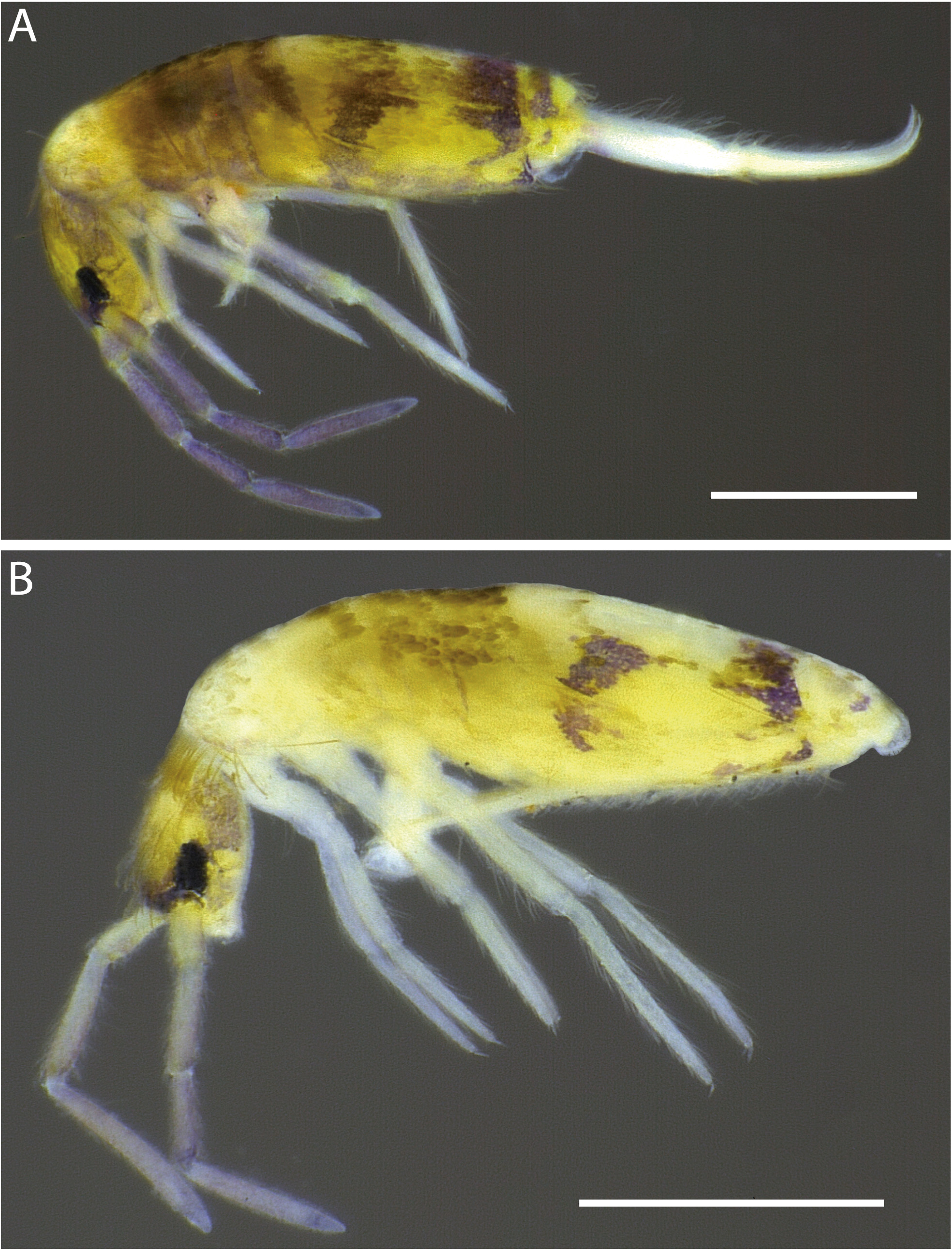
FIGURE 2A–B. Acanthocyrtus necropolitanus sp. nov., habitus of specimens fixed in ethanol (lateral view): A, specimen with weak pigments on trunk lateral; B, pigments absent on trunk lateral. Scale bars: 0.5mm.

FIGURE 3A–F. Acanthocyrtus necropolitanus sp. nov., antennae and dorsal head; A, apex of left Ant IV (dorsal view); B, Ant III distally (lateral view); C, chaetotaxy (mac and elongated sens) of left Ant III–I (dorsal view, from left to right), respectively; D, chaetotaxy of clypeus and prelabrum; E, labral papillae (ventral view); F, head dorsal chaetotaxy and eyes (dorsal view, left side).
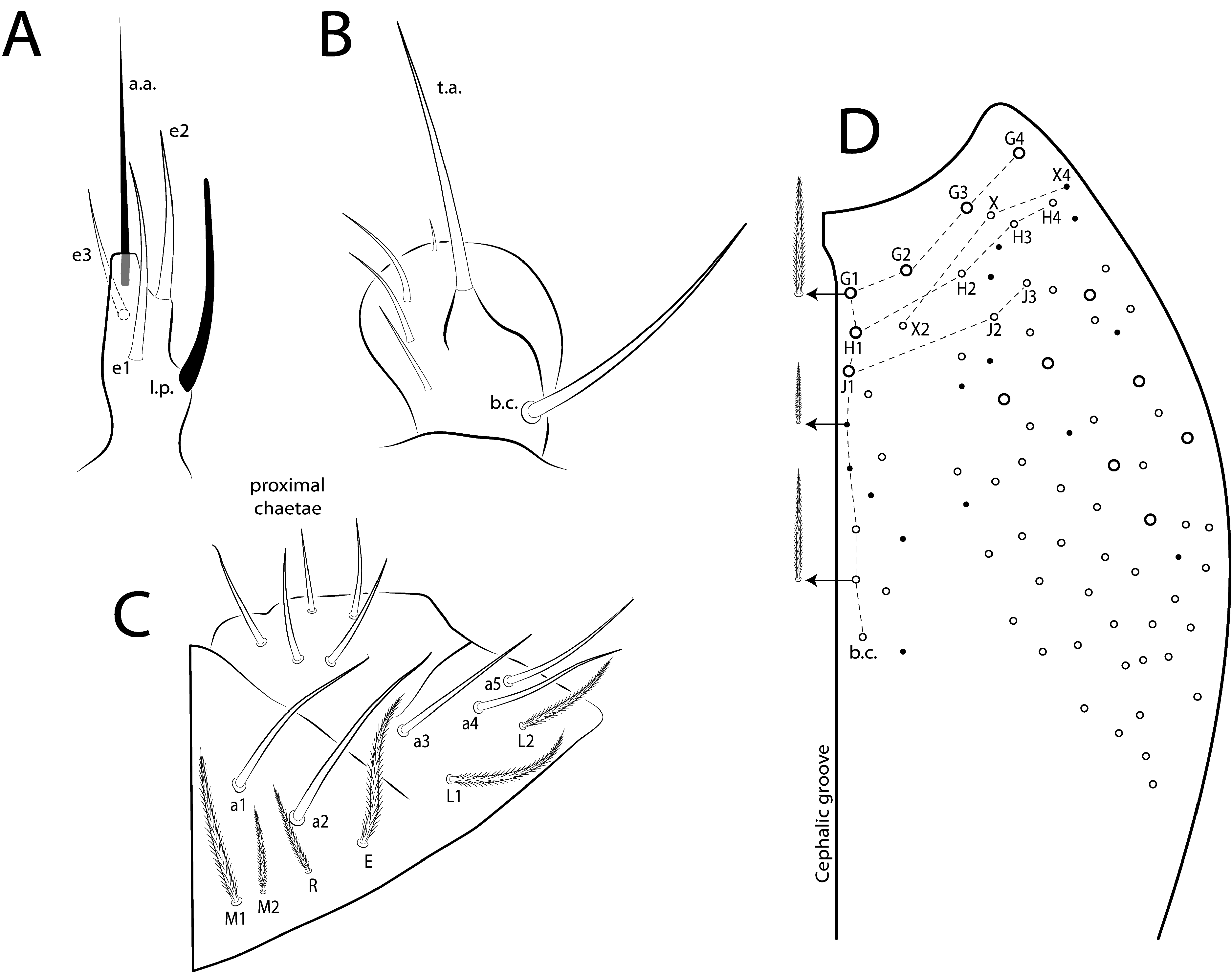
FIGURE 4A–D. Acanthocyrtus necropolitanus sp. nov., ventral head (right side); A, labial papillae E (right side); B, maxillary palp and sublobal plate (right side); C, basomedian and basolateral labial fields and proximal chaetae; D, complete postlabial chaetotaxy.

FIGURE 7A–E. Acanthocyrtus necropolitanus sp. nov., abdominal appendages; A, collophore (lateral view); B, distal manubrium (ventral view); C, manubrial plate (dorsal view), arrow indicates chaeta present or absent; D, dens spines chaetotaxy (dorsal view), E, distal dens and mucro (dorsal view).
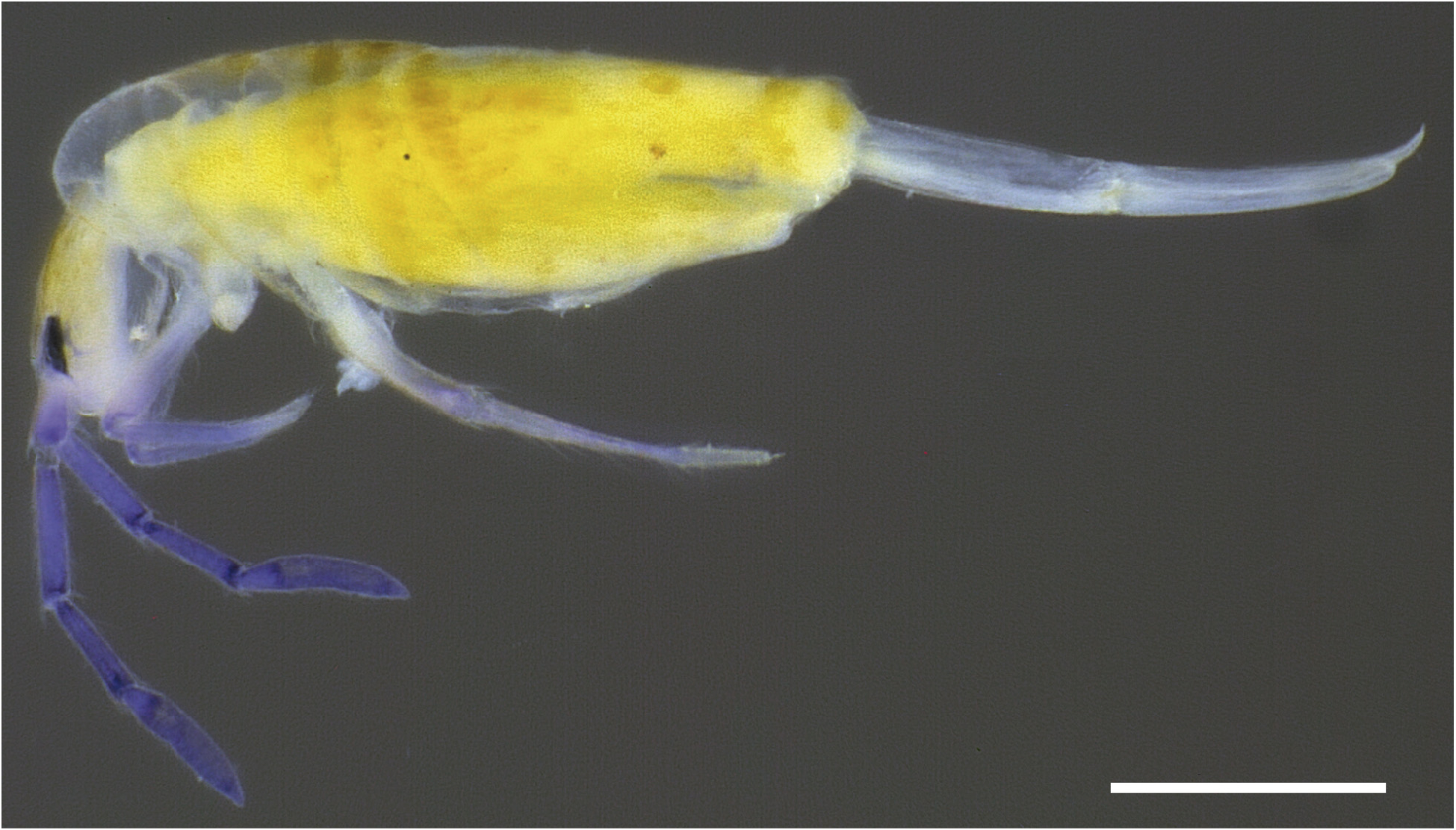
FIGURE 8. Acanthocyrtus pallidus sp. nov., habitus of specimens fixed in ethanol (lateral view). Scale bars: 0.5mm.
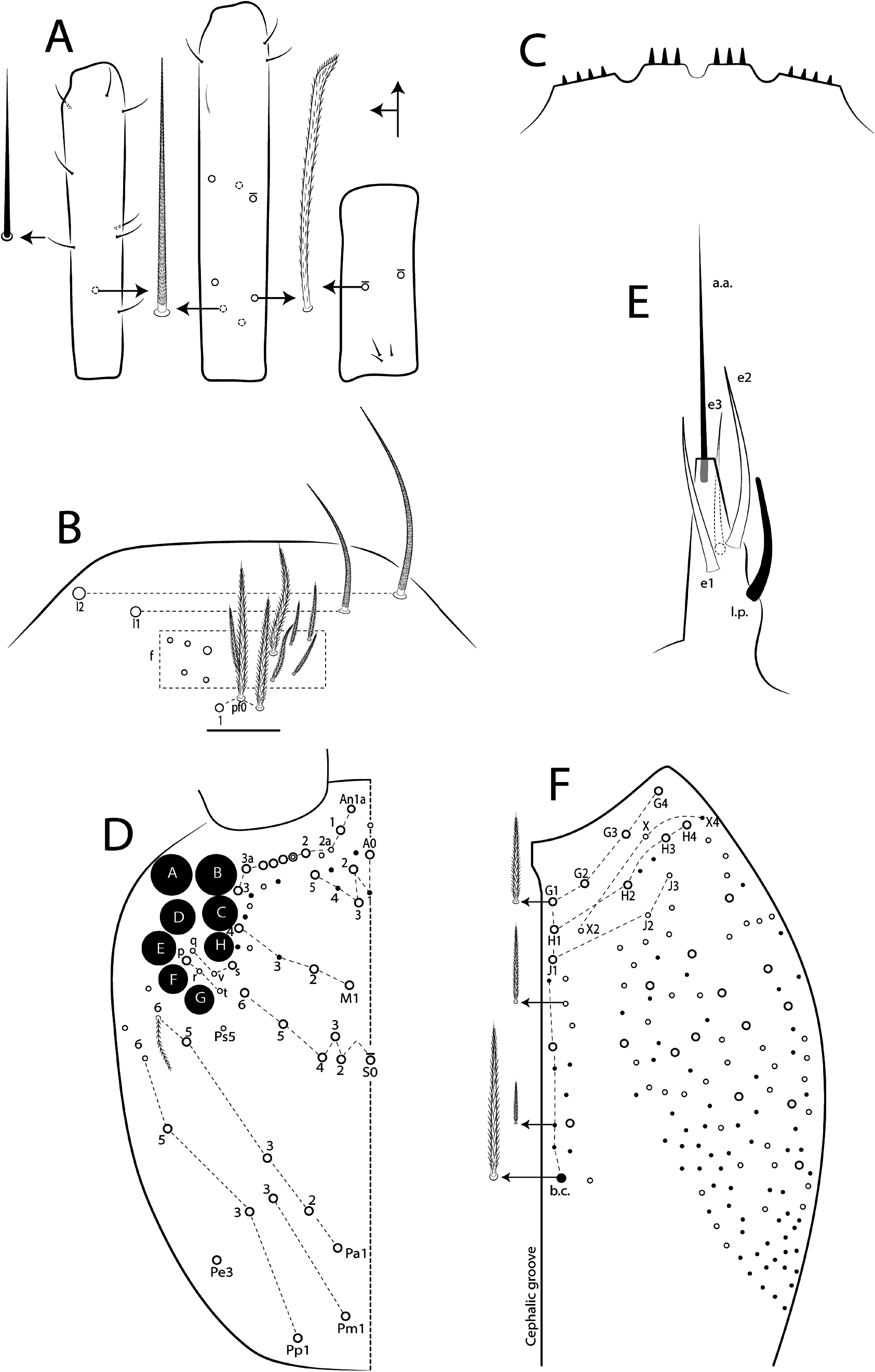
FIGURE 9A–F. Acanthocyrtus pallidus sp. nov., antennae and head; A, chaetotaxy (mac and elongated sens) of left Ant III–I (dorsal view, from left to right), respectively; B, chaetotaxy of clypeus (dorsal view); C, labral papillae (ventral view); D, head dorsal chaetotaxy and eyes (dorsal view, left side); E, labial papillae E (ventral view, right side); F, complete postlabial chaetotaxy (ventral view, right side).
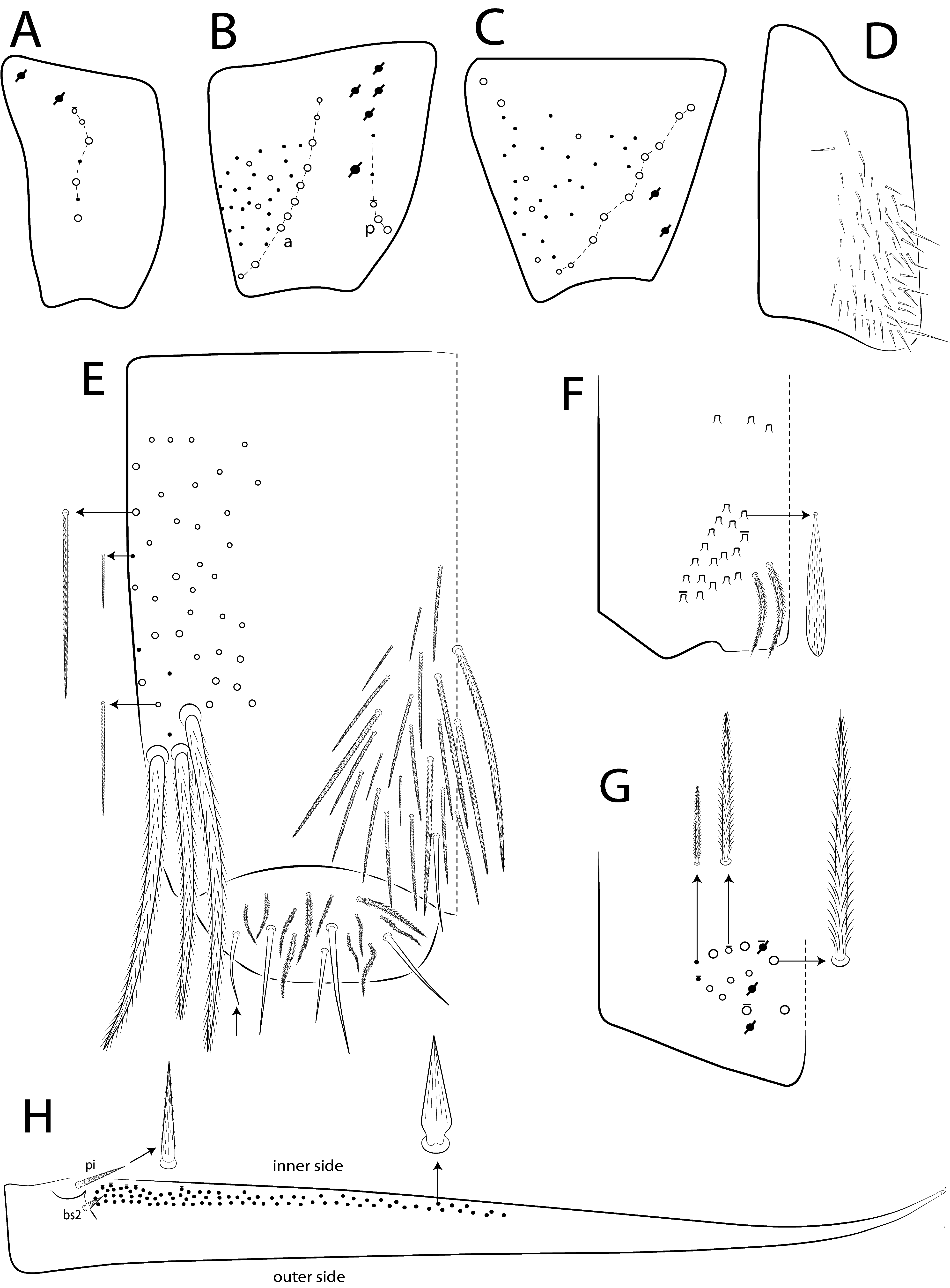
FIGURE 11A–H. Acanthocyrtus pallidus sp. nov., legs and abdominal appendages; A–C, chaetotaxy of left subcoxa I–III respectively (outer side); D, trochanteral organ (posterior view); E, collophore (lateral view), arrow on lateral flap indicate smooth chaeta present or absent; F, distal manubrium (ventral view); G, manubrial plate (dorsal view); H, dens spines chaetotaxy (dorsal view),
No known copyright restrictions apply. See Agosti, D., Egloff, W., 2009. Taxonomic information exchange and copyright: the Plazi approach. BMC Research Notes 2009, 2:53 for further explanation.
|
Kingdom |
|
|
Phylum |
|
|
Class |
|
|
Order |
|
|
Family |
|
|
SubFamily |
Entomobryinae |
1 (by plazi, 2022-04-04 06:40:43)
2 (by ExternalLinkService, 2022-04-04 08:13:01)
3 (by ExternalLinkService, 2022-04-04 19:58:01)
4 (by ExternalLinkService, 2022-04-04 20:04:02)
5 (by ExternalLinkService, 2022-04-04 21:23:37)
6 (by plazi, 2023-11-06 22:50:55)