
Physodera amplicollis
|
publication ID |
https://doi.org/10.11646/zootaxa.4243.2.3 |
|
publication LSID |
lsid:zoobank.org:pub:7393131D-564F-417C-817E-AC75C2BCD2C4 |
|
DOI |
https://doi.org/10.5281/zenodo.6046738 |
|
persistent identifier |
https://treatment.plazi.org/id/042587AE-3A04-FFEE-0E80-52C4FBEFFD11 |
|
treatment provided by |
Plazi (2017-03-15 08:23:58, last updated 2024-11-26 07:19:35) |
|
scientific name |
Physodera amplicollis |
| status |
|
amplicollis View in CoL -group
The amplicollis -group contains three species: P. amplicollis van de Poll , P. diglena Andrewes , and P. bousqueti Mateu.
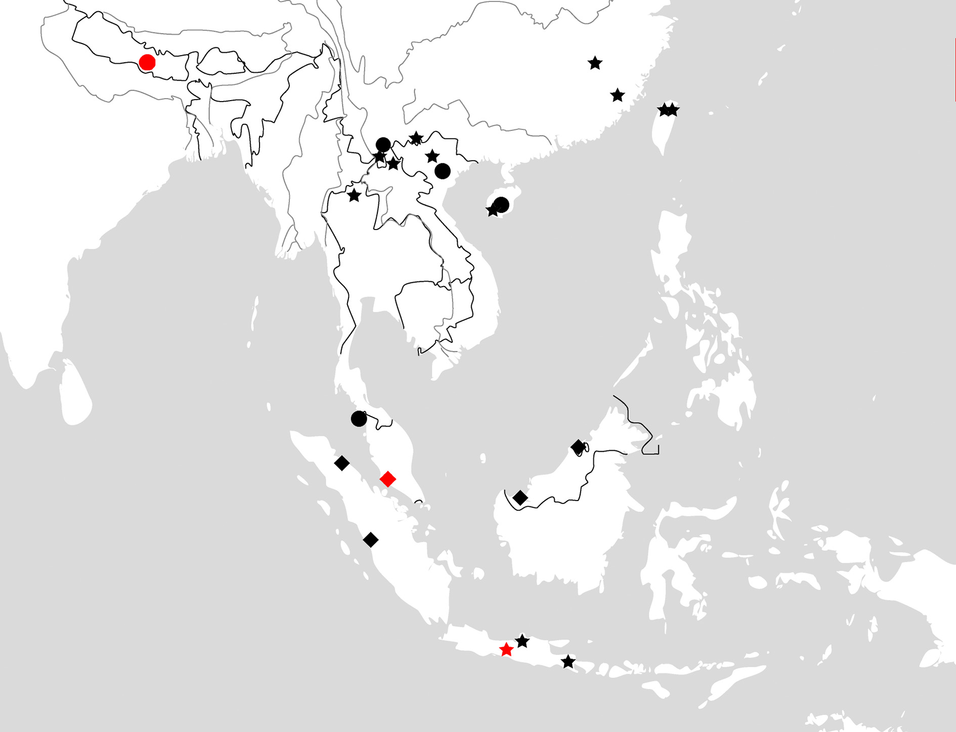
This species group distributes in southeastern Asia, including southern Chinese continent, Taiwan Island, Indo- China Peninsula, Malay Peninsula, Borneo, Java, and Sumatra ( Fig.63 View FIGURE 63 ).
The diagnostic characters of this species group are: Pronotum and elytra with distinct pattern; tergum VII yellowish, with distinct black pattern: three well defined spots or one central spot and vague lateral ones ( Figs. 45, 46). Terminal labial palpomeres securiform in both sexes (Fig. 61); male mesotarsus with adhesive hairs at least well developed on the first tarsomere; males with two pairs of setae on sternum VII. Aedeagus gently slender, with apical lamella large, strongly oblique to right side; internal sac with main flagellum not reaching apical orifice; trumpet-form expansion small, length about 0.3 times of the main flagellum; secondary flagellum long and strongly sclerotized; apical bursa distinct ( Figs. 34–36 View FIGURE 34 View FIGURE 35 View FIGURE 36 ).
These three species are close in the following similarities: (1) aedeagal apical lamella longer than any other species in Physodera ; (2) secondary flagellum of aedeagal internal sac about half as main flagellum, main flagellum apex not reaching apical orifice; (3) tergum VII with three dark spots (lateral spots vague in P. bousqueti ) ( Figs. 45 B, 46B); (4) the fifth elytral interval with only one basal setigerous pore.
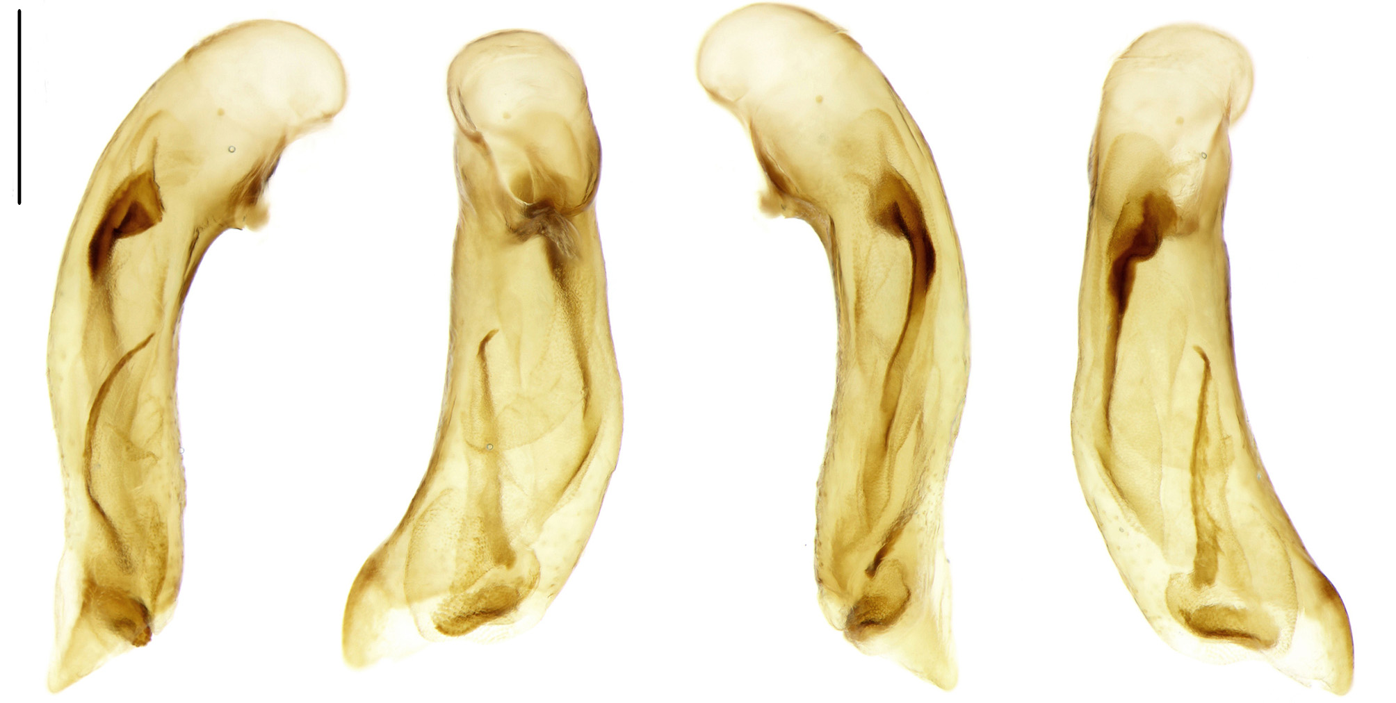
FIGURE 34. Median lobe of male genitalia, right-lateral, ventral, left-lateral, and dorsal view of P. amplicollis van de Poll, Taiwan, scale bar = 0.5 mm.

FIGURE 35. Median lobe of male genitalia, right-lateral, ventral, left-lateral, and dorsal view of P. bousqueti Mateu, Vietnam, scale bar = 0.5 mm.

FIGURE 36. Median lobe of male genitalia, right-lateral, ventral, left-lateral, and dorsal view of P. diglena Andrewes, Borneo, scale bar = 0.5 mm.
No known copyright restrictions apply. See Agosti, D., Egloff, W., 2009. Taxonomic information exchange and copyright: the Plazi approach. BMC Research Notes 2009, 2:53 for further explanation.
|
Kingdom |
|
|
Phylum |
|
|
Class |
|
|
Order |
|
|
Family |
|
|
Tribe |
Lebiini |
|
SubTribe |
Physoderina |
|
Genus |