
Didemnum flammacolor, Rocha, Rosana Moreira Da, Neves, Isabela Monteiro & Gamba, Gustavo Antunes, 2015
|
publication ID |
https://doi.org/10.11646/zootaxa.3905.3.4 |
|
publication LSID |
lsid:zoobank.org:pub:A6BB4C82-F905-46CC-BA7F-1D6EECD0455B |
|
DOI |
https://doi.org/10.5281/zenodo.6105537 |
|
persistent identifier |
https://treatment.plazi.org/id/03FEBD67-FFDF-FFE6-FF2D-FD97FAEDEE6D |
|
treatment provided by |
Plazi (2016-04-17 22:48:05, last updated 2024-11-27 14:02:14) |
|
scientific name |
Didemnum flammacolor |
| status |
sp. nov. |
Didemnum flammacolor sp. nov. Rocha & Neves
( Figs 4 View FIGURE 4 , 5 View FIGURE 5 )
Examined material: Holotype: MZUSP 0 0 0 90 one colony, Ondina, Salvador, Bahia, 13o00’33” S, 38o31’41” W, Col. R. M. Rocha, 0 6.06.2004.
Etymology. The name refers to the red color of the living colony.
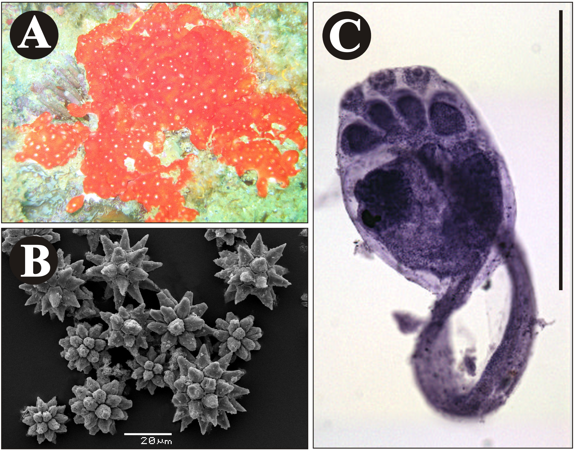
The encrusting colony is approximately 3 cm long and 1 mm thick. Colony surface is red and the rim of cloacal cavities is white due to the presence of spicules ( Fig. 4 View FIGURE 4 A); when fixed in formaldehyde, the color fades to pinkish. Spicules are abundant throughout the surface, giving the colony a firm and brittle consistency; spicules are less abundant in the middle and lower layers of the tunic. Spicules are stellate, approximately 30 Μm in diameter with eight tapered rays in cross section; some spicules are approximately 20 Μm in diameter and have 7–10 conical and truncated rays in optical transverse section ( Fig. 4 View FIGURE 4 B).
Zooids are about 1 mm long. The oral siphon has six small triangular lobes. The atrial aperture is wide open, exposing most of the pharynx and an atrial languet is absent. The muscular process projects from the esophagealrectal peduncle and is variable in length and may be absent in some zooids, and tends to be shorter than the abdomen. Thoracic organs are circular, 80 µm in length, protruding, at the level of the fourth row of pharyngeal slits. There are between 6 and 7 stigmata per row on each side of the pharynx ( Fig. 5 View FIGURE 5 A).
The abdomen is larger than the thorax. One small stolonic vessel was observed in some zooids. The esophagus is short. The location and shape of the stomach are typical; the duodenum is shorter than the stomach; the intestinal loop forms a deep secondary loop, covering the stomach. The single testis is spherical and surrounded by nine coils of the sperm duct ( Fig. 5 View FIGURE 5 B). No oocytes were found.
Larvae are oval, trunk is approximately 0.4 mm in length, around which the tail winds three-quarters of the way. Larvae are non-pigmented, have three linearly arranged adhesive papillae that are close together on short stalks, plus four pairs of rounded ectodermal ampullae. The sensory vesicle (including ocellus and otolith) is in the mid-dorsal region of the larval trunk ( Figs. 4 View FIGURE 4 C, 5C).
Remarks. The known Didemnum species with reddish colonies differ from D. flammacolor in many ways. Didemnum moseleyi ( Herdman, 1886) and D. pseudofulgens Médioni, 1970 have spicules with a greater number of short, conical rays ( Kott 2001; Médioni 1970). Didemnum rodriguesi colonies have a reticulate pattern on the surface of the tunic and a different number of larval ampullae ( Rocha & Monniot 1993; Monniot 2010). Didemnum membranaceum Sluiter, 1909 has larger spicules (0.1 mm) with 4–6 conical rays in transverse optical section ( Monniot & Monniot 2008). Didemnum madeleinae Monniot & Monniot, 2001 have larvae with 12 or more pairs of ampullae and the trunk is longer ( Monniot & Monniot 2001). Didemnum coccineum Von Drasche, 1883 and D. amourouxi Lafargue, 1976 also have a greater number of ampullae but only two adhesive papillae ( Lafargue & Wahl 1987). Some other species of Didemnum have 9 turns of sperm duct and larvae with 3 adhesive papillae and 4 pairs of ampullae. Didemnum crescente Kott, 2001 has larger spicules (70 µm), longer retractor muscles and 0.6 mm long larval trunk ( Kott 2001). Didemnum cuculiferum ( Sluiter, 1909) has longer retractor muscle and larger larvae (0.57 mm). Didemnum fragilis Sluite 1909 , has a fragile colony with circular spicules and larger larvae (0.78 mm) ( Monniot & Monniot 1987). Didemnum hiopaa Monniot & Monniot, 1987 form small and transparent colonies and has spicules with larger number of rays. Didemnum incantum ( Herdman, 1899) has longer retractor muscle, fewer pharyngeal stigmata in each row (4–6), and the spicules rays have a tetrahedral basis. Didemnum vulgare Kott, 2001 has larger spicules (60 µm) with more rays and longer retractor muscle ( Kott, 2001).
Herdman, W. A. (1886) Report on the Tunicata collected during the years 1873 - 1876. Part 2. Ascidiae Compositae. Zoological Challenger Expedition, 14 (38), 1 - 425.
Herdman, W. A. (1899) Descriptive catalogue of the Tunicata in the Australian Museum Sydney N. S. W. Australian. Australian Museum, Sydney- Catalogue, 17, 1 - 139. http: // dx. doi. org / 10.5962 / bhl. title. 1561
Kott, P. (2001) The Australian Ascidiacea. Part 4, Aplousobranchia (3), Didemnidae. Memoirs of the Queensland Museum, 47 (1), 1 - 407.
Lafargue, F. (1976) Revision taxonomique des didemnidae des cotes de France (Ascidies Composes). Les especes de Banyuls- Sur-Mer. Genre Didemnum. Deuxieme partie: Larves a deux ventouses. Annales de l'Institut Oceanographique, 52 (2), 259 - 281.
Lafargue, F. & Wahl, M. (1987) The didemnid ascidian fauna of France. Annales de l'Institut Oceanographique, 63 (I), 1 - 46.
Medioni, A. (1970) Ascidies du benthos rocheux de Banylus-Sur-Mer. Didemnidae (Ascidies Composees). Vie et Milieu, Series A, Biologie Marine, 21 (1 A), 25 - 48.
Monniot, C. & Monniot, F. (1987) Les ascidies de Polynesie Francaise. Memoires du Museum National d'Histoire Naturelle, Zoologie, 136, 1 - 155.
Monniot, F. & Monniot, C. (2001) Ascidians from the tropical western Pacific. Zoosystema, 23 (2), 201 - 383.
Monniot, F. & Monniot, C. (2008) Complements sur la diversite des ascidies (Ascidiacea, Tunicata) de l'ouest Pacifique tropical. Zoosystema, 30 (4), 799 - 872.
Monniot, F. (2010) Some new data on tropical western Pacific Ascidians. Zootaxa, 2561, 1 - 29.
Rocha, R. M. & Monniot, F. (1993) Didemnum rodriguesi sp. nov., a new didemnid tunicate common to southern Brazil and New Caledonia. Annales de l'Institut Oceanographique, 69 (2), 261 - 265.
Sluiter, C. P. (1909) Die Tunicaten der Siboga Expedition. Pt II. Die merosomen Ascidien. Siboga Expedition, 56 B, 1 - 112.
Von Drasche, R. (1883 - 1884) Die Synascidien der Bucht von Rovigno (Istrien). Ein Beitrag zur Fauna der Adria. Carl Gerolds Sohn, Wien, 41 pp.

| MZUSP |
Museu de Zoologia da Universidade de Sao Paulo |
No known copyright restrictions apply. See Agosti, D., Egloff, W., 2009. Taxonomic information exchange and copyright: the Plazi approach. BMC Research Notes 2009, 2:53 for further explanation.
|
Kingdom |
|
|
Phylum |
|
|
SubPhylum |
Tunicata |
|
Class |
|
|
Order |
|
|
Family |
|
|
Genus |
1 (by plazi, 2016-04-17 22:48:05)
2 (by ImsDioSync, 2017-01-13 23:18:51)
3 (by ImsDioSync, 2017-01-13 23:19:52)
4 (by ExternalLinkService, 2019-09-26 13:50:11)
5 (by ExternalLinkService, 2022-01-30 02:56:07)
6 (by ExternalLinkService, 2022-02-16 11:21:32)
7 (by plazi, 2023-10-29 04:30:55)