
Amplirhagada gibsoni, Köhler, 2010
|
publication ID |
https://doi.org/10.3853/j.0067-1975.62.2010.1554 |
|
publication LSID |
lsid:zoobank.org:pub:749A271A-6942-4D4C-B0DF-B968462BFF0C |
|
persistent identifier |
https://treatment.plazi.org/id/9CDFCE7C-399C-4473-95D7-E8C49F674AB5 |
|
taxon LSID |
lsid:zoobank.org:act:9CDFCE7C-399C-4473-95D7-E8C49F674AB5 |
|
treatment provided by |
Felipe (2021-08-21 19:30:25, last updated by Plazi 2023-11-04 06:11:23) |
|
scientific name |
Amplirhagada gibsoni |
| status |
sp. nov. |
Amplirhagada gibsoni View in CoL n.sp.
Type locality ( Fig. 1 View Figure 1 ). Western Australia, Kimberley, Bonaparte Archipelago, Boongaree Island , central section, 15°04'15"S 125°11'14"E; KIS 1-30. Dry vine thicket on west facing sandstone scree below escarpment, under rocks (leg. M. Shea, 09 August 2007) GoogleMaps .
Type material. Holotype WAM S34617 View Materials (Pl. 2.1) . Paratypes AMS C463709 (1 preserved specimen, as holotype), WAM S36650 View Materials (2 preserved specimens, as holotype), WAM S36468 View Materials (6 shells, 15°04'15"S 125°11'14"E) GoogleMaps , AMS C463708 (4 shells, 15°04'36"S 125°11'18"E), WAM S36469 View Materials (10 shells, 15°04'36"S 125°11'18"E) GoogleMaps , WAM S36651 View Materials (1 preserved specimen, 15°04'36"S 125°11'18"E) GoogleMaps .
Etymology. Named in honour of Lesley Gibson, Western Australian Department of Environment and Conservation, in recognition of her support for my work.
Shell ( Fig. 59A–C View Figure 59 , Pl. 2.1–2). Semi-globose to conical with medium high spire; thin (translucent) to solid. Periphery evenly rounded to slightly angulate; upper and basal sectors of whorls rounded. Umbilicus open, forming a narrowly winding chink, about 90 percent concealed by columellar reflection. Background colour light brownish to horn; uniform. Outer lip colour same as shell, inner lip colour white. Protoconch c. 2.2 mm in diameter, comprising 2 whorls, with fine and distinct axial lirae. Teleoconch with fine axial lirae, rounded in cross-section, regularly spaced, spaces equal to thickness of lirae, evenly distributed across shell surface, reduced underneath suture; across whorls of shell. Angle of aperture 30°; outer lip rounded, sharp to moderately thick, slightly expanded, slightly reflected; basal node absent or very weak, palatal node absent. Parietal wall of inner lip inconspicuous.
Pallial morphology. Pallial cavity deep, extending one whorl; mantle pigmentation mottled, black. Kidney extending about half of pallial cavity.
Genital morphology ( Figs. 57–58 View Figure 57 View Figure 58 ). Penis straight, more or less of same length as anterior part of oviduct. Vas deferens forms simple loop before entering penis. Penial retractor muscle as long as penis. Penial sheath evenly thick. Penial verge very long (c. 1 ⁄ 3 penial chamber), broad, with pointed tip. Penial wall pustules of normal size, randomly and densely arranged over entire length of inner penial wall. At base of penial chamber fused rows of pustules form 3–4 smooth longitudinal pilasters. Main stimulatory pilaster absent. Vas deferens entering penial sheath in upper third. Vagina tubular; inner vaginal wall with smooth longitudinal pilasters. Spermathecal duct moderately wide, internally with smooth longitudinal pilasters. Spermathecal head elongately inflated, connected with oviduct by connective tissue, internally smooth, with delicate wall. Free oviduct comprising about half of anterior part of oviduct, more or less straight. Spermoviduct longer than anterior part of oviduct. Talon embedded in albumen gland close to anterior end of albumen gland.
Radular morphology ( Fig. 59E–F View Figure 59 ). Rectangular. Only a partial fragment was studied with 32 rows per mm. Central teeth with sharply pointed, triangular mesocones, shorter than base of teeth. Central ectocones reduced. Lateral teeth with sharply pointed, triangular to ovate mesocones. Lateral ectocones tiny, endocones reduced. Marginals with triangular mesocones. Marginal ectocones smaller and narrower than mesocones, endocones smaller than ectocones.
Comparative remarks. Shell smaller in size, not as elevated in shape as in congeners from the same island. Large pustules and very large penial verge are also diagnostic. A lateral pocket off the penial chamber as mentioned by Solem (1991) was not observed. This species was referred to as “ Amplirhagada sp. 32” by Solem (1991).

Figure 1. Distribution of Amplirhagada species in the Kimberley region, northwestern Australia. (1) A. euroa n.sp., Adolphus Island. (2) A. solemiana n.sp., Middle Osborn Island. (3) A. indistincta n.sp., Southwest Osborn Island, Kidney Island. (4) A. combeana, Cassini Island. (5) A. mckenziei n.sp., Oliver Island. (6) A. ponderi n.sp., Kingsmill Island. (7) A. montesquieuana n.sp., Fenelon Island. (8) A. descartesana n.sp., Descartes Island. (9) A. katerana, Katers Island. (10) A. puescheli n.sp., unnamed island near Prudhoe Island. (11) A. decora n.sp., A. kessneri n.sp., Bigge Island. (12) A. berthierana n.sp., Berthier Island. (13) A. lamarckiana n.sp., Lamarck Island.(14) A. anderdonensis n.sp., unnamed island, Anderdon Islands. (15) A. tricenaria n.sp., Prince Frederick Harbour, north of Hunter River mouth. (16) A. regia n.sp., A. boongareensis n.sp., A. gibsoni n.sp., and Amplirhagada sp., Boongaree Island. (17) A. yorkensis n.sp., Coronation Island. (18) A. buffonensis n.sp., Buffon Island. (19) A. uwinsensis n.sp., Uwins Island. (20) A. sphaeroidea n.sp., St. Andrews Island. (21) A. basilica n.sp., and A. camdenensis n.sp., Augustus Island. (22) A. kimberleyana n.sp., NW of Wilson Point. (23) A. gemina n.sp., 1.5 km SE of Hall Point. (24) A. dubitabile n.sp., Steep Island. (25) A. napierana, north Napier ranges. (26) A. percita, Napier ranges. (27) A. castra, A. mitchelliana, and A. varia, Mitchell Plateau.
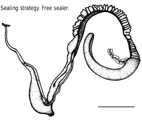
Figure 57. Genitalia of Amplirhagada gibsoni n.sp., paratype AMS C463709 (8 August, scale 10 mm). Compare with Fig. 3 for labelling of structures.

Figure 58. Interior of penial chamber of Amplirhagada gibsoni n.sp., paratype AMS C463709 (8 August, scale 5 mm) (drawing F.K.). Compare with Fig. 4 for labelling of structures.
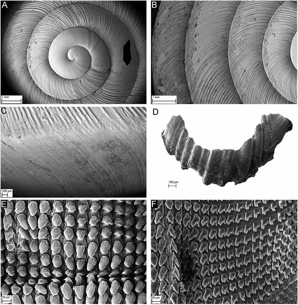
Figure 59. SEM photographs of Amplirhagada gibsoni n.sp. (A–D) Shell, paratype AMS C463708: (A) apical whorl viewed from above (scale 1 mm); (B) sculpture on second to forth whorl viewed from above (scale 1 mm); (C) close-up showing axial periphery of last whorl (Scale 200 µm); (D) Jaw, paratype AMS C463709 (scale 100 µm). (E–F) Radula, paratype AMS C463709: (E) central and inner lateral teeth (scale 20 µm); (F) outer lateral and inner marginal teeth (scale 20 µm).
| WAM |
Western Australian Museum |
No known copyright restrictions apply. See Agosti, D., Egloff, W., 2009. Taxonomic information exchange and copyright: the Plazi approach. BMC Research Notes 2009, 2:53 for further explanation.
|
Kingdom |
|
|
Phylum |
|
|
Class |
|
|
Order |
|
|
Family |
|
|
Genus |