
Afropselaphus tymficus Davranoglou, Hlaváč & Baňař, 2023
|
publication ID |
https://doi.org/10.11646/zootaxa.5351.5.4 |
|
publication LSID |
lsid:zoobank.org:pub:C6C7769C-94F6-496A-9293-2F0E1E201330 |
|
DOI |
https://doi.org/10.5281/zenodo.8400490 |
|
persistent identifier |
https://treatment.plazi.org/id/03F487CE-E47B-FFC6-FF23-5331FCDC441E |
|
treatment provided by |
Plazi (2023-09-29 13:10:06, last updated 2024-11-27 18:24:09) |
|
scientific name |
Afropselaphus tymficus Davranoglou, Hlaváč & Baňař |
| status |
sp. nov. |
Afropselaphus tymficus Davranoglou, Hlaváč & Baňař , sp. n.
( Figs 2B View FIGURE 2 – 6B View FIGURE 6 , 7C, D View FIGURE 7 )
Material studied. Holotype, ♁: GREECE, with one label: “GREECE [GR 014] 23-24.vi.2021 / OROs TYMFI, 0.5km W PAPINGO / N39°56´49.0´´ E20°41´07.2´´, 428 m / sifting in Platanus forest; along / Voidomatis river , GoogleMaps
Winkler app. extr. / P. Baňař, P. Hlaváč & A. Balázs lgt.” [white, printed] ( NMPC). Paratypes: 1 ♁♁, 2 ♀♀, 1 ex, same data as for holotype ( MMBC, PCPH) GoogleMaps ; 1 ♁, 11 exx, with one label: “ GREECE [GR 013] 23-24.vi.2021 / OROs TYMFI, 0.5km W PAPINGO / 39°57´55.0´´E20°42´39.2´´ / sifting in narrow canyon along / stream, 907 m, Winkler app. extr. / P.Baňař, P.Hlaváč & A. Balázs lgt.” [white, printed] ( MMBC, PCPH) .
Diagnosis. Head 1.4 times longer than wide, scape 2.3 times longer than wide, median gular ridge with a distinct median bulge ( Fig. 4B View FIGURE 4 ), tergite 1 (IV) 1.6–1.7 times broader than long, sternite 2 (IV) 2.3 times wider than long; metaventrite of male produced into distinct subtriangular spine ( Fig. 6B View FIGURE 6 ); central spine of internal sac of aedeagus with three simple branches adjacent to a denticle-bearing sclerite ( Fig. 7C, D View FIGURE 7 ); second spine of aedeagus simple, overlaying membrane bearing large number of denticles ( Fig. 7C, D View FIGURE 7 ).
Description. Body length 1.9 mm. Coloration largely reddish-brown, maxillary palpi and legs lighter, posterior margin of elytra much darker, almost black ( Fig. 2B View FIGURE 2 ). Pilosity of body sparse; head, legs, and antennae covered by sparse, adjacent golden setae; elytra with few rows of setae, which are denser on elytral marginal line; with row of setae on distal margin of elytra; proximal margin of tergite II with very dense pilosity ( Fig. 2B View FIGURE 2 ), remaining segment largely glabrous.
Head 1.4 times longer than wide, each eye composed of 7 ommatidia ( Figs. 3B View FIGURE 3 , 4B View FIGURE 4 ); median gular ridge with a distinct median protrusion ( Fig. 4B View FIGURE 4 ); antennae 0.9 mm long; scape cylindrical, 2.3 times longer than wide, and 2.3 times longer than pedicel; pedicel about as long as wide; pedicel and antennomeres 3–8 rounded, subequal in length; antennomeres 9–10 about 1.4 times longer than length of each antennomere 3–8; terminal antennomere (11) about 1.3 times as long as 9–10 combined, and 1.9 times longer than wide ( Fig. 5B View FIGURE 5 ). Maxillary palpomere 1 shortest, 2 2.1 times longer than 1; 3 extremely short and rounded; palpomere 4 1.9 times longer than 2, apex club-shaped.
Pronotum 0.4 mm wide, smooth, about 1.5 times longer than wide; slightly constricted proximally and distally, broadest medially; about 1.1 times longer than head; metaventrite produced into a distinct subtriangular protuberance ( Fig. 6B View FIGURE 6 ). Elytra about 1.4 times broader than long, and 1.1 times longer than pronotum
Abdomen about as broad as elytra; tergite 1 (IV) (excluding paratergites) 1.2 times wider than long; sternite 2 (IV) 1.9 times wider than long.
Aedeagus ( Fig. 7C, D View FIGURE 7 ) with central spine of internal sac of aedeagus with three simple branches adjacent to a denticle-bearing sclerite ( Fig. 7C, D View FIGURE 7 ); second spine of aedeagus simple, overlaying a membrane bearing a large number of denticles.
Measurements of holotype (in mm)—W = width, L = length. Body, L—1.87; head, L—0.37, W—0.26; antenna, L—0.9; scapus, L—0.16, W—0.07; pedicel, L—0.07, W—0.06; antennomere III, L—0.06; antennomere IV, L—0.05; antennomere V, L—0.06; antennomere VI, L—0.06; antennomere VII, L—0.05; antennomere VIII, L—0.05; antennomere IX, L—0.07; antennomere X, L—0.07; antennomere XI, L—0.19, W—0.1; maxillary palpomere I, L—0.09; maxillary palpomere II, L—0.19; maxillary palpomere III, L—0.05; maxillary palpomere IV, L—0.36; pronotum, L—0.4, W—0.27; elytra, L—0.45, W—0.31; tergite IV, L—0.45, W—0.69; tergite IV (excluding paratergites) W—0.56; ventrite IV, L—0.39, W—0.75.
Female. Similar to male, but with a smooth metaventrite.
Etymology. The new species is named after Mt. Tymfi, where it was discovered.
Habitat. Specimens were collected in dense leaf-litter accumulations from a mixed deciduous forest on Mt. Tymfi. ( Fig. 8B View FIGURE 8 ).
Distribution. Greece (so far endemic to Mt. Tymfi).
Remarks. The complex sclerites of the internal sac of aedeagus of A. tymficus sp. n. are strongly autapomorphic and no close parallels can be found in Balkan congeners.
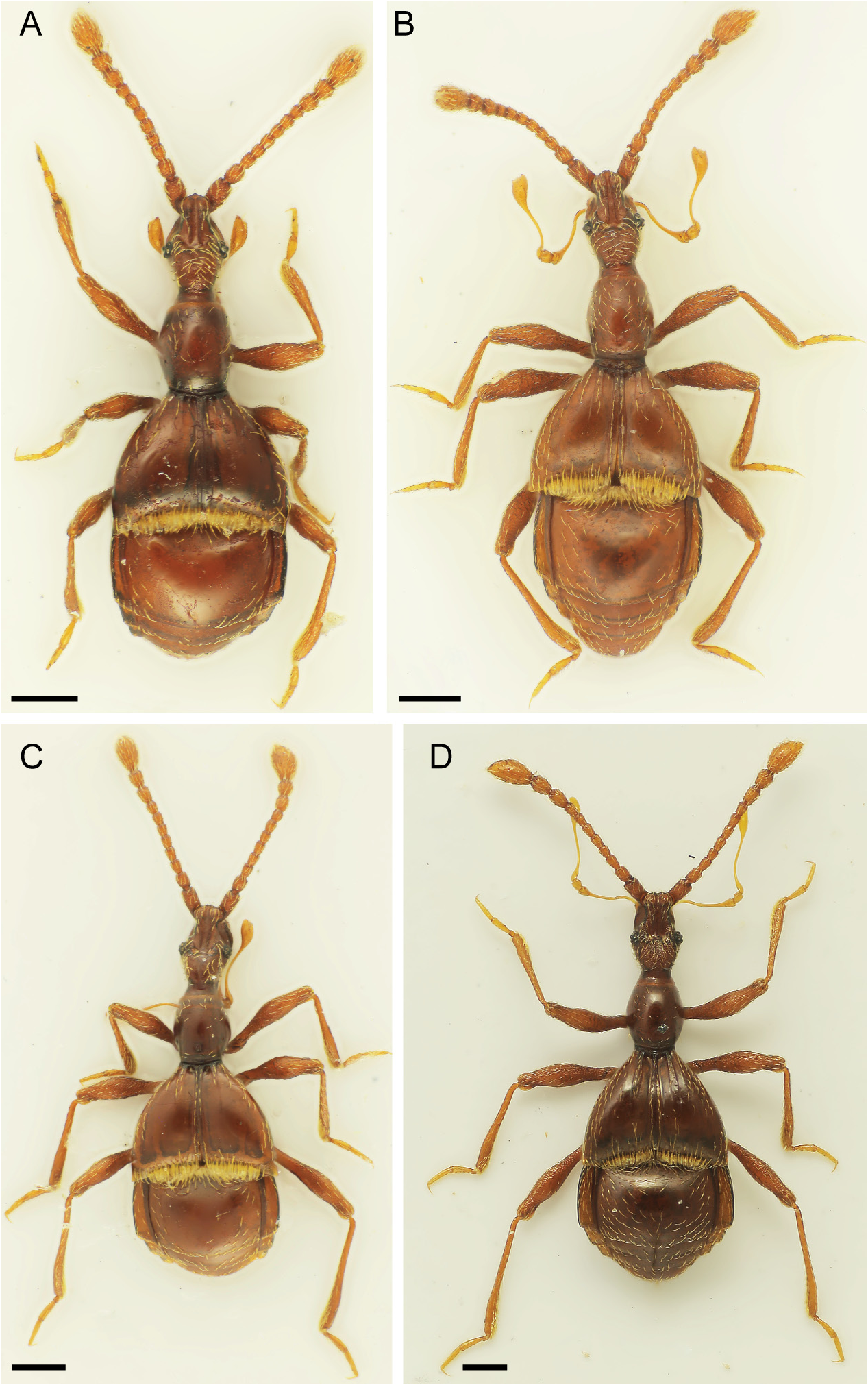
FIGURE 2. Habitus photos of examined species of Pselaphini.A) A. taygetensis sp. n.; B) A. tymficus sp. n.; C) A. euboicus sp. n.; D) Pselaphogenius treskanus (Karaman, 1940). Scale bars correspond to 250 um.
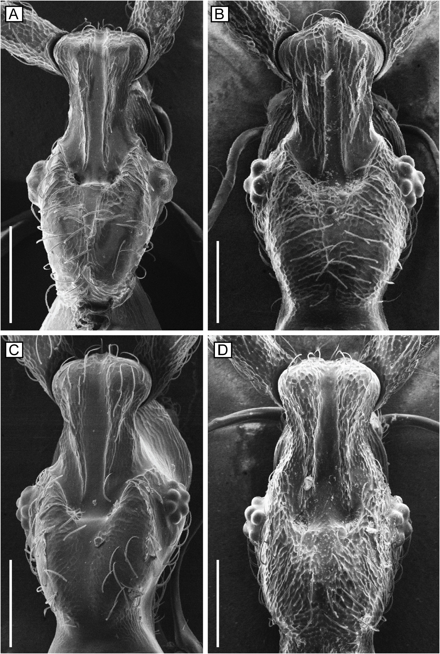
FIGURE 3. Scanning electron microscopy image of dorsal view of head in the examined Pselaphini.A) A. taygetensis sp. n.; B) A. tymficus sp. n.; C) A. euboicus sp. n.; D) Pselaphogenius treskanus (Karaman, 1940). Scale bars correspond to 100 um.
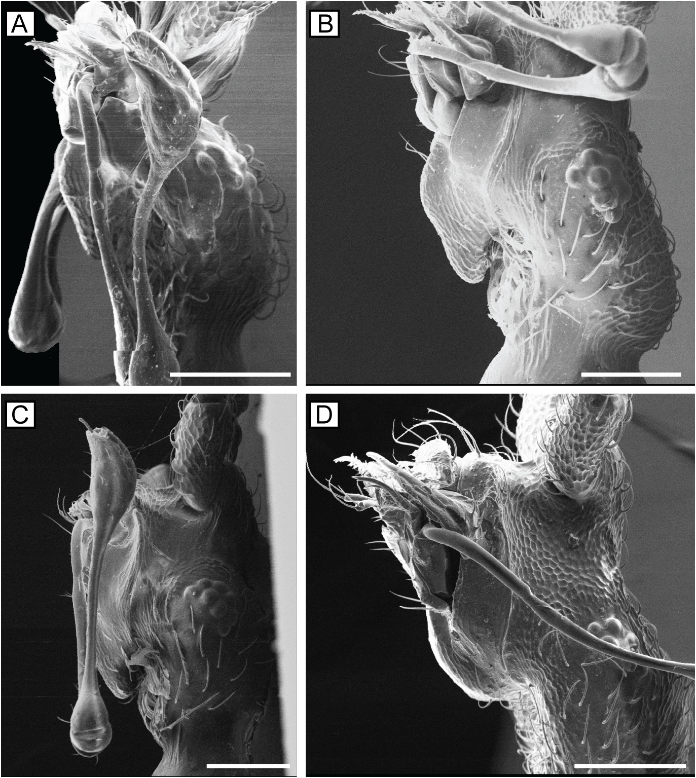
FIGURE 4. Scanning electron microscopy image of lateral view of head in the examined Pselaphini.A) A. taygetensis sp. n.; B) A. tymficus sp. n.; C) A. euboicus sp. n.; D) Pselaphogenius treskanus (Karaman, 1940). Scale bars correspond to 100 um.
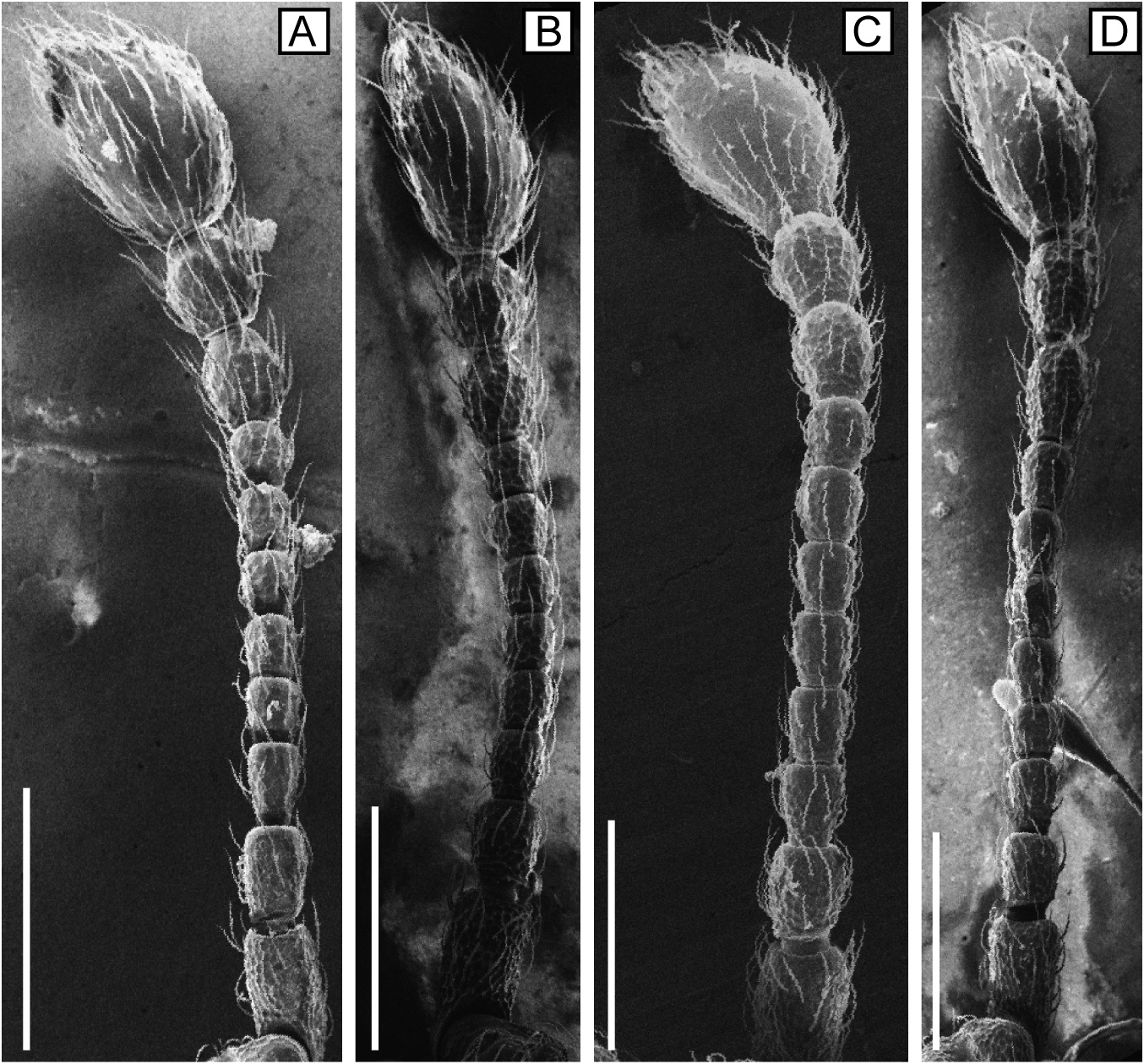
FIGURE 5. Scanning electron microscopy image of antenna in the examined Pselaphini.A) A. taygetensis sp. n.; B) A. tymficus sp. n.; C) A. euboicus sp. n.; D) Pselaphogenius treskanus (Karaman, 1940). Scale bars correspond to 200 um.
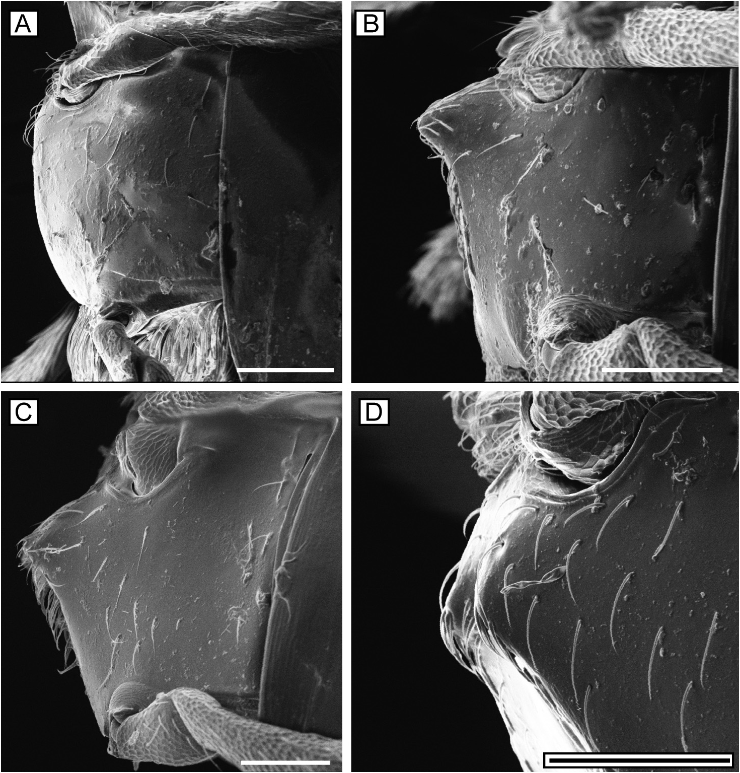
FIGURE 6. Lateral view of metaventrite in the examined Pselaphini, using scanning electron microscopy.A) A. taygetensis sp. n.; B) A. tymficus sp. n.; C) A. euboicus sp. n.; D) Pselaphogenius treskanus (Karaman, 1940). Scale bars correspond to 100 um.
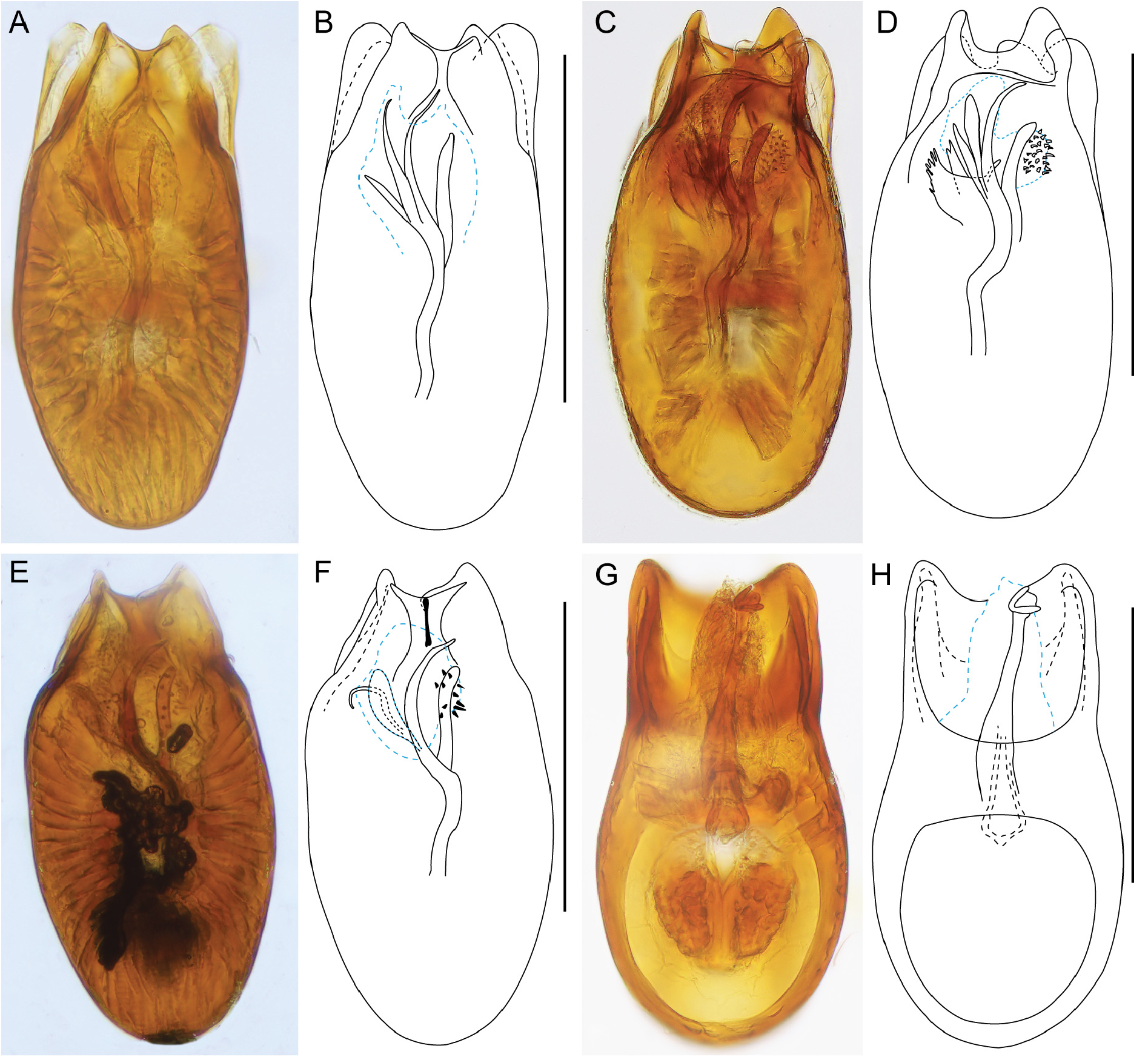
FIGURE 7. Photomicrographs showing the dorsal aedeagal surface of the examined Pselaphini. A) A. taygetensis sp. n.; B) same, schematic; C) A. tymficus sp. n.; D) same, schematic; E) A. euboicus sp. n.; F) same, schematic; G) Pselaphogenius treskanus (Karaman, 1940); F) same, schematic. Blue dashed lines indicate membranous structures. All scale bars correspond to 200 um.
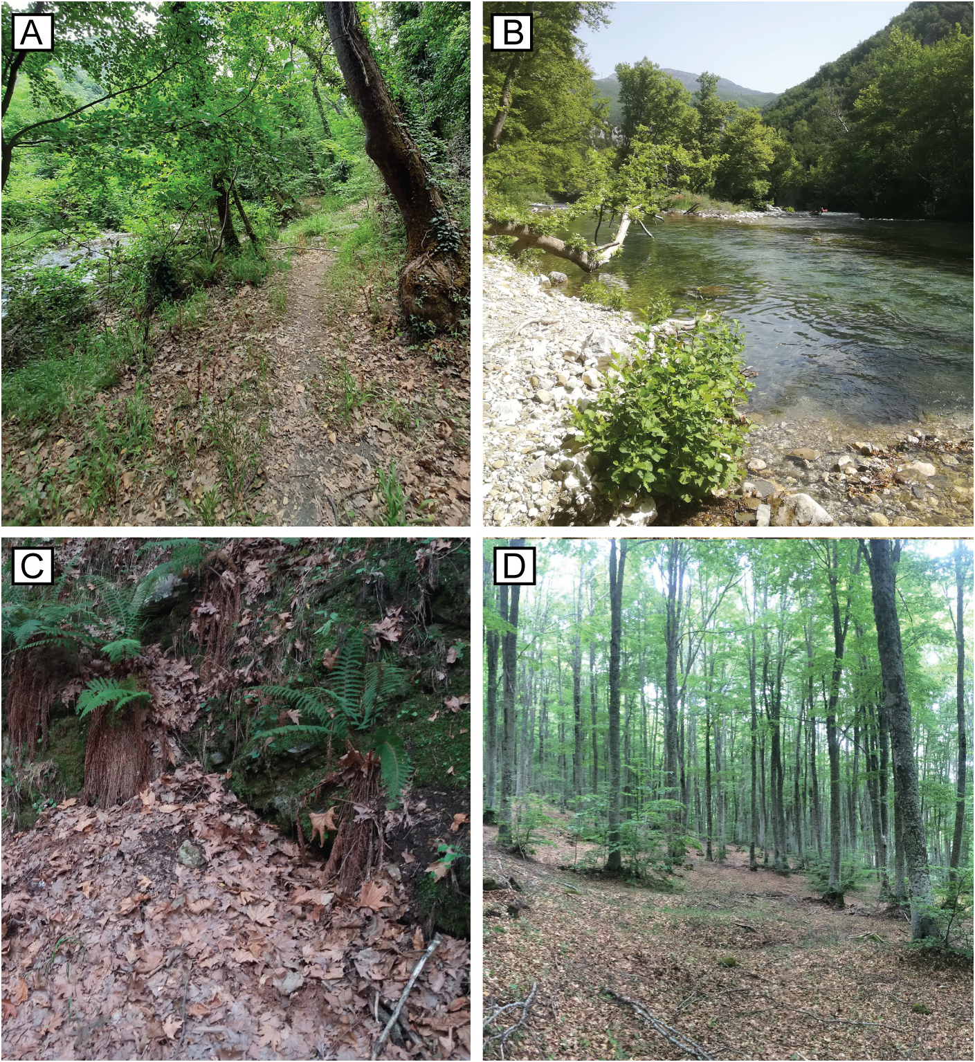
FIGURE 8. Habitat photos of examined species. A) Type locality of A. taygetensis sp. n. in Kastoreion; B) type locality of A. tymficus sp. n.; in Mt Tymfi; C) leaf litter where A. euboicus sp. n. was collected from in Mt Dirfis, the type locality; D) habitat in Mt Vitsi, where Pselaphogenius treskanus (Karaman, 1940) was collected from.
No known copyright restrictions apply. See Agosti, D., Egloff, W., 2009. Taxonomic information exchange and copyright: the Plazi approach. BMC Research Notes 2009, 2:53 for further explanation.
|
Kingdom |
|
|
Phylum |
|
|
Class |
|
|
Order |
|
|
Family |
|
|
SubFamily |
Pselaphinae |
|
Genus |