
Platyozius laevis ( Borradaile, 1902 )
|
publication ID |
https://doi.org/10.11646/zootaxa.2375.1.1 |
|
persistent identifier |
https://treatment.plazi.org/id/03F487A8-390F-427C-7D8C-FB23F645F9D1 |
|
treatment provided by |
Felipe (2021-08-22 21:11:18, last updated by Plazi 2023-11-04 10:04:21) |
|
scientific name |
Platyozius laevis ( Borradaile, 1902 ) |
| status |
|
Platyozius laevis ( Borradaile, 1902) View in CoL
( Figs. 33A–F View FIGURE 33 ; 34A–F View FIGURE 34 ; 35A–G View FIGURE 35 ; 38A–C View FIGURE 38 )
Pseudozius (Platyozius) laevis Borradaile, 1902: 243 , fig. 45. — Garth 1971: 185 [in list].
Platyozius laevis View in CoL — Rathbun 1906: 861, pl. 11, fig. 7 [Hawaiian Is.].
Eucrate sulcatifrons View in CoL — Edmondson 1962: 4, figs. 1 b, 2a-c [Hawaiian Is.] — McLaughlin et al. 2005: 257 [in list]. [not Eucrate sulcatifrons ( Stimpson, 1858) View in CoL = Eucrate crenata (De Haan, 1835) View in CoL ]
Eucrate laevis View in CoL — Dai et al. 1996: 247, fig. 10 [South China Sea]. — Ng et al. 2008: 78 [in list]. — Yang et al. 2008: 770 [in list] [ China].
Eucrate sp. — Kawamoto & Okuno 2003: 149 [colour photograph] [ Japan].
Type material. Male holotype, 5.0 mm × 6.0 mm ( UMZC) ( Fig. 34A View FIGURE 34 ) ; 1 female, 7.5 mm × 9.5, 1 male, 7.0 mm × 8.5 mm ( UMZC) not mentioned in the description .
Type locality. Maldives, Suvadiva Atoll , 36 m.
Material examined. Madagascar. North of Nosy Be , 12°26’N, 48°16’E, 200 m, G. Casellato coll., 10.1971: 1 male ( MNHN-B30524 ) GoogleMaps .
Seychelles. REVES 2: stn.18, 05.09.1980: 1 female, 4.4 mm × 4.8 mm ( MNHN-B30539 ) ; stn. 32, 57 m, 09.09.1980: 1 female, 4.2 mm × 4.3 mm ( MNHN-B30535 ) ; stn. 51, 42 m, coral and sand, 15.09.1980: 1 male 6.4 mm × 7.7 mm, 1 female, 5.3 mm × 6.2 mm ( MNHN-B30537 ) ; stn. 60, 41 m, 19.09.1960: 1 male, 6.0 mm × 7.5 mm, 4 pre-adults females, 6.3 mm × 7.7 mm, 6.4 mm × 7.7 mm, 6.0 mm × 7.2 mm, cw 7.8 mm, 1 preadult 4.5 mm × 5.2 mm ( MNHN-B30534 ) .
Maldives. Suvadiva Atoll , 36 m.: male holotype, 5.0 mm × 6.0 mm; same location: 1 female, 7.5 mm × 9.5, 1 male, 7.0 mm × 8.5 mm ( UMZC). – Laamu Atoll, under boulders in reef, A. Anand J. K. coll., 1 male, 6.1 mm × 7.8 mm ( ZRC 2007.0707 View Materials ) .
Japan. Ogasawara Is., off Chichi-jima I., 27°06.35’N, 142°10.48’E, 59– 55 m, coral sand and rock, TRV Shin’yo-maru, 1997 cruise, stn. 10, T. Komai coll.: 2 males, 5.0 mm × 5.6 mm, 4.6 mm × 5.2 mm ( CBMZC 6531 ) GoogleMaps .
Hong Kong. No data: 1 female ( SWIMS SML-Z-159.1 ) .
Philippines. Sulu Archipelago , Basilan Straits, SW Malanipa I., 48–51 m, Pele, B. R. Wilson coll., 12.02.1964: 1 male, 5.4 mm × 7.0 mm ( MNHN-B10342 ) .
Bohol, Panglao I., Balicasag I., tangle nets of local fishermen, 12.2003: 1 female, 11.0 mm × 14.0 mm ( ZRC 2004.0806 View Materials ); 02.05.2004 : 1 female, 9.5 mm × 11.9 mm ( ZRC 2004.0732 View Materials ) .
Indonesia. Ceram , Mariel King Memorial Expedition: stn. CP I, haul 1, Piru Bay, 03°15’S, 128°8’E, 59– 64 m, 01.06.1970: 1 male, 4.5 mm × 4.8 mm ( MNHN-B30773 ); stn. CP III, 01.06.1970: 1 male, 5.7 mm × 6.6 mm ( MNHN-B30774 ) GoogleMaps .
Kai Is., Mariel King Memorial Expedition: stn. KR VI, haul 3–10, north of Du Rowa I., 05°32’S, 132°41’E, 27–37 m, 11.06.1970: 1 female, 5.1 mm × 6.3 mm ( MNHN-B30840 ) GoogleMaps .
Solomon Is. SALOMON 1: stn. DW 1822, 09°51.8’S, 160°51.8’E, 51–54 m: 1 male, 4.2 mm × 4.7 mm ( MNHN-B30777 ); 1 male, 4.6 mm × 4.8 mm ( MNHN-B32011 ) GoogleMaps .
Vanuatu. SANTO 2006: stn. DB29, west of Malo I., 15°38.9’S, 167°05.1’E, 15 m, 17.09.2006: 1 male, 6.9 mm × 8.5 mm ( ZRC) GoogleMaps .
Australia. Queensland, Wreck Reef near Porpoise Cay , 22°11’S, 155°20’E, outer reef flat under dead coral boulders at base of live coral heads, low tide, J. Short & S. Mullens coll., 14.05.1988: 1 female, 12.3 mm × 15.0 mm ( QM W15174) GoogleMaps . – Flinders Reef, off Cape Moreton , 26°59’S, 155°29’E, fringing reef under rock, scuba, 20 m, N. Coleman coll., 09.07.1997: 1 male, 10.9 mm × 13.5 mm ( QM W24144) GoogleMaps .
Chesterfield Is. CORAIL 2: stn.DW1, 20°55.9’S, 161°40.7’E, 59 m, 20.07.1988: 1 female, 6.5 mm × 7.7 mm ( MNHN-B30801 ). – Stn. DW 11, 20°59’S, 161°41’E, 58 m, 20.07.1988: 1 male, 5.5 mm × 6.9 mm ( MNHN-B30779 ) GoogleMaps .
New Caledonia. LAGON: stn. 229, Ile Ouen, Baie du Prony, 22°39’S, 166°39’E, 41 m: 1 male, 6.6 mm × 8.0 mm ( MNHN-B30778 ) GoogleMaps .
LAGON EST: stn. 748, 21°16.9’S, 165°49.9’E, 35 m, 06.01.1987: 1 pre-adult female, 6.1 mm × 7.4 mm, photograph CB 392 ( MNHN-B30782 ) GoogleMaps .
MUSORSTOM 5: stn. DW 264, 25°19.69’S, 159°44.33’E, 56 m, 08.10.1986: 1 male, 7.0 mm × 8.5 mm, photograph ( MNHN-B30780 ) GoogleMaps
NORFOLK 2: stn. DW 2135, 23°02’S, 168°21’E, 295–330 m, 03.11.2003: 1 female, 5.4 mm × 6.7 mm ( MNHN-B30781 ) GoogleMaps .
Loyalty Is. Ouvéa I., Mouli, diving, 11 m, J.-P. Menou coll., 13.11.1991: 1 male, 10.1 mm × 12.5 mm, photograph CB 1070 ( MNHN-B30608 ) .
Micronesia. Yap I., Y–251, R. Hiatt coll., 1952: 1 male, 8.5 mm × 10.7 mm ( MNHN-B24496 ) .
Marshall Is. Enewetak Atoll, #090351-B: 1 male, 1 pre-adult female, 1 female ( LACM) .
Hawaiian Is. Northwest Hawaiian Is, French Frigate Shoals, stn. FFS 114, 23.699°N, 166.058°W, fore reef, 16.8 m, G. Paulay et al. coll., 19.10.2006: 1 male, photograph 1JMO166, 6.9 mm × 8.6 mm ( LACM) GoogleMaps ; 23.699°N, 166.0575°W, under coral rubble, 16.7 m: 1 male, photograph, 5.0 mm × 6.5 mm ( UF 12194 ) GoogleMaps . – Stn FFS 192, 23.699°N, 166.058°W, fore reef, under coral rubble, 25.7 m, G. Paulay et al. coll., 26.10.2006: 1 male, photograph, 7.7 mm × 9.7 mm ( UF 12283 ) GoogleMaps ; 23.8633°N, 166.1877°W: 1 male, photograph, 4.4 mm × 4.9 mm ( UF 12284 ) GoogleMaps . – Stn FFS 193, 23.8445°N, 166.3348°W, back reef, under coral rubble, 18.1 m, G. Paulay et al. coll., 26.10.2006: 1 male, photograph, 13.5 mm × 10.4 mm ( UF 12282 ) GoogleMaps .
Oahu, Halona Blowhole, 17 m, 21°26’N, 157°58’W, under surface of rubble, R. Holcom coll., 2.08.1997: 1 male, 7.7 mm × 9.5 mm (QM W28380) GoogleMaps .
French Polynesia. Tuamotu Archipelago , Rangiroa Atoll , ca. 1 km S of NW point of atoll off Motu Maeherahonae, stn. BRNG–31, 14°55.72’S, 147°51.47’W, reef flat, under coral rubble, Liu et al. coll., 13.10.2001: 2 females, 9.5 mm × 12.1 mm, 12.5 mm × 16.0 mm, photographs GP909: 5–7 and 912: 9–11 ( UF 1534 ) GoogleMaps .
Society Is., Moorea , offshore between Motu Tiahura and Fareone, outer reef slope, 17.4848°N, 149.9172°W, S. McKeon et al. coll., 14.10.2008: 1 ovigerous female, 7.8 mm × 9.4 mm ( UF 15510 ) GoogleMaps .
Diagnosis. Anterolateral borders of carapace each with three short, triangular teeth ( Figs. 33A–E View FIGURE 33 ; 34 View FIGURE 34 ); postorbital ridge along entire anterior portion of carapace ( Fig. 34E View FIGURE 34 ); obtuse outer orbital angle ( Figs. 34 View FIGURE 34 ; 35A–C View FIGURE 35 ). Dorsal surface of carapace with brightly coloured pattern of irregular lines ( Fig. 33 View FIGURE 33 ).
Remarks. Unique among euryplacids is a postorbital ridge that curves along the entire anterior portion of the carapace between the second anterolateral teeth and parallel to the front and orbits of Platyozius . In small individuals (e.g. male, 5.5 mm × 6.9 mm, MNHN-B30779; male, 7.0 mm × 8.5 mm MNHN-B30780; female, 5.4 mm × 6.7 mm, MNHN-B30781) the ridge, as well as the third anterolateral teeth, are only slightly evident.
Also diagnostic is the unique colour pattern ( Fig. 33 View FIGURE 33 ). The details of the colour pattern (see below) vary, even among specimens collected from the same locality. This relative variation is not correlated with any morphological differences suggesting that these differences are not species specific.
Other details of the morphology of the species are given in the diagnosis of Platyozius (see above) and in Table 1.
Platyiozius laevis shows a very wide geographical distribution across the Indo-West Pacific , wider than any known euryplacid. It has been collected from under coral rubble or rocks at low tide in the Maldives, Australia, the Hawaiian Islands, and French Polynesia as well as dredged in water as deep as 200 m.
Colour pattern. The colourful pattern diagnostic of P. laevis rapidly disappears in alcohol-preserved specimens. It is still discernible in a photograph of a specimen from the Hawaiian Is. given by Rathbun (1906: pl. 11, fig. 7). A colour photograph of a specimen from the type locality, the Maldives (male, 6.1 mm × 7.8 mm; ZRC 2007.0707), shows irregular, red-brown spots and wavy lines across the carapace, an almost continuous, wavy, thin, red-brown band around the anterior and lateral borders of the carapace, and a large irregular spot of the same red-brown colour on the gastric region. There are three red-brown bands across each of the ambulatory legs and one on dactylus of the chelipeds. All carapace and pereopod markings are on a light pink background, except on the anterior half of the carapace, which is yellowish orange. A colour photograph of a specimen from the Ryukyu Is., Japan ( Kawamoto & Okuno 2003: 149, as Eucrate sp. ) shows an almost identical colour pattern to that of the Maldives specimen. There are variations of this colour pattern, however. A specimen from New Caledonia (male, 7.0 mm × 8.5 mm; MNHN-B30780) shows the dark red-brown pattern across the anterior and anterolateral portions of the carapace and a cross-like pattern along the front, bound by two pink circles. The rest of the carapace and the pereopods are pink, with red-brown bands on the ambulatory legs. The colour pattern of two females from French Polynesia (9.5 mm × 12.1 mm, 12.5 mm × 16.0 mm; UF 1534; Fig. 33A, B View FIGURE 33 ) is similar to that of the New Caledonia male except that the red-brown markings on the carapace show slightly different patterns, the carapace having an orange background except six greyish spots, the bands on the ambulatory legs thicker and four in number, and there are irregular spots on each of the chelipeds. Specimens from the Northwest Hawaiian Islands ( Fig. 33C, E, F View FIGURE 33 ) and Vanuatu ( Fig. 33D View FIGURE 33 ) show an orange or white carapace background and variations in the spotting of the chelipeds and banding of the ambulatory legs.
Distribution. Wide distribution across the Indo-West Pacific region: Indian Ocean from Madagascar, Seychelles, the Maldives, and India (unpublished manuscript by M. Deb, Zoological Survey of India); western Pacific from southern Japan to Indonesia (photographic evidence from Sulawesi), New Caledonia, and Queensland, Australia; Micronesia, Marshall Is., Hawaiian Is., and French Polynesia. Depth: intertidal to 200 m.
Genus Psopheticoides Sakai, 1969
Psopheticoides Sakai, 1969: 272 View in CoL ; 1976: 523 [in key], 528. — Serène & Vadon 1981: 127 [discussion]. - Karasawa & Kato 2003a: 151 [in list]; 2003b: 139 [in list]. — Castro 2007: 618, 620 [discussion]. — Ng & Castro 2007: 44 [in list]. — Ng et al. 2008: 78, 79 [in list]. — De Grave et al. 2009: 33 [in list].
Diagnosis. Carapace ( Figs. 36A View FIGURE 36 ; 39C View FIGURE 39 ) trapezoidal, almost as wide as long, dorsal surface smooth without clear indication of regions, large orange-red, circle on cardiac region (remains visible in preserved specimens); anterolateral borders arched; front wide, straight with small median notch (absent in large individuals), transverse sulcus along margin. Two teeth posterior to triangular outer orbital angle, first short, elongated with rounded outline following arched anterolateral border, second acute, dorsally salient. Orbits long, wide, spherical ( Fig. 36B View FIGURE 36 ), 2 weak notches on supraorbital border, raised lobe immediately before inner notch; small, low inner suborbital tooth on broad, suborbital border ( Fig. 36C View FIGURE 36 ); eye peduncles short, large reniform (dorsoventrally flattened) corneas ( Fig. 36B, C View FIGURE 36 ). Basal antennal article slightly mobile, completely closing orbital hiatus so that antennal flagellum is excluded from orbit ( Fig. 36B, C View FIGURE 36 ). Anteroexternal margin of third maxilliped merus auriculiform ( Fig. 36C View FIGURE 36 ). Cheliped fingers ( Fig. 36D View FIGURE 36 ) stout, slightly longer than swollen propodus, light in colour; carpus with tooth on inner margin, merus with acute anterodorsal tooth; row of short setae along anterior margin of carpus. Dorsal margins of ambulatory legs (P2–P5) meri, carpi, propodi unarmed, dactyli slender, smooth, setose; P5 propodus, dactylus proportionally short, flattened ( Fig. 36A View FIGURE 36 ), fringed with many short setae. Thoracic sternum ( Fig. 36E, G View FIGURE 36 ) wide; thoracic suture 2/3 complete, straight ( Fig. 36E View FIGURE 36 ); 3/4 deep, short, interrupted; 4/5, 6/7, 7/8 interrupted, 5/6 complete ( Fig. 36G View FIGURE 36 ); median groove on thoracic sternites 7, 8. Sterno-abdominal cavity of male deep, nearly reaching anterior margin of sternite 4 ( Fig. 36E View FIGURE 36 ). Press-button of male abdominal-locking mechanism as large tubercle near thoracic suture 4/5 (presence in pre-adult females unknown). Male abdomen narrow, slender (T-shaped), lateral margins of somites 4–6 abruptly narrowing from somite 3 to transversely narrow, pointed telson ( Fig. 36E View FIGURE 36 ); somite 3 reaching inner margins of P5 coxae; no portions of thoracic sternite 8 exposed by closed abdomen, somite 2 transversely slightly shorter than somite 3 ( Fig. 36F View FIGURE 36 ), somite 1 conspicuous. G1 long, slender, slightly sinuous, arrowhead-shaped apex, relatively large denticles ( Fig. 38D, E View FIGURE 38 ); G2 less than one-third of G1, straight, apex with 2 processes: one long, tip obtuse; second much shorter, tip obtuse ( Fig. 38F View FIGURE 38 ). Male genital opening (gonopore) coxal; coxosternal disposition of long penis with greatly expanded proximal portion, protected by concave posterior portion of thoracic sternite 7. Vulva crescent shaped, extending across anterior, median portion of sternite 6 close to median axis of thorax ( Fig. 36G View FIGURE 36 ); thick, transverse, ventrally projecting sternal vulvar cover on outer margin in large females ( Fig. 36G View FIGURE 36 ).
Type species. Psopheticoides sanguineus Sakai, 1969 View in CoL (by original designation, gender masculine).
Remarks. The inclusion of the genus in the subfamily Euryplacinae of the Goneplacidae was first suggested (conduisent à l’inclure) by Serène & Vadon (1981: 127), although Sakai (1969: 272) had noted the similarities in the male abdomen and gonopods of his new species with those of Eucrate . Karasawa & Kato (2003a: 151; 2003b: 139) formally included Psopheticoides in the Euryplacinae and ultimately in the Euryplacidae ( Castro 2007: 618, 620; Ng et al. 2008: 78, 79).
Unusual among euryplacids are the dorsoventrally flattened reniform eyes in this species ( Fig. 36B View FIGURE 36 ), a character also found in Frevillea ( Figs. 18E, F View FIGURE 18 ; 19A, B View FIGURE 19 ) and in some members of the Goneplacidae ( Castro 2007) . The G1 is unusual in featuring an arrowhead-shaped apex ( Fig. 38D, E View FIGURE 38 ; Sakai 1969: fig. 17a, a’). Also unusual among euryplacids is the presence of a fully sclerotised sternal vulvar cover ( Fig. 36G View FIGURE 36 ). Psopheticoides shares such a structure with only one euryplacid genus, Trissoplax n. gen. ( Fig. 41G View FIGURE 41 ). The typical euryplacid vulva has simple margins and the opening itself is covered by a soft membrane (e.g. Figs. 3G View FIGURE 3 , 19H View FIGURE 19 ). The role of such a sclerotised vulvar cover, in goneplacoids remains unknown (see Castro 2007: 755).
Species included. Psopheticoides sanguineus Sakai, 1969 The genus is restricted to the Indo-West Pacific region.
Borradaile, L. A. (1902) The Xanthidae and some other crabs. III. Marine crustaceans. In: Gardiner, J. S. (Ed.), The Fauna and Geography of the Maldive and Laccadive Archipelagoes Being the Account of the Work Carried on and of the Collections Made by an Expedition During the Years 1899 and 1900, vol. 1, part 3. Cambridge University Press, Cambridge, pp. 237 - 271.
Castro, P. (2007) A reappraisal of the family Goneplacidae MacLeay, 1838 (Crustacea, Decapoda, Brachyura) and revision of the subfamily Goneplacinae, with the description of 10 new genera and 18 new species. Zoosystema, 29 (4), 609 - 774.
Dai, A, Cai, Y. & Yang, S. (1996) New species and new records of crabs (Crustacea; Decapoda; Brachyura) from Nansha Islands, China. In: Studies on Marine Fauna and Flora and Biogeography of the Nansha Islands and Neighbouring Waters, 2, 234 - 257. Academia Sinica, Beijing. [In Chinese with English abstract and descriptions]
De Grave, S., Pentcheff, N. D., Ahyong, S. T., Chan, T-Y, Crandall, K. A., Dworschak, P. C., Felder, D. L., Feldmann, R. M., Fransen, C. H. J. M., Goulding, L. Y. D., Lemaitre, R., Low, M. E. Y., Martin, J. W., Ng, P. K. L., Schweitzer, C. E., Tan, S. H., Tshudy, D. & Wetzer, R. (2009) A classification of living and fossil genera of decapod crustaceans. Raffles Bulletin of Zoology, Supplement 21, 1 - 109.
Edmondson, C. H. (1962) Hawaiian Crustacea: Goneplacidae, Pinnotheridae, Cymopoliidae, Ocypodidae, and Gecarcinidae. Occasional Papers of Bernice P. Bishop Museum, 23 (1), 1 - 27.
Garth, J. S. (1971) Borradaile's Maldivian collections revisited. Journal of the Marine Biological Association of India, 11 [1969], 182 - 190.
Karasawa, H. & Kato, H. (2003 a) The phylogeny and fossil record of the Goneplacidae MacLeay (Crustacea, Decapoda, Brachyura) revisited. Contributions to Zoology (The Hague), 72, 147 - 152.
Karasawa, H. & Kato, H. (2003 b) The family Goneplacidae MacLeay, 1838 (Crustacea: Decapoda: Brachyura): systematics, phylogeny, and fossil records. Paleontological Research, 7 (2), 129 - 151.
Kawamoto, T. & Okuno, J. (2003) Shrimps and Crabs of Kume Island, Okinawa. Hankyu Publications, Tokyo, 174 pp.
McLaughlin, P. A., Camp, D. K., Angel, M. V., Bousfield, E. L., Brunel, P., Brusca, R. C., Cadien, D., Cohen, A. C., Conlan, K., Eldredge, L. G., Felder, D. L., Goy, J. W., Haney, T., Hann, B., Heard, R. W., Hendrickx, E. A., Hobbs, H. H., Holsinger, J. R., Kensley, B., Laubitz, D. R., LeCroy, S. E., Lemaitre, R., Maddocks, R. F., Martin, J. W., Nikkelsen, P., Nelson, E., Newman, W. A., Overstreet, R. M., Poly, W. J., Price, W. W., Reid, J. W., Robertson, A., Rogers, D. C., Ross, A., Schotte, M., Schram, F. R., Shih, C. - T., Watling, L., Wilson, G. D. F. & Turgeon, D. D. (2005) Common and scientific names of aquatic invertebrates from the United States and Canada: Crustaceans. American Fisheries Society, Special Publication 31, xiii + 545 pp., Bethesda, Maryland.
Ng, P. K. L. & Castro, P. (2007) On a new genus and species of euryplacid crab (Crustacea: Decapoda: Brachyura: Goneplacoidea) from the Philippines. Zootaxa, 1549, 43 - 53.
Ng, P. K. L., Guinot, D. & Davie, P. (2008) An annotated checklist of extant brachyuran crabs of the world. Systema Brachyurorum, Part I. Raffles Bulletin of Zoology, Supplement 17, 1 - 286.
Rathbun, M. J. (1906) The Brachyura and Macrura of the Hawaiian Islands. United States Fish Commission Bulletin, 1903 (3), 827 - 930, i - viii, pls. 1 - 24.
Sakai, T. (1969) Two new genera and twenty-two new species of crabs from Japan. Proceedings of the Biological Society of Washington, 82, 243 - 280.
Sakai, T. (1976) Crabs of Japan and the Adjacent Seas. Kodansha, Tokyo, vol. 1 [English text], xxix + 773 pp., figs. 1 - 379, maps 1 - 3; vol. 2 [Japanese text], 461 pp., figs. 1, 2; vol. 3 [plates], 16 pp. + pls. 1 - 251.
Serene, R. & Vadon, C. (1981) Crustaces Decapodes: Brachyoures. Liste preliminaire, description de formes nouvelles et remarques taxonomiques. In: Resultats des Campagnes MUSORSTOM. I. Philippines (18 - 28 mars 1976), vol. 1. Memoires ORSTOM 91, 117 - 140.
Stimpson, W. (1858) Crustacea Ocypodoidea. Prodromus descriptionis animalium evertebratorum, quae in Expeditione ad Oceanum Pacificum Septentrionalem, a Republica Federata missa, Cadwaladaro Ringgold et Johanne Rodgers Ducibus, observavit et descripsit W. Stimpson, Pars V. Proceedings of the Academy of Natural Sciences of Philadelphia, 1858 [10], 93 - 110 (39 - 56 in separate).
Yang, S., Chen, H. & Jiang, W. (2008) Crustacea: Decapoda Brachyura. In: Checklist of MarineBiota of China Seas. Lui, J. Y. (Ed.). Institute of Oceanology, Qingdao, pp. 761 - 810.
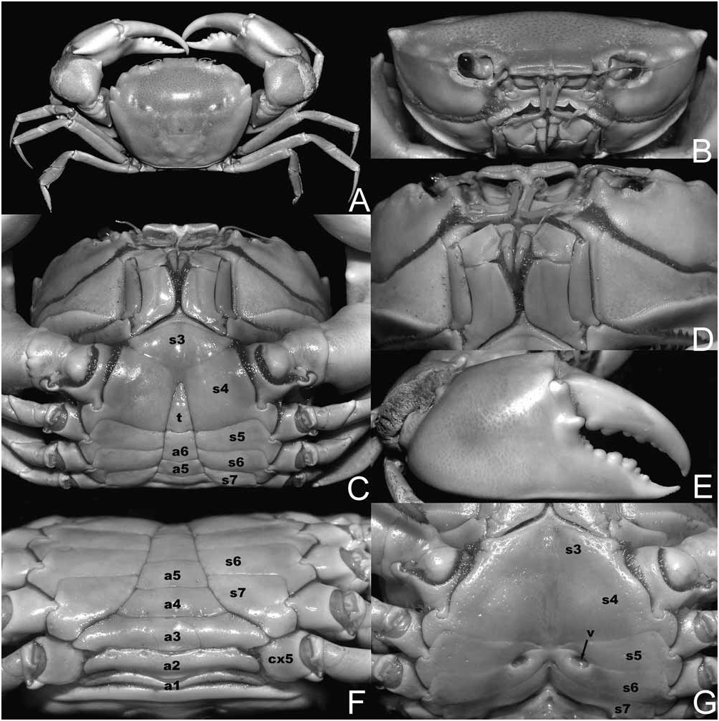
FIGURE 3. Eucrate crenata (De Haan, 1835); A–F, male neotype of Pilumnoplax sulcatifrons Stimpson, 1858 (34.0 mm × 49.1 mm) (QM W27449), Hong Kong, New Territories, Tolo Channel; G, female (22.1 mm × 27.1 mm) (ZRC 2002.0493), China, Qingdao. A, overall view; B, frontal view showing orbits and antennae; C, anterior portion of male thoracic sternum and abdomen; D, third maxillipeds, antennules, antennae and orbits; E, outer surface of right chela; F, posterior portion of male thoracic sternum and abdomen; G, female thoracic sternum and vulvae. Abbreviations: a1–a6 = male abdominal somites 1–6, respectively; cx5 = coxa of fifth pereopod (P5); s3–s7 = thoracic sternites 3 to 7, respectively; t = telson; v = vulva.

FIGURE 18. A, B, Euryplax nitida Stimpson, 1859; A, female (USNM 276579), Gulf of Mexico, off Florida, ventral view of carapace; B, female (10.1 mm × 7.1 mm) (LACM), Colombia, Atlantic coast, ventral view of carapace; C, D, Euryplax polita Smith, 1870; male (18.9 mm × 33.8 mm) (LACM), Perú, Tumbes; E, F, Frevillea hirsuta (Borradaile, 1916); E, male lectotype of Goneplax hirsutus Borradaile, 1916 (10.5 mm × 15.2 mm) (NHM 1917.1.29.148–9); F, female paralectotype of Goneplax hirsutus Borradaile, 1916 (12.0 mm × 17.8 mm) (NHM 1917.1.29.148–9). A, ventral view; B, D, female sterno-abdominal cavity; C-F, overall view; D; male sterno-abdominal cavity. Abbreviations: a3 = male abdominal somite 3; G1 = first male pleopod; G2 = second male pleopod; pb = press-button of male abdominallocking mechanism; s4–s8 = thoracic sternites 4 to 8, respectively; v = vulva. Photo credits: B, C, D by J. Martin (LACM); E, F by P. Crabb (NHM).
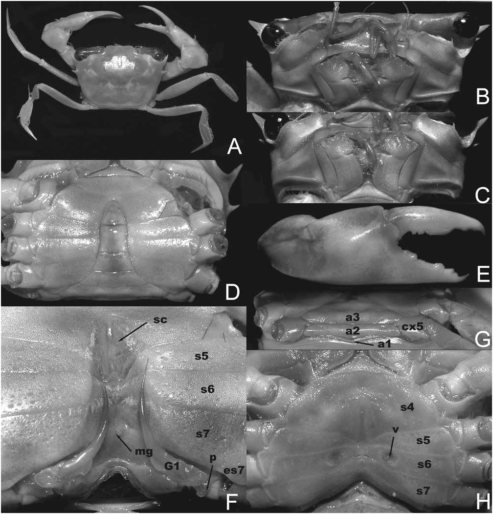
FIGURE 19. Frevillea hirsuta (Borradaile, 1916); A, H, female (13.3 mm × 19.9 mm) (MNHN-B30601), West Indies, Guadeloupe, off Basse Terre; B–G, male (16.6 mm × 24.9 mm) (MNHN-B30601), West Indies, Guadeloupe, off Basse Terre. A, overall view; B, frontal view showing orbits and antennae; C, third maxillipeds; D, anterior portion of male thoracic sternum and abdomen; E, outer view of right chela; F, male sterno-abdominal cavity, G1, and penis; G, posterior portion of male abdomen; H, female thoracic sternum and vulvae. Abbreviations: a1–a3 = male abdominal somites 1, 2, 3, respectively; cx5 = coxa of fifth pereopod (P5); es7 = episternite 7; G1 = first male pleopod; mg = median groove; p = penis; pb = press-button of male abdominal-locking mechanism; s4–s7 = thoracic sternites 4 to 7, respectively; sc = sterno-abdominal cavity.

FIGURE 33. Platyozius laevis (Borradaile, 1902), colours in life. A, female (12.5 mm × 16.0 mm) (UF 1534), French Polynesia, Tuamotu Archipelago, Rangiroa Atoll; B, female (9.5 mm × 12.1 mm) (UF 1534), same location; C, specimen not located (LACM), Hawaiian Is., Northwest Hawaiian Islands, French Frigate Shoals; D, specimen not located, male, Vanuatu; E, F, male (6.9 mm × 8.6 mm) (LACM), Hawaiian Is., Northwest Hawaiian Islands, French Frigate Shoals. A– E, overall views; F, ventral view. Photo credits: A, B by Florida Museum of Natural History (UF); C–F by J. Martin (LACM).
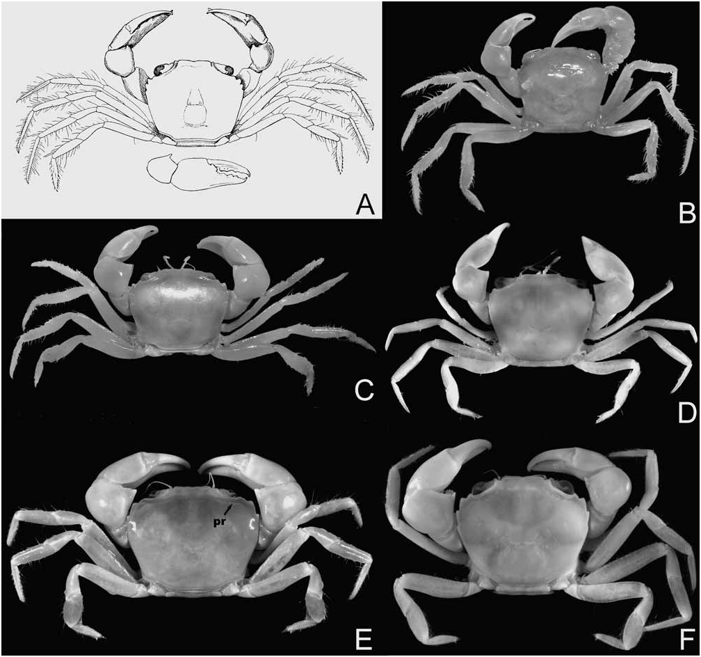
FIGURE 34. Platyozius laevis (Borradaile, 1902); A, male holotype of Pseudozius (Platyozius) Borradaile, 1902 (5.0 mm × 6.0 mm) (UMZC), Maldives, Suvadiva Atoll (after Borradaile 1902: fig. 45); B, male holotype of Pseudozius (Platyozius) Borradaile, 1902 (5.0 mm × 6.0 mm) (UMZC), Maldives, Suvadiva Atoll; C, topotypic female of Pseudozius (Platyozius) Borradaile, 1902 (7.5 mm × 9.5 mm) (UMZC), Maldives; D, male (6.0 mm × 7.5 mm) (MNHN- B30534), Seychelles; E, female (12.5 mm × 16.0 mm) (UF 1534), French Polynesia, Tuamotu Archipelago, Rangiroa Atoll; F, female (6.3 mm × 7.7 mm) (MNHN-B30534), Seychelles. Abbreviation: pr = postorbital ridge. Photo credits: B, C by T.-Y. Chan (NTOU).

FIGURE 35. Platyozius laevis (Borradaile, 1902); A, female (11.0 mm × 14.0 mm) (ZRC 2004.0806), Bohol, Panglao Is., Balicasag I.; B, C, F, male (7.7 mm × 9.5 mm) (QM W28380), Hawaiian Is., Oahu, Halona Blowhole; D, E male (6.0 mm × 7.5 mm) (MNHN-B30534), Seychelles; F, female (12.5 mm × 16.0 mm) (UF 1534), French Polynesia, Tuamotu Archipelago, Rangiroa Atoll. A, frontal view showing orbits and antennae; B, outer surface of left chela; C, third maxillipeds; D, anterior portion of male thoracic sternum and abdomen; E, posterior portion of male thoracic sternum and abdomen; F, male sterno-abdominal cavity, G1, and penis; G, female thoracic sternum and vulvae. Abbreviations: a2, a3 = male abdominal somites 2 and 3, respectively; cx5 = coxa of fifth pereopod (P5); es7 = episternite 7; G1 = first male pleopod; p = penis; s4–s7 = thoracic sternites 4 to 7, respectively; v = vulva.
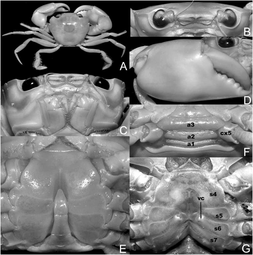
FIGURE 36. Psopheticoides sanguineus Sakai, 1969; male (21.3 mm × 26.5 mm) (ZRC 2004.0752), Philippines, Bohol, Panglao. A, overall view; B, frontal view showing orbits and antennae; C, third maxillipeds; D, outer surface of right chela; E, anterior portion of male thoracic sternum and abdomen; F, posterior portion of male thoracic sternum and abdomen; G. female sternum and vulvae. Abbreviations: a1–a3 = male abdominal somites 1, 2, 3, respectively; cx5 = coxa of fifth pereopod (P5); s4–s7 = thoracic sternites 4 to 8, respectively; vc = sternal vulvar cover.

FIGURE 38. Gonopods of Platyozius, Psopheticoides, and Systroplax; carapace of Systroplax. A–C, Platyozius laevis (Borradaile, 1902), male (6.1 mm × 7.8 mm) (ZRC 2007.0707), Maldives, Laamu Atoll; D–F, Psopheticoides sanguineus Sakai, 1969, male (21.9 mm × 27.8 mm) (ZRC 2008.1357), Philippines, Bohol, Balicasag I.; G–J, Systroplax angusta (Rathbun, 1914); G, G1 (after Guinot 1989: fig. 39D); H, male (32.3 mm × 37.2 mm) (MNHN-B29377), Fiji; I, J, variation in anterolateral borders of carapace (after Guinot 1989: fig. 39A, C). A, B, D, E, right G1, dorsal view; C, F, H, right G2, dorsal view. Scale bars: A, D, 1.0 mm; B, E, 0.5 mm; C, F, H, 0.2 mm; G, 0.1 mm.
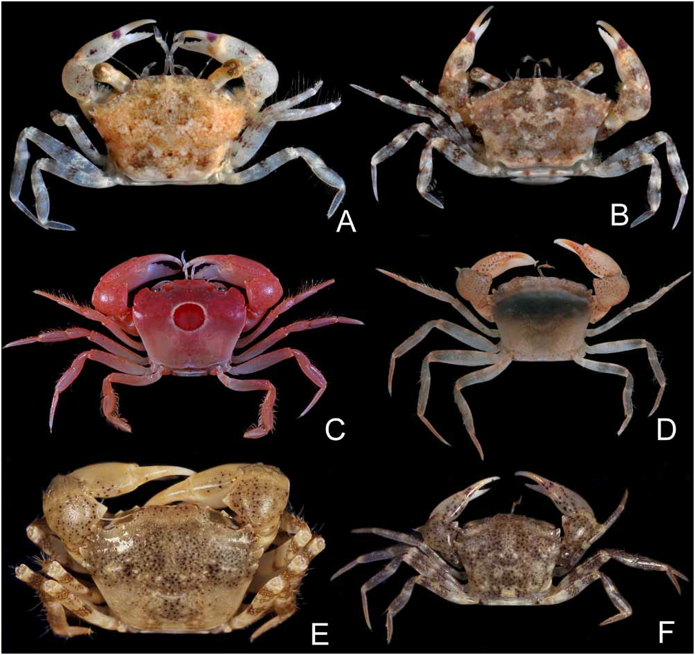
FIGURE 39. Colours in life. A, Euryplax nitida Stimpson, 1871; male neotype (7.1 mm × 11.3 mm) (UF 15166), Gulf of Mexico; B, Euryplax nitida Stimpson, 1871; male (4.4 mm × 7.0 mm) (UF 15168), Gulf of Mexico; C, Psopheticoides sanguineus Sakai, 1969, male (22.2 mm × 26.7 mm) (MNHN-B29728), Philippines, Bohol, Panglao; D, Trissoplax dentata (Stimpson, 1858), male (6.2 mm × 8.3 mm) (NHM), Thailand, Andaman Sea; E, Trissoplax dentata (Stimpson, 1858), young male, not preserved, Singapore, Changi; F, Trissoplax dentata (Stimpson, 1858), female (8.7 mm × 11.5 mm) (ZRC 2000.2240), Singapore. Photo credits: A, B by Florida Museum of Natural History (UF); D by P. F. Clark (NHM).

FIGURE 41. Trissoplax dentata (Stimpson, 1858); A–D, F, male neotype of Heteroplax dentatus Stimpson, 1858 (16.4 mm × 21.5 mm) (QM W27400), Hong Kong, Tolo Harbour; E, male (20.0 mm × 26.2 mm), Singapore, Changi Point; G, female (8.7 mm × 11.5 mm) (ZRC 2000.2240) (ZRC 2008.1236), Singapore. A, frontal view showing third maxillipeds; B, frontal view showing orbits and antennae; C, anterior portion of male thoracic sternum; D, posterior portion of male thoracic sternum and abdomen; E, outer surface of right chela; F, male sterno-abdominal cavity, G1 and penis; G, female thoracic sternum and vulvae. Abbreviations: a1–a3 = male abdominal somites 1, 2, 3, respectively; cx5 = coxa of fifth pereopod (P5); es7 = episternite 7; G1 = first male pleopod; G2 = second male pleopod; mg = median groove; p = penis; s4–s7 = thoracic sternites 4 to 7, respectively; vc = sternal vulvar cover.
No known copyright restrictions apply. See Agosti, D., Egloff, W., 2009. Taxonomic information exchange and copyright: the Plazi approach. BMC Research Notes 2009, 2:53 for further explanation.
|
Kingdom |
|
|
Phylum |
|
|
Class |
|
|
Order |
|
|
Family |
|
|
Genus |
Platyozius laevis ( Borradaile, 1902 )
| CASTRO, PETER & NG, PETER K. L. 2010 |
Eucrate sp.
| Kawamoto, T. & Okuno, J. 2003: 149 |
Eucrate laevis
| Ng, P. K. L. & Guinot, D. & Davie, P. 2008: 78 |
| Yang, S. & Chen, H. & Jiang, W. 2008: 770 |
| Dai, A & Cai, Y. & Yang, S. 1996: 247 |
Psopheticoides
| De Grave, S. & Pentcheff, N. D. & Ahyong, S. T. & Chan, T-Y & Crandall, K. A. & Dworschak, P. C. & Felder, D. L. & Feldmann, R. M. & Fransen, C. H. J. M. & Goulding, L. Y. D. & Lemaitre, R. & Low, M. E. Y. & Martin, J. W. & Ng, P. K. L. & Schweitzer, C. E. & Tan, S. H. & Tshudy, D. & Wetzer, R. 2009: 33 |
| Ng, P. K. L. & Guinot, D. & Davie, P. 2008: 78 |
| Castro, P. 2007: 618 |
| Ng, P. K. L. & Castro, P. 2007: 44 |
| Karasawa, H. & Kato, H. 2003: 151 |
| Serene, R. & Vadon, C. 1981: 127 |
| Sakai, T. 1976: 523 |
| Sakai, T. 1969: 272 |
Eucrate sulcatifrons
| McLaughlin, P. A. & Camp, D. K. & Angel, M. V. & Bousfield, E. L. & Brunel, P. & Brusca, R. C. & Cadien, D. & Cohen, A. C. & Conlan, K. & Eldredge, L. G. & Felder, D. L. & Goy, J. W. & Haney, T. & Hann, B. & Heard, R. W. & Hendrickx, E. A. & Hobbs, H. H. & Holsinger, J. R. & Kensley, B. & Laubitz, D. R. & LeCroy, S. E. & Lemaitre, R. & Maddocks, R. F. & Martin, J. W. & Nikkelsen, P. & Nelson, E. & Newman, W. A. & Overstreet, R. M. & Poly, W. J. & Price, W. W. & Reid, J. W. & Robertson, A. & Rogers, D. C. & Ross, A. & Schotte, M. & Schram, F. R. & Shih, C. - T. & Watling, L. & Wilson, G. D. F. & Turgeon, D. D. 2005: 257 |
| Edmondson, C. H. 1962: 4 |
Platyozius laevis
| Rathbun, M. J. 1906: 861 |
Pseudozius (Platyozius) laevis
| Garth, J. S. 1971: 185 |
| Borradaile, L. A. 1902: 243 |