
Clubiona Wagner, 1887
|
publication ID |
https://doi.org/10.11646/zootaxa.4353.1.1 |
|
publication LSID |
lsid:zoobank.org:pub:7D342E35-8D42-4C3D-9903-49E5F08D7D34 |
|
DOI |
https://doi.org/10.5281/zenodo.6020646 |
|
persistent identifier |
https://treatment.plazi.org/id/03EF9555-0907-FF98-1DD1-FA54177DFA81 |
|
treatment provided by |
Plazi (2017-11-22 10:31:57, last updated 2024-11-26 07:26:33) |
|
scientific name |
Clubiona Wagner, 1887 |
| status |
|
Genus Clubiona Wagner, 1887 View in CoL View at ENA
Type species: Clubiona pallidula (Clerck, 1757) .
The genevensis View in CoL group. Mikhailov (1992; 1995) accommodated some Clubiona View in CoL species into the comta View in CoL group, which also comprised the genevensis View in CoL subgroup. According to him, these species differed from the others by some characters linked to the embolus in the males and to the copulatory apparatus of the females. All species but Clubiona comta View in CoL were part of the genevensis View in CoL subgroup. Actually, the representatives of the last group are well distinguished by conspicuous characters, and are treated here as belonging to a species group of their own. Clubiona comta View in CoL remains therefore the only representative of the comta View in CoL group, and the newly erected genevensis View in CoL group includes therefore eight species: Clubiona decora Blackwall, 1859 View in CoL , C. diniensis Simon 1878 View in CoL , C. genevensis L. Koch, 1866 View in CoL , C. leucaspis Simon, 1932 View in CoL , C. minor Wunderlich, 1987 View in CoL , C. pseudominor Wunderlich, 1987 View in CoL , C. vegeta, Simon, 1918 View in CoL , C. wunderlichi Mikhailov, 1992 View in CoL . Some of these species are not always easy to distinguish and often have broadly overlapping distributions ( Helsdingen 1979). Two species described from China, i.e. C. parallela Hu & Li, 1987 View in CoL and C. zhangmuensis Hu & Li, 1987 View in CoL , and originally included in the genevensis View in CoL group ( Mikhailov 1995), do not fit the diagnosis below. The strongly enlarged and protruding tegulum of the male palp and characteristics of the female epigyne and vulva of those two species fit perfectly with the diagnosis of the “ corticalis View in CoL group” (subgenus Paraclubiona), to which they must be transferred.
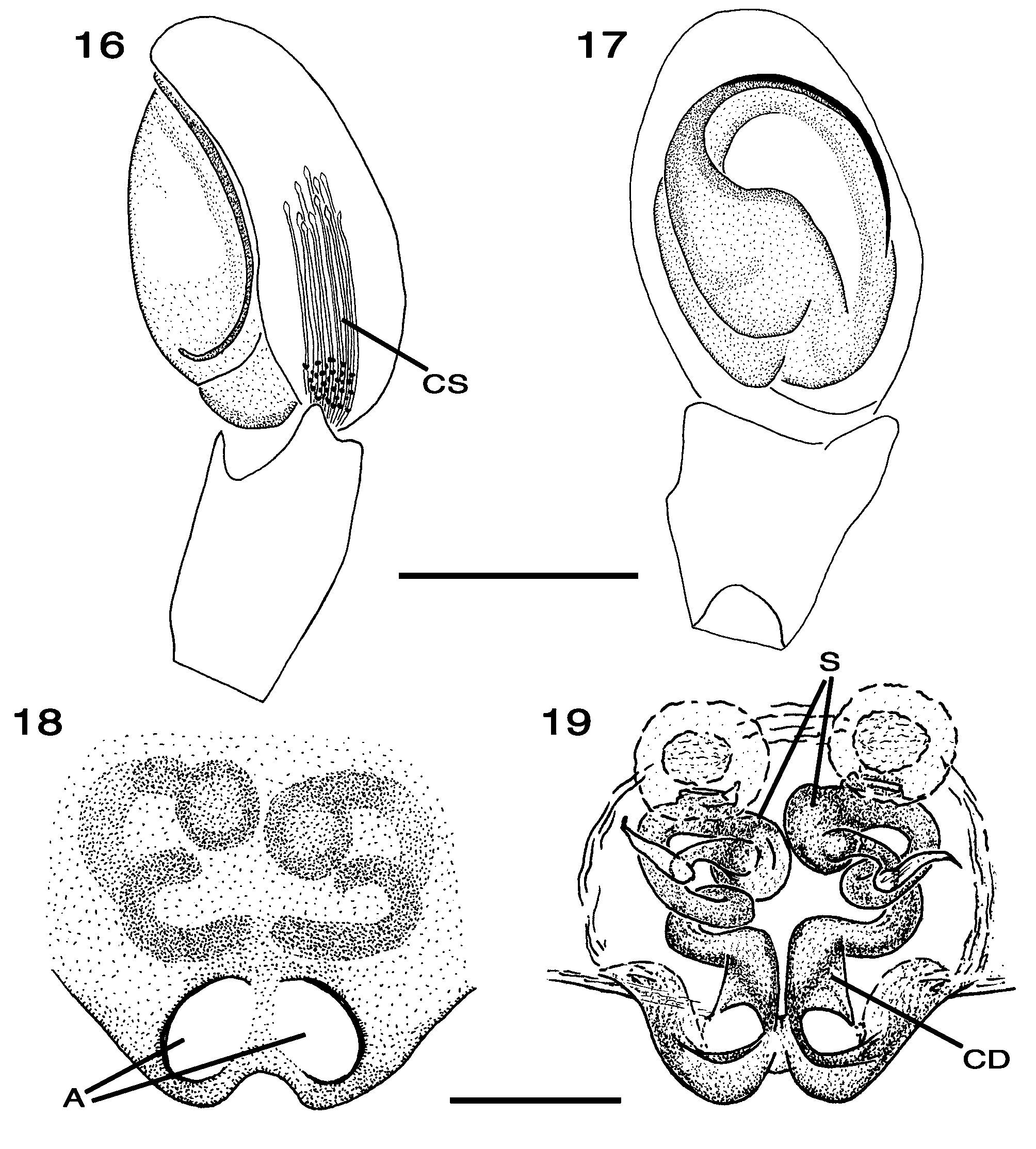
Diagnosis. Representatives of the genevensis group are characterized in the males by the palp with an almost rounded or ovoid bulbus ( Figs 4 View FIGURES 1–8 , 12 View FIGURES 9–15 , 17 View FIGURES 16–19 , 25 View FIGURES 20–25 , 38 View FIGURES34–41 , 46 View FIGURES 45–51 ) and bearing a group of modified dark setae on the retrolateral face of the cymbium ( Figs 3–5 View FIGURES 1–8 , 11 View FIGURES 9–15 , 16 View FIGURES 16–19 , 24 View FIGURES 20–25 , 37 View FIGURES34–41 , 45 View FIGURES 45–51 , 68–79 View FIGURES68–79 ). The females have an epigyne characterized by wide atria (or copulatory openings) and a deep notch in its ventral margin ( Figs 6 View FIGURES 1–8 , 13 View FIGURES 9–15 , 18 View FIGURES 16–19 , 26–28 View FIGURES 26–33 , 39 View FIGURES34–41 , 47 View FIGURES 45–51 ).
Remarks. The examined species of the genevensis group, i.e. Clubiona diniensis , C. genevensis , C. leucaspis and C. vegeta , are provided with modified setae situated on the retrolateral face of the cymbium ( Figs 11 View FIGURES 9–15 , 16 View FIGURES 16–19 , 24 View FIGURES 20–25 , 37 View FIGURES34–41 , 45 View FIGURES 45–51 , 52, 53, 56, 57, 60, 61, 64 View FIGURES 52–67 , 71, 77 View FIGURES68–79 ). They also occur in C. decora (see Figs 3–5 View FIGURES 1–8 ) but were not examined here with SEM. The group also includes C. minor from Tenerife and La Gomera (the Canaries), C. pseudominor from La Palma (the Canaries) ( Wunderlich, 1987, 1991; World Spider Catalog 2017) and C. wunderlichi from Mongolia ( Mikhailov 1992; World Spider Catalog 2017) but these were not examined. The special setae are inserted near the base of the cymbium and extend up to half its length ( Figs 3, 4 View FIGURES 1–8 , 11 View FIGURES 9–15 , 52 View FIGURES 52–67 ) or less ( Figs 24 View FIGURES 20–25 , 37 View FIGURES34–41 , 45 View FIGURES 45–51 , 56, 60, 64 View FIGURES 52–67 ). The basal part is slightly barbed and is provided with a sub-basal enlargement that can be spherical ( Figs 70, 76 View FIGURES68–79 ) or plate-shaped ( Figs 73, 79 View FIGURES68–79 ). The extremity is needle like and barbed ( Figs 57, 61 View FIGURES 52–67 , 71, 77 View FIGURES68–79 ) or flattened and lanceolate ( Figs 16 View FIGURES 16–19 , 53 View FIGURES 52–67 , 68 View FIGURES68–79 ). The retrolateral tibial apophysis (RTA) is sometimes provided with a thin, translucent, needle-like extension at its apex. It was observed in one specimen of C. decora ( Fig. 5 View FIGURES 1–8 ), one specimen of C. vegeta ( Fig. 65 View FIGURES 52–67 ) and on one specimen of C genevensis (P. Oger, pers. comm.; see also on http://arachno.piwigo.com/). This structure appears to be fragile (e.g. it could break during copulation) and it was lacking on another C. vegeta male observed with SEM ( Fig. 66 View FIGURES 52–67 ). This character may be informative but was not used in the key since its presence in other species may be overlooked.
Inside the genevensis group, two well-defined subgroups (here called the genevensis subgroup and the decora subgroup) can be recognized. The genevensis subgroup includes Clubiona genevensis , C. leucaspis and C. vegeta . These species are characterized in the males by the palp with the embolus originating basally and directed laterally ( Figs 25 View FIGURES 20–25 , 38 View FIGURES34–41 , 46 View FIGURES 45–51 ), by the basolateral extension of the bulbus and cymbium much longer than wide ( Figs 59, 63, 67 View FIGURES 52–67 ), and by the modified short cymbial setae (length less than half length of cymbium) ( Figs 24 View FIGURES 20–25 , 37 View FIGURES34–41 , 45 View FIGURES 45–51 , 56, 60, 64 View FIGURES 52–67 ). The females have an epigyne with large, tightened and strongly convoluted copulatory ducts connected inferiorly to the atrio-spermathecal part ( Figs 29–32 View FIGURES 26–33 , 40, 41 View FIGURES34–41 , 48–51 View FIGURES 45–51 ). The decora subgroup includes Clubiona decora , C. diniensis , C. minor , C. pseudominor and C. wunderlichi . Males of this subgroup are well characterized by the palp with embolus originating in distal or median part of bulbus and directed anteriorly ( Figs 4 View FIGURES 1–8 , 12 View FIGURES 9–15 , 17 View FIGURES 16–19 ) (origin of embolus basal and directed laterally in the other species of the genevensis group: Figs 25 View FIGURES 20–25 , 38 View FIGURES34–41 , 46 View FIGURES 45–51 ), short basolateral extension of bulbus and cymbium ( Figs 54, 55 View FIGURES 52–67 ) (much longer in the other species: Figs 59, 63, 67 View FIGURES 52–67 ) and long modified cymbial setae ( Figs 3, 4 View FIGURES 1–8 , 11 View FIGURES 9–15 , 52 View FIGURES 52–67 ) (shorter in the others: Figs 24 View FIGURES 20–25 , 37 View FIGURES34–41 , 45 View FIGURES 45–51 , 56, 60, 64 View FIGURES 52–67 ). Females are characterized by the epigyne with copulatory ducts loose, slightly coiled and connected laterally to the atriospermathecal part ( Figs 6–8 View FIGURES 1–8 , 13–15 View FIGURES 9–15 ) (in the other species of the genevensis group, the tightened and strongly convoluted copulatory ducts are connected inferiorly to the atrio-spermathecal part: Figs 29–32 View FIGURES 26–33 , 40, 41 View FIGURES34–41 , 48–51 View FIGURES 45–51 ).
Blackwall, J. (1859) Descriptions of newly discovered spiders captured by James Yate Johnson Esq., in the island of Madeira. Annals and Magazine of Natural History, 3 (4), 255 - 267.
Helsdingen, P. J. van (1979) Remarks concerning Clubionidae. Bulletin of the British Arachnological Society, 4, 298 - 302.
Koch, L. (1866) Die Arachniden-Familie der Drassiden. Hefte 1 - 6. Lotzbeck, Nurnberg, 304 pp.
Mikhailov, K. G. (1992) The spider genus Clubiona Latreille, 1804 (Arachnida Aranei Clubionidae) in the USSR fauna: a critical review with taxonomical remarks. Arthropoda Selecta, 1 (3), 3 - 34.
Mikhailov, K. G. (1995) Erection of infrageneric groupings within the spider genus Clubiona Latreille, 1804 (Aranei Clubionidae): a typological approach. Arthropoda Selecta, 4 (2), 33 - 48.
Simon, E. (1878) Les arachnides de France. Tome 4. Librairie Encyclopedique de Roret, Paris, 334 pp.
Simon, E. (1932) Les arachnides de France. Tome VI. Encyclopedie Roret, Paris, 205 pp.
World Spider Catalog (2017) World Spider Catalog. Version 18.0. Natural History Museum Bern. Available from: http: // wsc. nmbe. ch (accessed 1 March 2017)
Wunderlich, J. (1987) Die Spinnen der Kanarischen Inseln und Madeiras: Adaptive Radiation, Biogeographie, Revisionen und Neubeschreibungen. Triops, Langen, 435 pp.
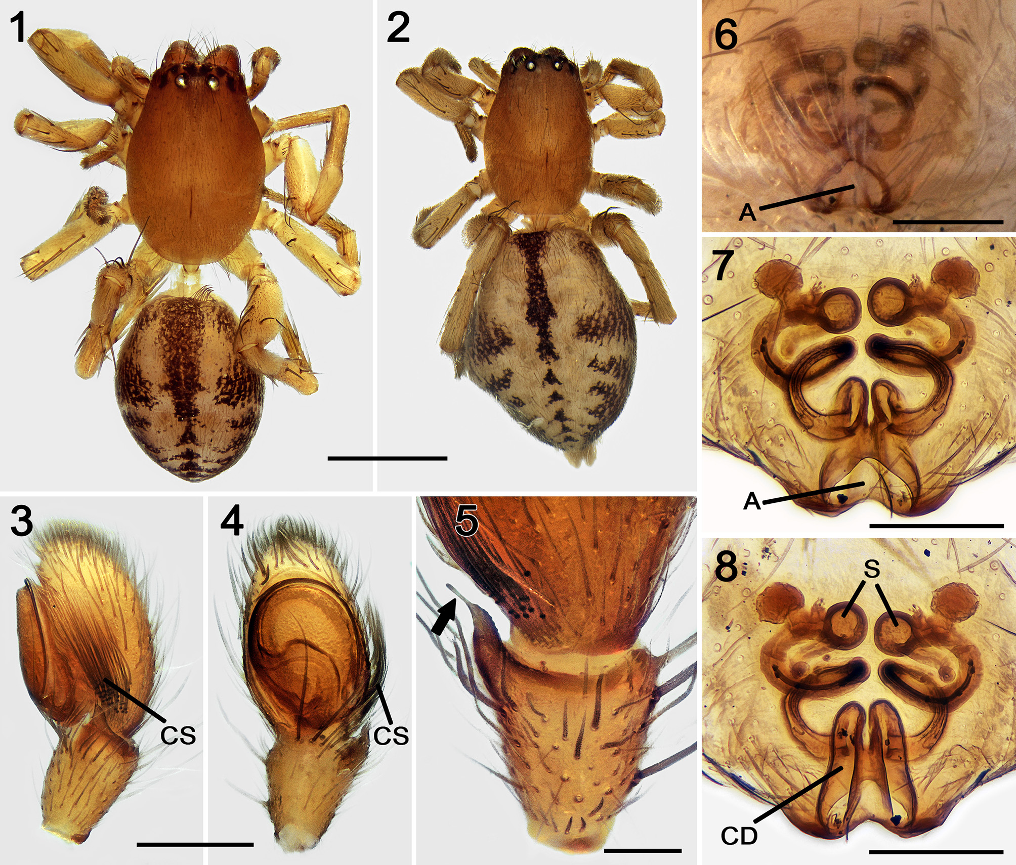
FIGURES 1–8. Clubiona decora Blackwall, 1859: male and female (from Madeira); 1 male habitus, dorsal view; 2 female habitus, dorsal view; 3 male palp, retrolateral view; 4 same, ventral view. 5 detail of the retrolateral tibial apophysis (RTA) of the male palp, dorsal view (arrow on needle-like extension of RTA); 6 epigyne, ventral view; 7 vulva, ventral view; 8 same, dorsal view. Abbreviatures: A = atrium, CD = longitudinal copulatory duct, CS = cymbial modified setae, S = spermathecae. Scale bars:1, 2 = 1 mm; 3, 4, 6–8) = 0.2 mm; 5 = 0.1 mm.
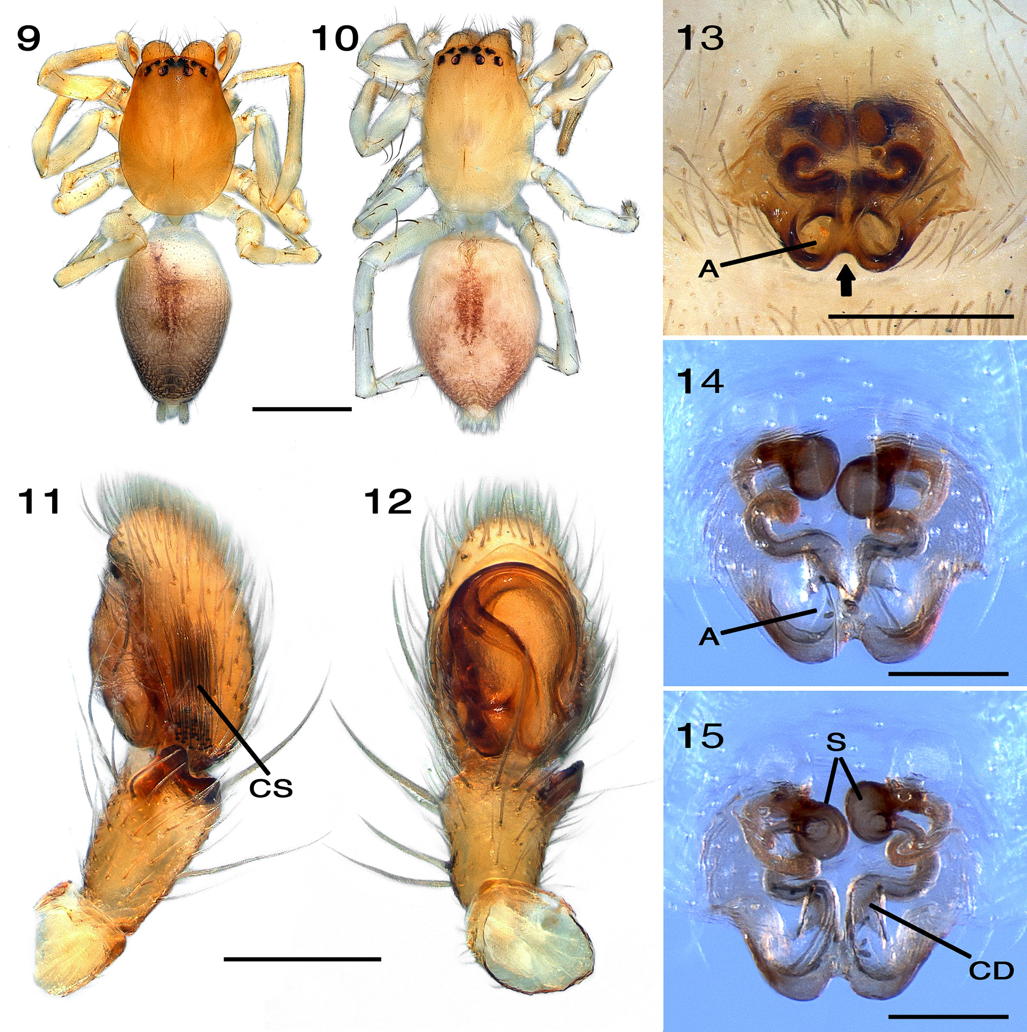
FIGURES 9–15. Clubiona diniensis Simon, 1878: 9 male (from Spain) habitus, dorsal view; 10 female (from Spain) habitus, dorsal view; 11 male (from Algeria) palp, retrolateral view; 12 same, ventral view; 13 Female (from Algeria) epigyne, ventral view (arrow on deep ventral notch of the epigynal margin); 14 female (from France) vulva, ventral view; 15 same, dorsal view. Abbreviatures: A = atrium, CD = longitudinal copulatory duct, CS = cymbial modified setae, S = spermathecae. Scale bars: 9, 10 = 1 mm; 11–13 = 0.2 mm; 14, 15 = 0.1 mm.
FIGURES 16–19. Clubiona diniensis Simon, 1878: 16 male (from Algeria) palp, retrolateral view; 17 same, ventral view; 18 female (from Algeria) epigyne, ventral view; 19 female (from France) vulva, dorsal view. Abbreviatures: A = atrium, CD = longitudinalcopulatory duct, CS = cymbial modified setae, S = spermathecae.Scale bars: 16, 17 = 0.2 mm; 18,19 = 0.1 mm.
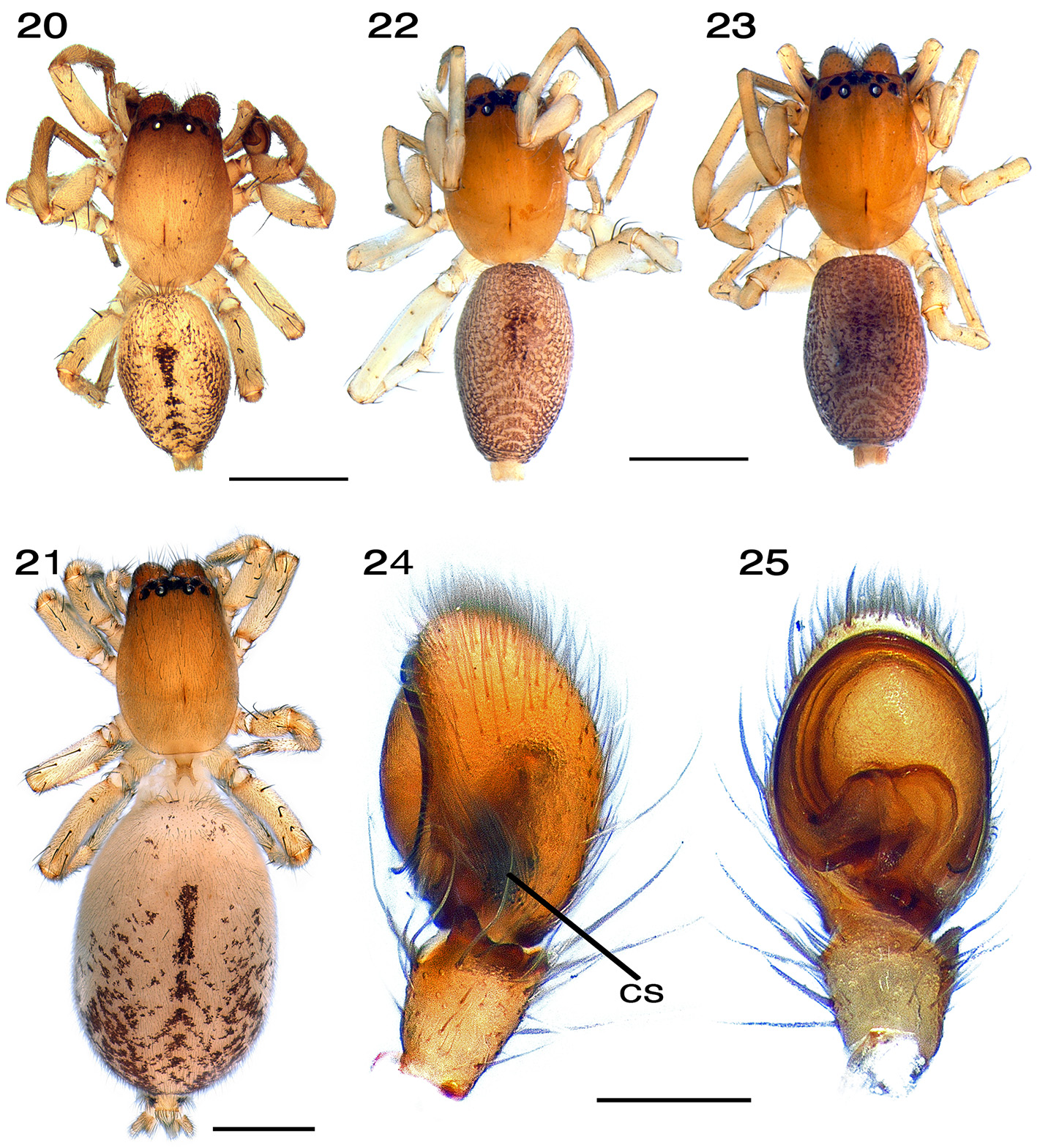
FIGURES 20–25. Clubiona genevensis L. Koch, 1866: 20 male (from France) habitus, dorsal view; 21 female (from France) habitus, dorsal view; 22, 23 males (from Greece) habitus, dorsal view; 24 male (from Greece) palp, retrolateral view; 25 same, ventral view. Abbreviature:CS = cymbial modified setae.Scale bars: 20–23 = 1 mm; 24, 25 = 0.2 mm.
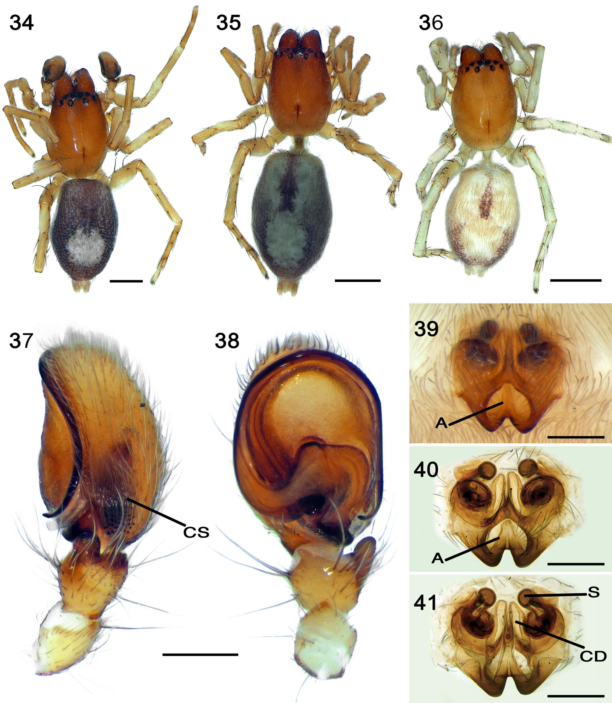
FIGURES34–41. Clubiona leucaspis Simon, 1932:34 male (from Spain) habitus, dorsal view; 35, 36 same, females; 37 male (from Spain) palp, retrolateral view; 38 same, ventral view; 39 female (from Spain) epigyne, ventral view; 40 female (from France) vulva, ventral view; 41 same, dorsal view. Abbreviatures: A = atrium, CD = longitudinal copulatory duct, CS = cymbial modified setae, S = spermathecae.Scale bars: 34–36 = 1 mm; 37–41 = 0.2 mm.
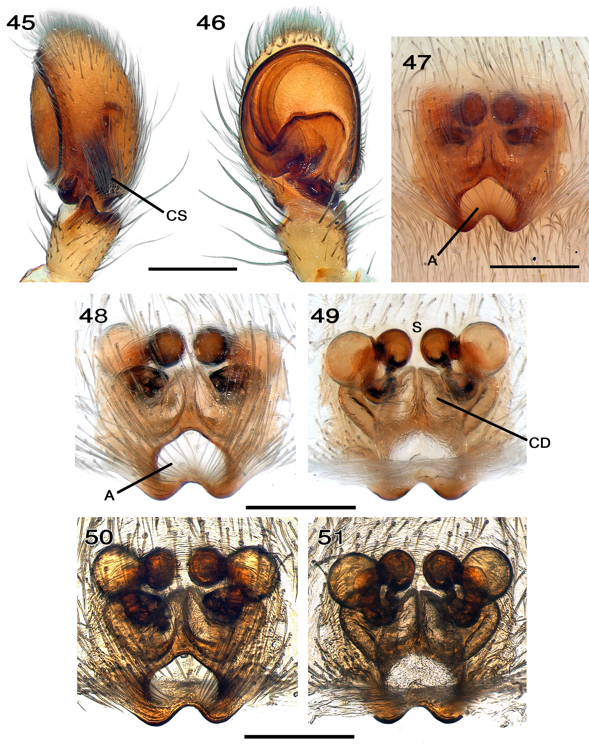
FIGURES 45–51. Clubiona vegeta Simon, 1918, male and female (from Tunisia): 45 male palp, retrolateral view; 46 same, ventral view; 47 epigyne, ventral view; 48 vulva, ventral view, reflected light; 49 same, dorsal view; 50 vulva, ventral view, transmitted light; 51 same, dorsal view.Abbreviatures:A = atrium, CD = longitudinal copulatory duct,CS = cymbial modified setae, S = spermathecae.Scale bars = 0.2 mm.
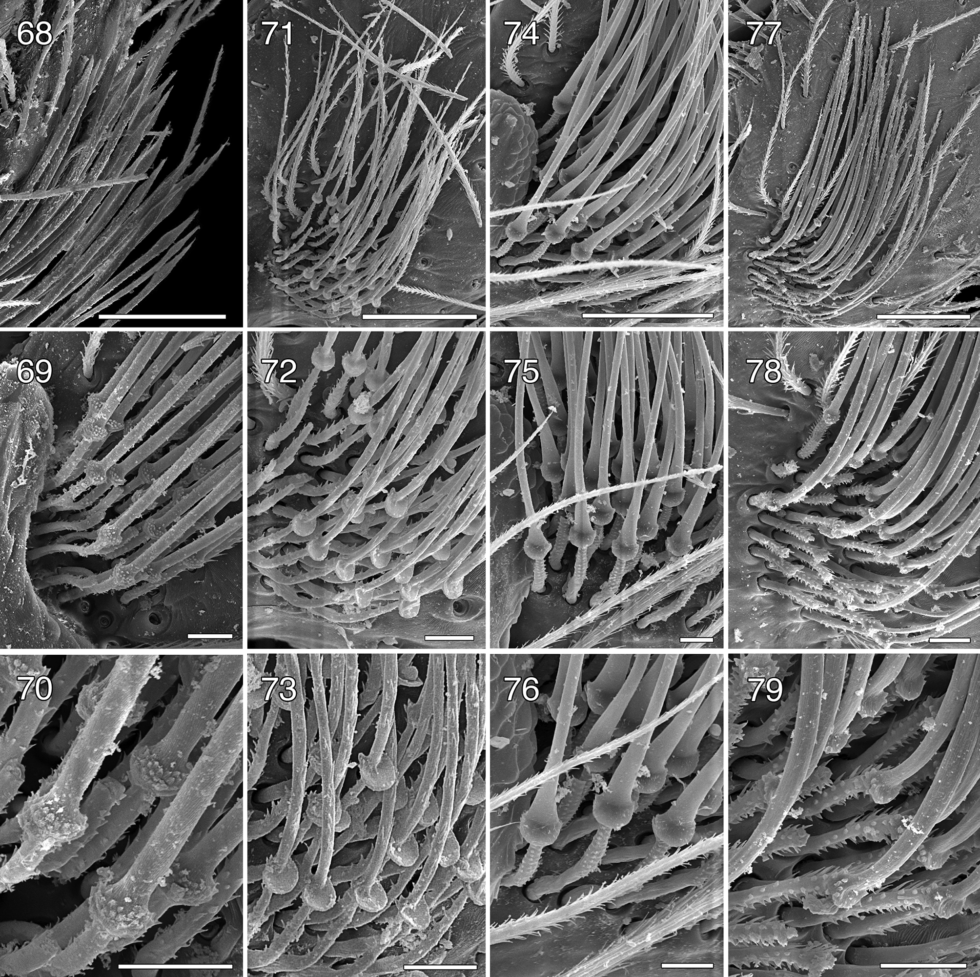
FIGURES68–79. Clubiona spp., male palp, details on the modifiedcymbialsetae, retrolateral view:68–70 Clubiona diniensis Simon,1878(from Algeria); 71–73 C.genevensis L.Koch, 1866(from Greece); 74–76 C. leucaspis Simon, 1932(from Spain); 77–79 C. vegeta Simon, 1918 (from Tunisia). 68 detail of setal apices; 69, 72, 74, 75, 78 detail of setal bases; 70, 73, 76, 79 detail of the swellings; 71, 77 global view. Scale bars: 68, 71, 74, 77 = 50 µm; 69, 70, 72, 73, 75, 76, 78, 79 = 10 µm.

FIGURES 26–33. Clubiona genevensis L. Koch, 1866, female genitalia: 26 epigyne (from France), ventral view; 27, 28 same (from Greece); 29 vulva (from France), ventral view, reflected light; 30 same, dorsal view; 31 same, ventral view; 32 vulva (from France), dorsal view; 33 same, ventro-dorsal view, transmitted light. Abbreviatures: A = atrium, CD = longitudinal copulatory duct, S = spermathecae.Scale bars = 0.2 mm.

FIGURES 52–67. Clubiona spp., male palp: 52–55 Clubiona diniensis Simon, 1878 (from Algeria); 56–59 C. genevensis L. Koch, 1866 (from Greece); 60–63 C. leucaspis Simon, 1932 (from Spain); 64–67 C. vegeta Simon, 1918 (from Tunisia). 52, 56, 60, 64 leftpalp, retrolateral view; 53, 57, 58, 61, 62, 65, 66 detail of RTA and base of cymbium, retrolateral view (65, white arrow, RTA needle-like extension present; 66, black arrow, RTA needle-like extension absent, probably broken); 54, 59, 63, 67 detail of bulbus and basolateral extension of cymbium (stars), retrolateral view; 55 same, ventral view. Scale bars: 52, 56, 60, 64 = 0.1 mm; 53, 57, 59, 61, 63, 67 = 50 µm; 54, 55, 58, 62, 65, 66 = 20 µm.
No known copyright restrictions apply. See Agosti, D., Egloff, W., 2009. Taxonomic information exchange and copyright: the Plazi approach. BMC Research Notes 2009, 2:53 for further explanation.
|
Kingdom |
|
|
Phylum |
|
|
Class |
|
|
Order |
|
|
Family |
Clubiona Wagner, 1887
| Bosmans, Robert, Henrard, Arnaud, Benhalima, Souâd & Kherbouche-Abrous, Ourida 2017 |
C. wunderlichi
| Mikhailov 1992 |
C. minor
| Wunderlich 1987 |
C. pseudominor
| Wunderlich 1987 |
C. parallela
| Hu & Li 1987 |
C. zhangmuensis
| Hu & Li 1987 |
C. leucaspis
| Simon 1932 |
C. vegeta
| , Simon 1918 |
Clubiona
| Wagner 1887 |
diniensis
| Simon 1878 |
genevensis
| L. Koch 1866 |
genevensis
| L. Koch 1866 |
genevensis
| L. Koch 1866 |
genevensis
| L. Koch 1866 |
C. genevensis
| L. Koch 1866 |
genevensis
| L. Koch 1866 |
Clubiona decora
| Blackwall 1859 |
comta
| C. L. Koch 1839 |
Clubiona comta
| C. L. Koch 1839 |
Clubiona comta
| C. L. Koch 1839 |
comta
| C. L. Koch 1839 |
1 (by plazi, 2017-11-22 10:31:57)
2 (by ImsDioSync, 2017-11-22 10:33:23)
3 (by ExternalLinkService, 2019-09-26 02:12:46)
4 (by ExternalLinkService, 2022-01-29 18:20:01)
5 (by ExternalLinkService, 2022-02-09 08:13:02)
6 (by GgImagineBatch, 2022-04-30 04:00:15)
7 (by plazi, 2023-10-28 13:49:48)