
Clubiona, Wagner, 1887
|
publication ID |
https://doi.org/10.11646/zootaxa.4353.1.1 |
|
publication LSID |
lsid:zoobank.org:pub:7D342E35-8D42-4C3D-9903-49E5F08D7D34 |
|
DOI |
https://doi.org/10.5281/zenodo.6020652 |
|
persistent identifier |
https://treatment.plazi.org/id/03EF9555-0904-FF99-1DD1-FA781790FB83 |
|
treatment provided by |
Plazi (2017-11-22 10:31:57, last updated 2024-11-26 07:26:33) |
|
scientific name |
Clubiona |
| status |
|
Key to the Clubiona View in CoL View at ENA species of the genevensis group
Clubiona minor , C. pseudominor and C. wunderlichi were not examined and, for these species, the key is based on characters found in literature.
1 Males (those of C. pseudominor View in CoL and C. wunderlichi View in CoL unknown).................................................. 2
- Females............................................................................................. 7
2(1) Embolus originating in distal or median part of bulbus and directed anteriorly ( Figs 4 View FIGURES 1–8 , 12 View FIGURES 9–15 , 17 View FIGURES 16–19 ; Wunderlich 1987: fig. 640), basolateral extension of bulbus and cymbium short, as long as wide ( Figs 54, 55 View FIGURES 52–67 ); modified cymbial setae long: as long as half the length of cymbium or more ( Figs 3, 4 View FIGURES 1–8 , 11 View FIGURES 9–15 , 16 View FIGURES 16–19 , 52 View FIGURES 52–67 )..........................................3 ( decora View in CoL subgroup)
- Embolus originating more basally and directed laterally ( Figs 25 View FIGURES 20–25 , 38 View FIGURES34–41 , 46 View FIGURES 45–51 ); basolateral extension of bulbus and cymbium much longer than wide ( Figs 59, 63, 67 View FIGURES 52–67 ); modified cymbial setae shorter, length less than half length of cymbium ( Figs 24 View FIGURES 20–25 , 37 View FIGURES34–41 , 45 View FIGURES 45–51 , 56, 60, 64 View FIGURES 52–67 )5........................................................................ ( genevensis View in CoL subgroup)
3(2) Palpal tibia nearly twice as long as wide ( Fig. 11 View FIGURES 9–15 ); RTA tip more rounded ( Fig. 11 View FIGURES 9–15 ); origin of embolus in distal part of bulbus ( Figs 12 View FIGURES 9–15 , 17 View FIGURES 16–19 )................................................................................ C. diniensis View in CoL
- Palpal tibia slightly longer than wide ( Fig. 3 View FIGURES 1–8 ; Wunderlich 1987: fig. 641); RTA with sharp tip ( Fig. 3 View FIGURES 1–8 ; Wunderlich 1987: fig. 641); origin of embolus in median part of bulbus ( Fig. 4 View FIGURES 1–8 ; Wunderlich 1987: fig. 640)................................ 4
4(3) RTA thin, triangular, not broadened basally ( Fig. 3 View FIGURES 1–8 ).................................................... C. decora View in CoL
- RTA with broad base extending dorsally ( Wunderlich 1987: figs 641, 642).................................. C. minor View in CoL
5(2) Retrolateral tibial apophysis (RTA) rounded, slightly longer than wide at its base ( Figs 37 View FIGURES34–41 , 62 View FIGURES 52–67 ); ventral profile of anterior part of bulbus concave ( Figs 37 View FIGURES34–41 , 60 View FIGURES 52–67 ); modified cymbial setae with spherical sub-basal swelling ( Figs 74–76 View FIGURES68–79 ); abdomen dorsally with pale spot on posterior half ( Fig. 34 View FIGURES34–41 ).......................................................... C. leucaspis View in CoL
- RTA roughly triangular or bluntly pointed ( Figs 24 View FIGURES 20–25 , 45 View FIGURES 45–51 , 57, 58, 65, 66 View FIGURES 52–67 ); ventral profile of bulbus convex ( Figs 24 View FIGURES 20–25 , 45 View FIGURES 45–51 , 56, 64 View FIGURES 52–67 ); modified cymbial setae with flattened sub-basal swelling ( Figs 73, 79 View FIGURES68–79 ); abdomen without postero-dorsal white spot ( Figs 20– 23 View FIGURES 20–25 , 42, 43 View FIGURES 42–44 )........................................................................................... 6
6(5) Chelicerae not enlarged ( Figs 20, 22, 23 View FIGURES 20–25 ); RTA roughly triangular, with blunt, rounded tip ( Figs 24 View FIGURES 20–25 , 57, 58 View FIGURES 52–67 ); modified cymbial setae with well developed, plate shaped sub-basal swelling ( Figs 72, 73 View FIGURES68–79 )............................... C. genevensis View in CoL
- Chelicerae dark brown, strongly protruding ( Figs 43, 44 View FIGURES 42–44 ); RTA triangular with sharper tip ( Figs 45 View FIGURES 45–51 , 65, 66 View FIGURES 52–67 ); sub-basal swelling of modified cymbial setae reduced, crescent shaped ( Fig. 79 View FIGURES68–79 )............................................ C. vegeta View in CoL
7(1) Copulatory ducts loose, slightly coiled or not, connected laterally to the atrio-spermathecal part ( Figs 6–8 View FIGURES 1–8 , 13–15 View FIGURES 9–15 ; Wunderlich 1987: figs 643–645; Mikhailov 1992: figs 2A, B)............................................ 8 ( decora View in CoL subgroup)
- Copulatory ducts tightened, strongly convoluted, connected inferiorly to the atrio-spermathecal part ( Figs 29–32 View FIGURES 26–33 , 40, 41 View FIGURES34–41 , 48– 51 View FIGURES 45–51 )............................................................................. 12 ( genevensis View in CoL subgroup)
8(7) Copulatory ducts straight forward, not coiled ( Wunderlich 1987: figs 643–645, 645a)..................................................................................................................................... 9
- Copulatory ducts coiled ( Figs 6–8 View FIGURES 1–8 , 13–15 View FIGURES 9–15 ; Mikhailov 1992: figs 2A, B).......................................... 10
9(8) Fertilization duct well developed, conical ( Wunderlich 1987: fig. 644)...................................... C. minor View in CoL
- Fertilization duct thinner, tube-shaped ( Wunderlich 1987: fig. 645a)................................. C. pseudominor View in CoL
10(8) Epigyne with two distinct oval atria ( Figs 13, 14 View FIGURES 9–15 , 18 View FIGURES 16–19 )................................................ C. diniensis View in CoL
- Epigyne with only one, roughly inverted heart-shaped atrium ( Figs 6–8 View FIGURES 1–8 ; Mikhailov 1992: figs 2A, B).................. 11
11(10) Spermathecae far away from the anterior part of longitudinal copulatory ducts ( Figs 6– 8 View FIGURES 1–8 )..................... C. decora View in CoL
- Anterior part of longitudinal copulatory ducts almost reaching the inferior part of spermathecae ( Mikhailov 1992: figs 2A, B)......................................................................................... C. wunderlichi View in CoL
12(7) Abdomen dorsally with short anterior median stripe and with contrasting pale oval area on posterior half ( Figs 35, 36 View FIGURES34–41 ); spermathecae small, separated by their diameter or more ( Figs 39–41 View FIGURES34–41 )...................................... C. leucaspis View in CoL
- Abdomen usually with longer, sometimes interrupted median stripe, posteriorly without dorsal pale spot ( Figs 21 View FIGURES 20–25 , 42 View FIGURES 42–44 ); spermathecae larger, separated by less than their diameter ( Figs 26–33 View FIGURES 26–33 , 47–51 View FIGURES 45–51 )....................................... 13
13(12) Dorsal median stripe most often absent in anterior third of abdomen, fading into V-shaped stripes in posterior third ( Fig. 21 View FIGURES 20–25 ); copulatory opening often narrower, longer than wide ( Figs 26, 27 View FIGURES 26–33 ); vulva with narrower atrium ( Figs 30, 32 View FIGURES 26–33 )... C. genevensis View in CoL
- Dorsal median stripe often reaching anterior margin of abdomen, tapered posteriorly and fading in posterior third ( Fig. 42 View FIGURES 42–44 ; better preserved in the male Fig. 43 View FIGURES 42–44 ); copulatory opening wider, at least as wide as long ( Figs 47, 48 View FIGURES 45–51 ); vulva with wider atrium ( Figs 49, 51 View FIGURES 45–51 ).................................................................................. C. vegeta View in CoL
Mikhailov, K. G. (1992) The spider genus Clubiona Latreille, 1804 (Arachnida Aranei Clubionidae) in the USSR fauna: a critical review with taxonomical remarks. Arthropoda Selecta, 1 (3), 3 - 34.
Wunderlich, J. (1987) Die Spinnen der Kanarischen Inseln und Madeiras: Adaptive Radiation, Biogeographie, Revisionen und Neubeschreibungen. Triops, Langen, 435 pp.
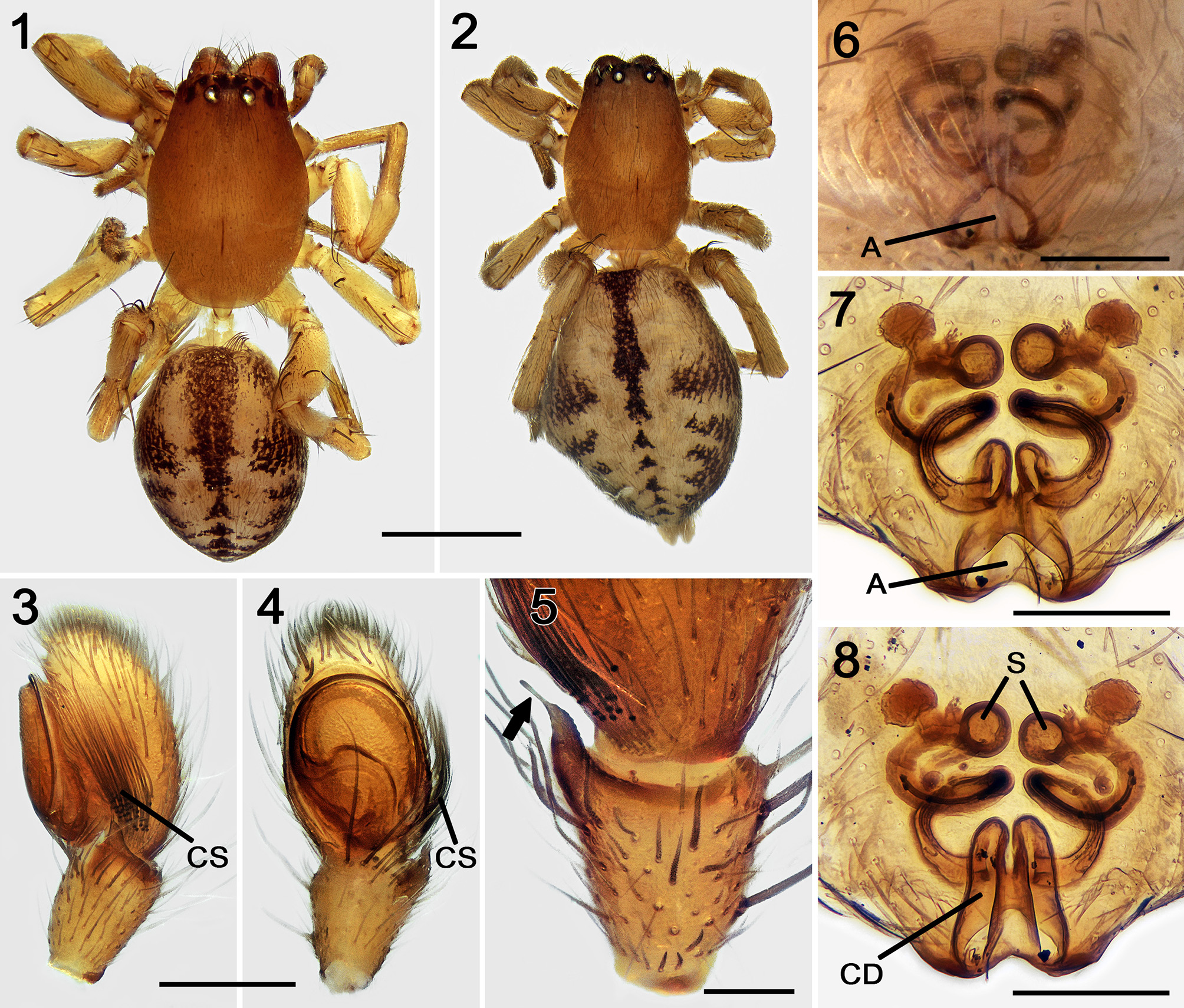
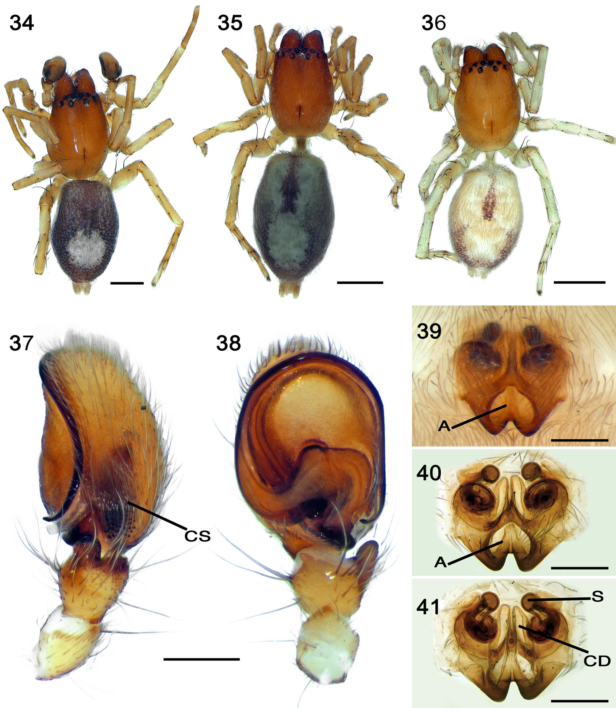
FIGURES 1–8. Clubiona decora Blackwall, 1859: male and female (from Madeira); 1 male habitus, dorsal view; 2 female habitus, dorsal view; 3 male palp, retrolateral view; 4 same, ventral view. 5 detail of the retrolateral tibial apophysis (RTA) of the male palp, dorsal view (arrow on needle-like extension of RTA); 6 epigyne, ventral view; 7 vulva, ventral view; 8 same, dorsal view. Abbreviatures: A = atrium, CD = longitudinal copulatory duct, CS = cymbial modified setae, S = spermathecae. Scale bars:1, 2 = 1 mm; 3, 4, 6–8) = 0.2 mm; 5 = 0.1 mm.
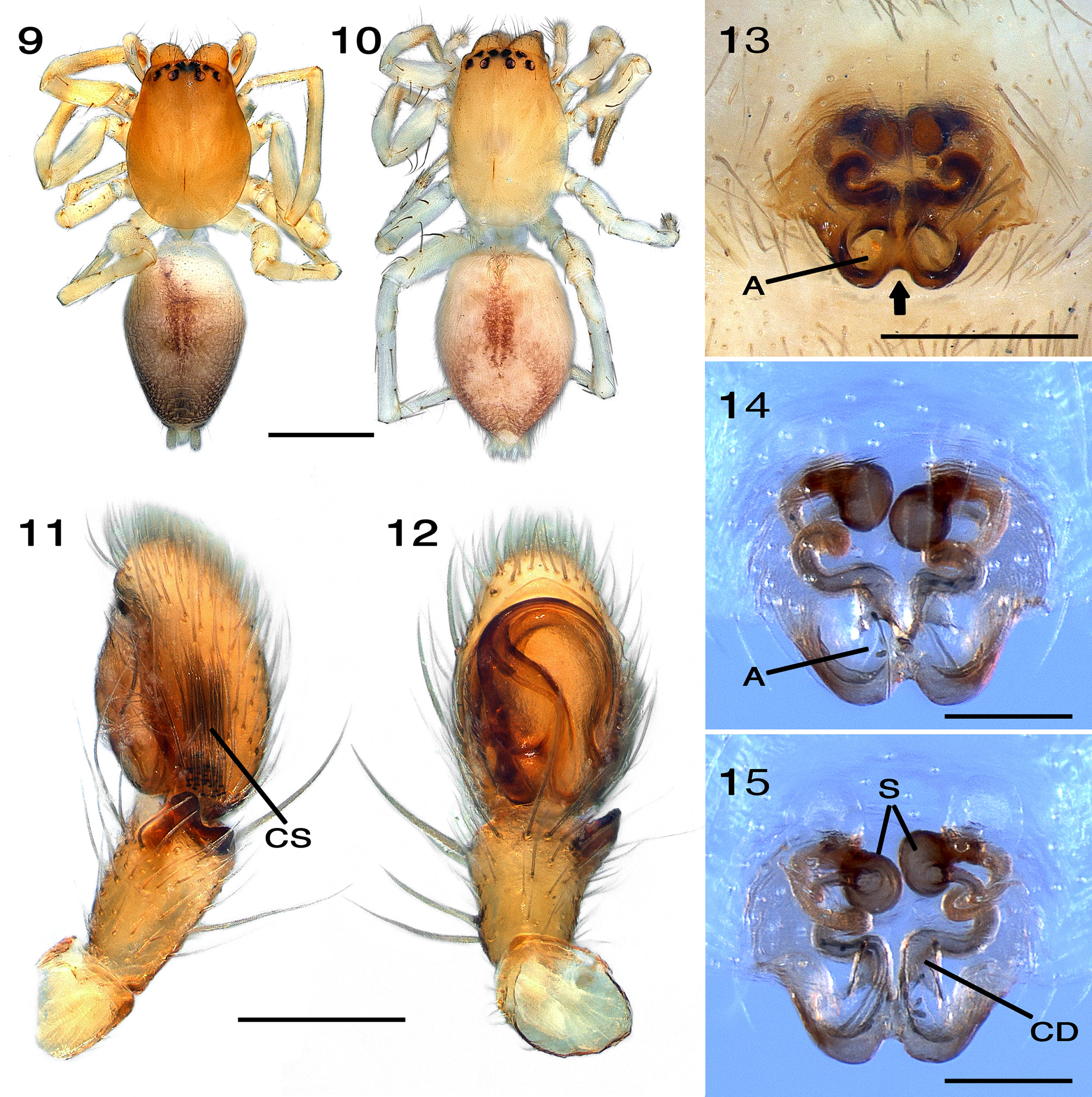
FIGURES 9–15. Clubiona diniensis Simon, 1878: 9 male (from Spain) habitus, dorsal view; 10 female (from Spain) habitus, dorsal view; 11 male (from Algeria) palp, retrolateral view; 12 same, ventral view; 13 Female (from Algeria) epigyne, ventral view (arrow on deep ventral notch of the epigynal margin); 14 female (from France) vulva, ventral view; 15 same, dorsal view. Abbreviatures: A = atrium, CD = longitudinal copulatory duct, CS = cymbial modified setae, S = spermathecae. Scale bars: 9, 10 = 1 mm; 11–13 = 0.2 mm; 14, 15 = 0.1 mm.
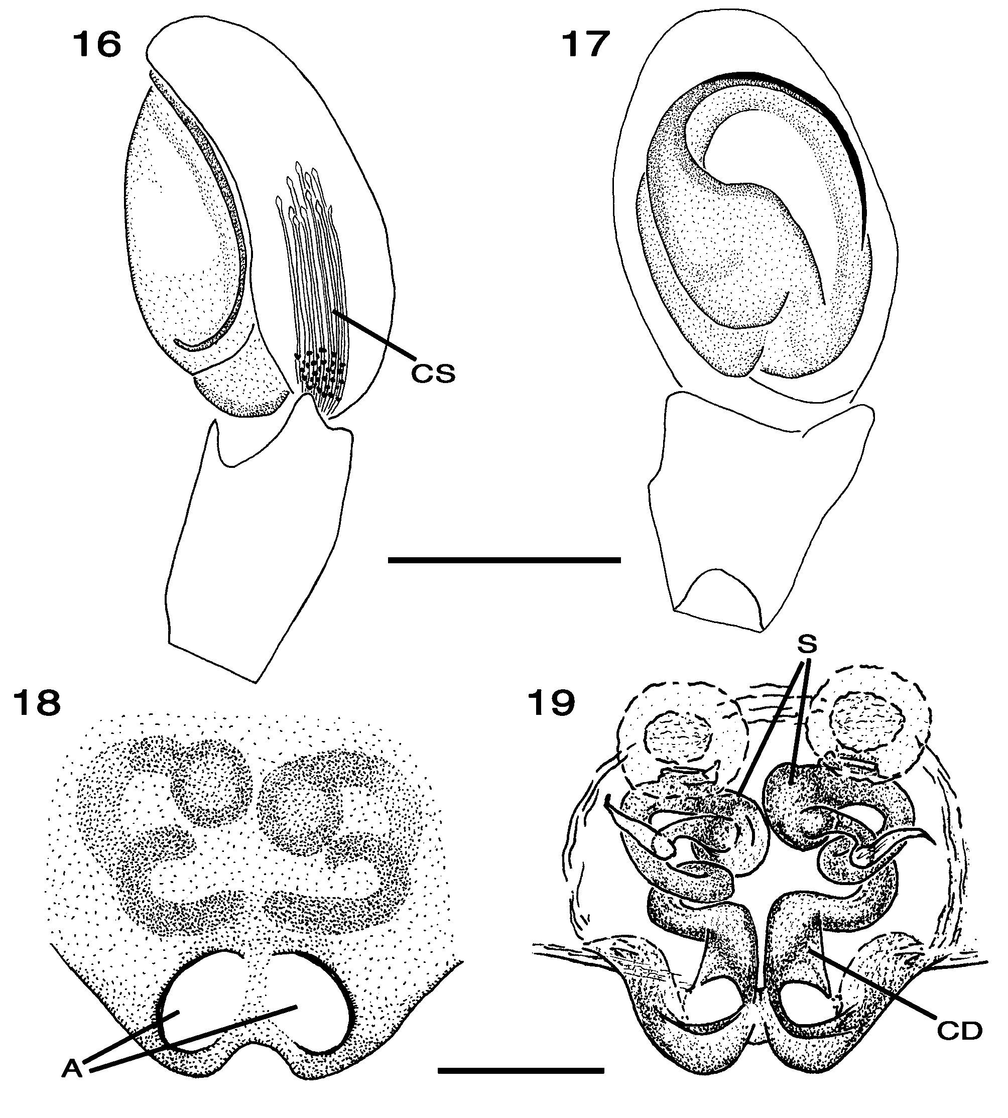
FIGURES 16–19. Clubiona diniensis Simon, 1878: 16 male (from Algeria) palp, retrolateral view; 17 same, ventral view; 18 female (from Algeria) epigyne, ventral view; 19 female (from France) vulva, dorsal view. Abbreviatures: A = atrium, CD = longitudinalcopulatory duct, CS = cymbial modified setae, S = spermathecae.Scale bars: 16, 17 = 0.2 mm; 18,19 = 0.1 mm.

FIGURES 52–67. Clubiona spp., male palp: 52–55 Clubiona diniensis Simon, 1878 (from Algeria); 56–59 C. genevensis L. Koch, 1866 (from Greece); 60–63 C. leucaspis Simon, 1932 (from Spain); 64–67 C. vegeta Simon, 1918 (from Tunisia). 52, 56, 60, 64 leftpalp, retrolateral view; 53, 57, 58, 61, 62, 65, 66 detail of RTA and base of cymbium, retrolateral view (65, white arrow, RTA needle-like extension present; 66, black arrow, RTA needle-like extension absent, probably broken); 54, 59, 63, 67 detail of bulbus and basolateral extension of cymbium (stars), retrolateral view; 55 same, ventral view. Scale bars: 52, 56, 60, 64 = 0.1 mm; 53, 57, 59, 61, 63, 67 = 50 µm; 54, 55, 58, 62, 65, 66 = 20 µm.
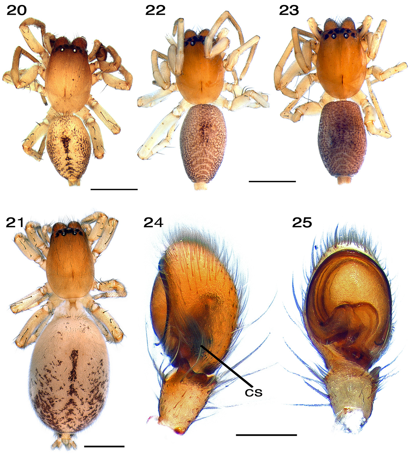
FIGURES 20–25. Clubiona genevensis L. Koch, 1866: 20 male (from France) habitus, dorsal view; 21 female (from France) habitus, dorsal view; 22, 23 males (from Greece) habitus, dorsal view; 24 male (from Greece) palp, retrolateral view; 25 same, ventral view. Abbreviature:CS = cymbial modified setae.Scale bars: 20–23 = 1 mm; 24, 25 = 0.2 mm.
FIGURES34–41. Clubiona leucaspis Simon, 1932:34 male (from Spain) habitus, dorsal view; 35, 36 same, females; 37 male (from Spain) palp, retrolateral view; 38 same, ventral view; 39 female (from Spain) epigyne, ventral view; 40 female (from France) vulva, ventral view; 41 same, dorsal view. Abbreviatures: A = atrium, CD = longitudinal copulatory duct, CS = cymbial modified setae, S = spermathecae.Scale bars: 34–36 = 1 mm; 37–41 = 0.2 mm.
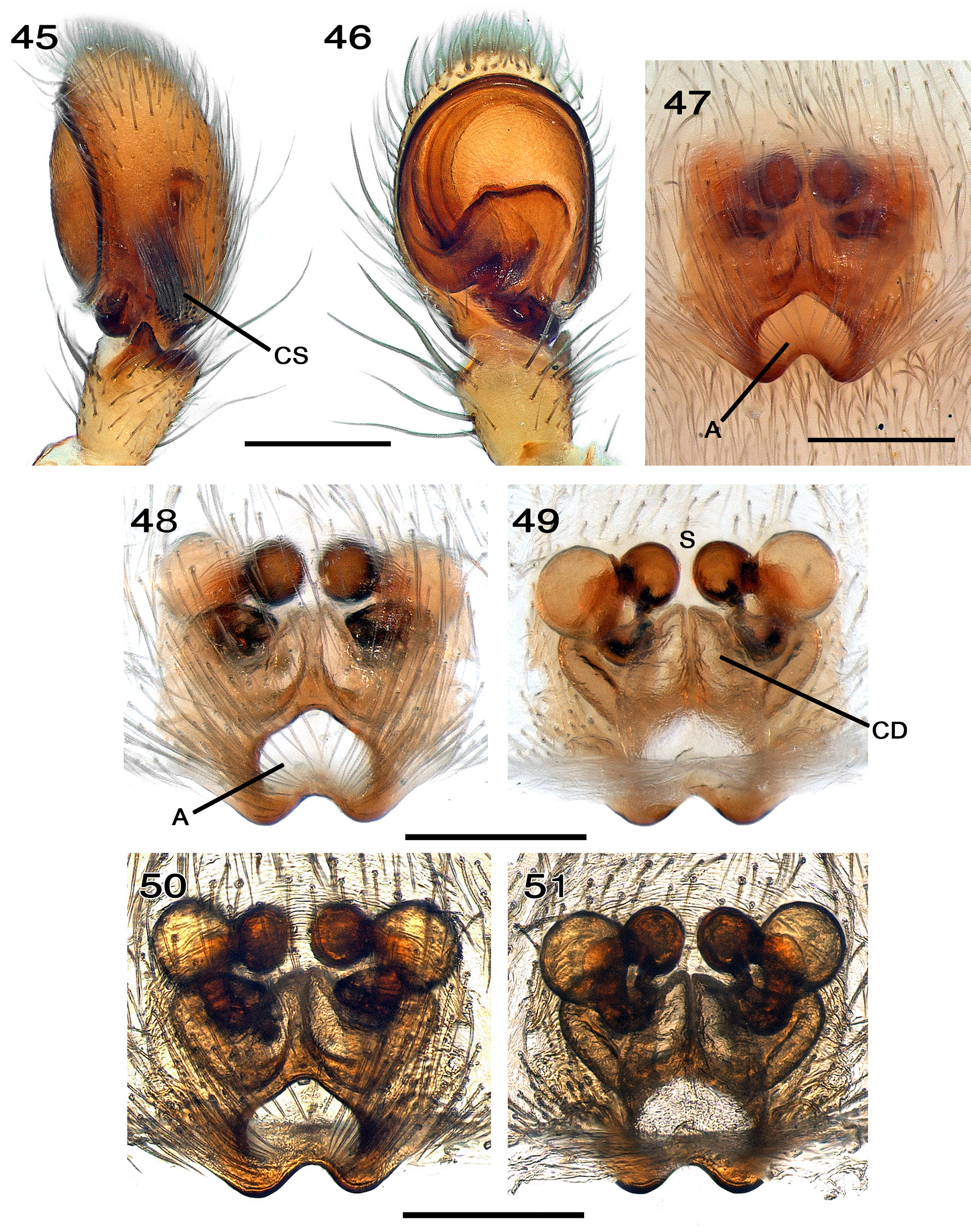
FIGURES 45–51. Clubiona vegeta Simon, 1918, male and female (from Tunisia): 45 male palp, retrolateral view; 46 same, ventral view; 47 epigyne, ventral view; 48 vulva, ventral view, reflected light; 49 same, dorsal view; 50 vulva, ventral view, transmitted light; 51 same, dorsal view.Abbreviatures:A = atrium, CD = longitudinal copulatory duct,CS = cymbial modified setae, S = spermathecae.Scale bars = 0.2 mm.
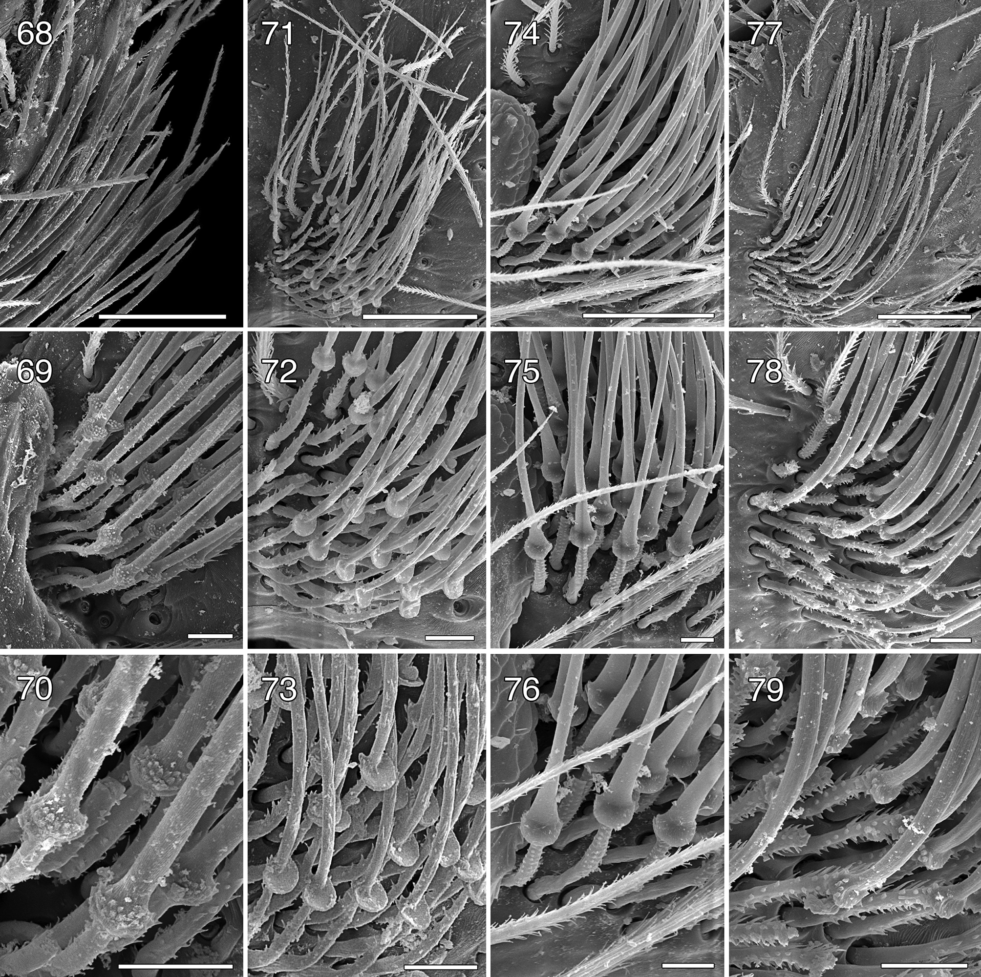
FIGURES68–79. Clubiona spp., male palp, details on the modifiedcymbialsetae, retrolateral view:68–70 Clubiona diniensis Simon,1878(from Algeria); 71–73 C.genevensis L.Koch, 1866(from Greece); 74–76 C. leucaspis Simon, 1932(from Spain); 77–79 C. vegeta Simon, 1918 (from Tunisia). 68 detail of setal apices; 69, 72, 74, 75, 78 detail of setal bases; 70, 73, 76, 79 detail of the swellings; 71, 77 global view. Scale bars: 68, 71, 74, 77 = 50 µm; 69, 70, 72, 73, 75, 76, 78, 79 = 10 µm.

FIGURES 42–44. Clubiona vegeta Simon, 1918, male and female (from Tunisia): 42 female habitus, dorsal view; 43 male habitus, dorsal view; 44 same, detail of the prosoma,ventral view.Scale bars = 1mm.

FIGURES 26–33. Clubiona genevensis L. Koch, 1866, female genitalia: 26 epigyne (from France), ventral view; 27, 28 same (from Greece); 29 vulva (from France), ventral view, reflected light; 30 same, dorsal view; 31 same, ventral view; 32 vulva (from France), dorsal view; 33 same, ventro-dorsal view, transmitted light. Abbreviatures: A = atrium, CD = longitudinal copulatory duct, S = spermathecae.Scale bars = 0.2 mm.
No known copyright restrictions apply. See Agosti, D., Egloff, W., 2009. Taxonomic information exchange and copyright: the Plazi approach. BMC Research Notes 2009, 2:53 for further explanation.
|
Kingdom |
|
|
Phylum |
|
|
Class |
|
|
Order |
|
|
Family |
|
|
Genus |