
Yoruba ibadanus, Rodrigues & Rheims, 2020
|
publication ID |
https://doi.org/10.11646/zootaxa.4799.1.1 |
|
publication LSID |
lsid:zoobank.org:pub:E295AAC9-09C8-48A3-8C5F-402117381B80 |
|
DOI |
https://doi.org/10.5281/zenodo.5920132 |
|
persistent identifier |
https://treatment.plazi.org/id/03EE8789-3B1B-A13A-6AEC-F974ADEDF967 |
|
treatment provided by |
Plazi (2020-06-16 11:50:33, last updated 2024-11-24 23:54:06) |
|
scientific name |
Yoruba ibadanus |
| status |
sp. nov. |
Yoruba ibadanus View in CoL sp. nov.
Figs 54 View FIGURE 54 B–F, 55A–F, 56A–F, 57A–F, 58A–F, 59A–F
Type material. Male holotype from Nigeria, Ibadan [7°23’47”N 3°55’E], 1 April 1973, A. Russell-Smith leg., deposited in MRAC 245546 View Materials . GoogleMaps Paratypes from the same vial as holotype: 13 males and 3 females GoogleMaps .
Etymology. The specific name refers to the type locality, adjective.
Diagnosis. Males of Yoruba ibadanus sp. nov. are distinguished from those of Y. toubensis sp. nov. by the presence of a screw-shaped embolus (laminar and without folds in Y. toubensis sp. nov.) and straight VTA (pointing retrolaterally in ventral view in Y. toubensis sp. nov.) ( Figs 58D View FIGURE 58 , 59B, C View FIGURE 59 ); females differ of Y. toubensis sp. nov. by the presence of an epigynal scape (absent in Y. toubensis sp. nov.) and the slender connecting ducts, with proximal part curving behind the copulatory duct ( Fig. 59E View FIGURE 59 ).
Description. Male (holotype). Total length: 2.78. Carapace 1.03 long, 0.70 wide; Abdomen 1.46 long, 0.68 wide; Sternum 0.68 long, 0.55 wide; Spinnerets: ALS 0.22 long, 0.14 wide. Eye diameters: AME 0.05, ALE 0.045, PME 0.04, PLE 0.04; interdistances: AME–AME 0.03, PME–PME 0.02. Chelicerae 0.27 long. Leg measurements: I: 3.30 (0.90, 0.66, 0.68, 0.56, 0.50); II: 2.46 (0.70, 0.40, 0.51, 0.45, 0.40); III: 2.28 (0.58, 0.30, 0.50, 0.46, 0.44); IV: 3.38 (0.92, 0.49, 0.75, 0.66, 0.56). Leg spination: I—femur d1-0-0. II—femur d1-0-0. III—femur d1-1-0; tibia v1-0-1, r0-0-1, p0-0-1; metatarsus v2-0-1, r1-0-1, p0-0-1. IV—femur 1-1-0; tibia v1-2-2, r1-0-1, p1-0-1; metatarsus v1-1-2, p1-0-1, r0-1-0. Palp: RTA short and tapered; embolus arising from center of tegulum at 9 o’clock position ( Figs 61 View FIGURE 61 D–F, 62A–C).
Female (Paratype). Total length: 2.21. Carapace 0.87 long, 0.62 wide; Abdomen 1.31 long, 0.78 wide; Sternum 0.62 long, 0.50 wide; Spinnerets ALS 0.20 long, 0.12 wide. Eye diameters: AME 0.04, ALE 0.03, PME 0.04, PLE 0.03; interdistances: AME–AME 0.01, PME–PME 0.02. Chelicerae 0.26 long. Leg measurements: I: 2.87 (0.78, 0.56, 0.59, 0.52, 0.42); II: 1.97 (0.55, 0.30, 0.40, 0.37, 0.35); III: 1.75 (0.47, 0.28, 0.36, 0.32, 0.32); IV: 3.01 (0.79, 0.45, 0.66, 0.60, 0.51). Leg spination: I—femur d1-1-0. II—femur d1-1-0. III—femur d1-1-0; tibia v0-1-1, p1-0-1; metatarsus v1-0-1, r0-1-0, p0-0-1. IV—femur 1-1-0; tibia v1p-1p-2, r1-0-1, p1-0-1; metatarsus v1-0-2, p0-0-1, r0- 1-0. Vulva: copulatory ducts with constrictions laterad; primary spermathecae rounded ( Fig. 59E View FIGURE 59 ).
Variation. Total length (14 males): 1.97–2.40; (3 females): 2.12–2.70.
Distribution. Known only from type locality, Ibadan, Nigeria ( Fig. 61 View FIGURE 61 ).

FIGURE 54. Yoruba toubensis sp. nov., male: (A) habitus, dorsal view. Y. ibadanus sp. nov., male: (B) habitus, dorsal view; (C) eyes, frontal view; (D) sternum and endites; (E) spinnerets, lateral view; (F) spinnerets, ventral view
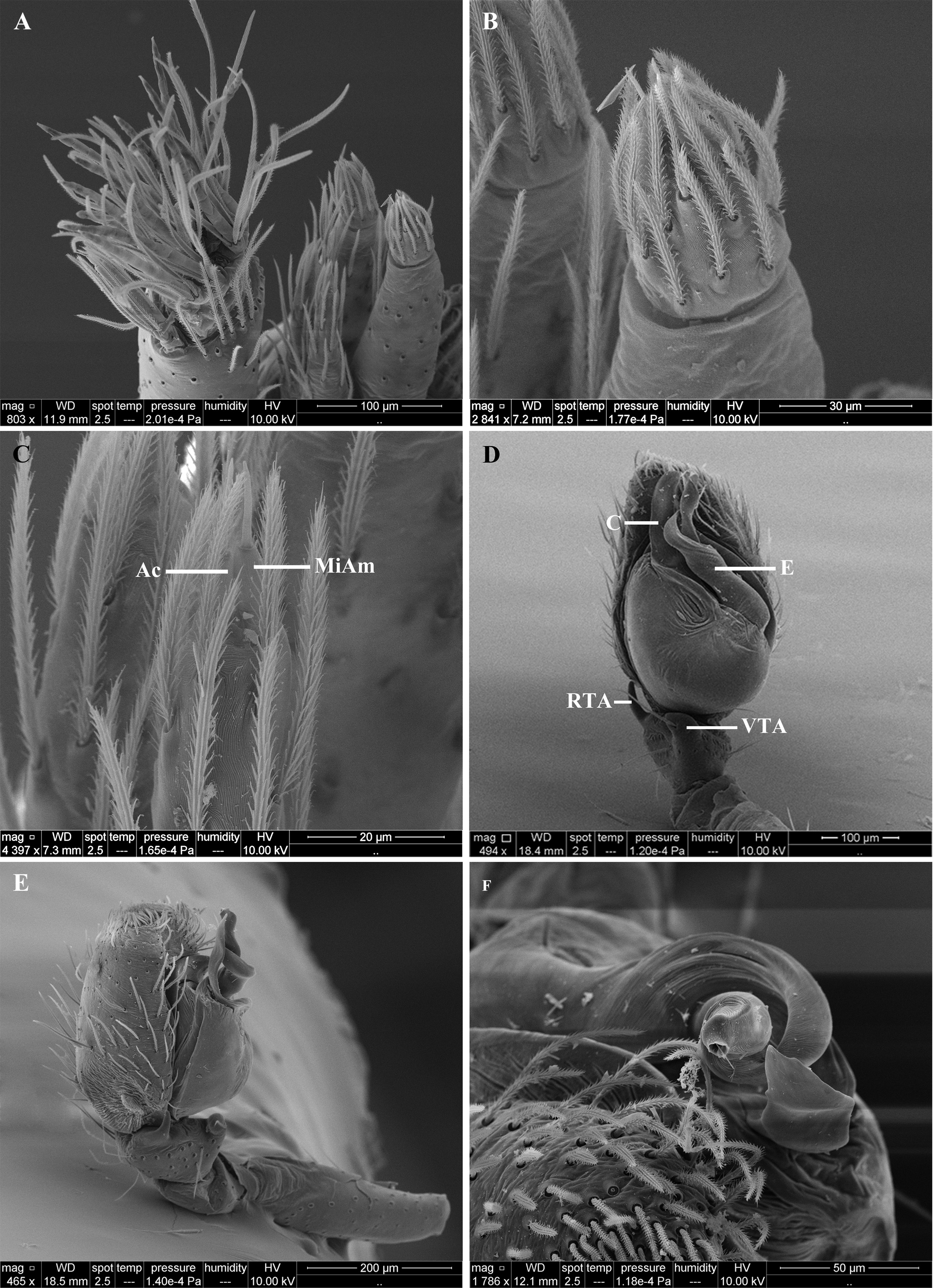
FIGURE 58. Yoruba ibadanus sp. nov., male: (A) spinnerets; (B) spinnerets, PLS; (C) spinnerets, PMS; (D) palp, ventral view; (E) palp, retrolateral view; (F) palp, detail embolus, apical view. Ac—aciniform gland spigots; MiAm—minor ampulate gland spigots; C—conductor; E—embolus; RTA—retrolateral tibial apophysis; VTA—ventral tibial apophysis.

FIGURE 59. Yoruba ibadanus sp. nov. Male palp: (A) prolateral view; (B) ventral view; (C) retrolateral view. Female: (D) epigyne, ventral view; (E) vulva, dorsal view; (F) vulva, schematic course of internal duct system. C—conductor; CD—copulatory duct; CO—copulatory opening; CoD—connecting duct; E—embolus; FD—fertilization duct; PS—primary spermathecae; SS—secondary spermathecae; RTA—retrolateral tibial apophisys; VTA—ventral tibial apophisys. Scale bars: 0.2 mm.

No known copyright restrictions apply. See Agosti, D., Egloff, W., 2009. Taxonomic information exchange and copyright: the Plazi approach. BMC Research Notes 2009, 2:53 for further explanation.
|
Kingdom |
|
|
Phylum |
|
|
Class |
|
|
Order |
|
|
Family |
|
|
Genus |
1 (by plazi, 2020-06-16 11:50:33)
2 (by ExternalLinkService, 2020-06-16 13:11:59)
3 (by admin, 2020-06-17 00:11:26)
4 (by ExternalLinkService, 2020-06-17 00:22:33)
5 (by ExternalLinkService, 2022-01-29 08:46:30)
6 (by ExternalLinkService, 2022-01-30 12:14:19)
7 (by plazi, 2023-10-31 17:58:14)