
Breda Peckham & Peckham, 1894
|
publication ID |
https://doi.org/ 10.11646/zootaxa.3664.4.1 |
|
publication LSID |
lsid:zoobank.org:pub:8E88DF35-70E0-4C21-BC8B-F4F5C85E307D |
|
DOI |
https://doi.org/10.5281/zenodo.6151361 |
|
persistent identifier |
https://treatment.plazi.org/id/03EC879B-FFB8-FF8F-7B85-909F27EED9BF |
|
treatment provided by |
Plazi |
|
scientific name |
Breda Peckham & Peckham, 1894 |
| status |
|
Breda Peckham & Peckham, 1894 View in CoL View at ENA
Breda Peckham & Peckham, 1894: 92 (Type species: Marpissa milvina C.L. Koch , by original designation); Platnick, 2013.
Paradescanso Vellard, 1924: 33 [Type species: Paradescanso fallax Vellard (= Breda apicalis Simon ), by original designation]; Platnick, 2013. Syn. nov.
Oserictops Mello-Leitão, 1941 b: 222 (Type species: Oserictops o serictops Mello-Leitão, by original designation). Synonymized by Galiano (1980: 36).
Thianioides Mello-Leitão, 1941a: 190 (Type species: Thianioides spinimanu Mello-Leitão [= Breda modesta (Taczanowski) syn. nov.], by original designation). Synonymized by Galiano (1981: 12).
Bredops Mello-Leitão, 1944: 378 [Type species: Bredops extraordinarius Mello-Leitão (= Breda tristis Mello-Leitão ), by original designation]. Synonymized by Galiano (1981: 15).
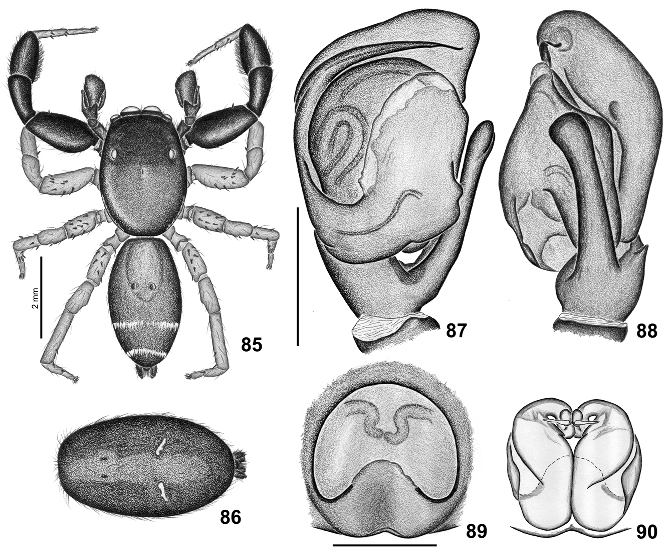
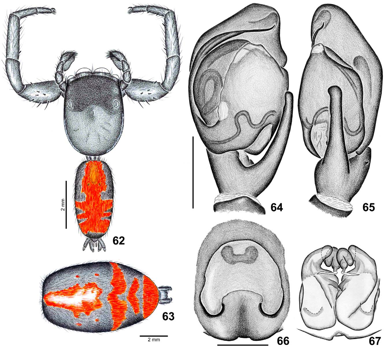
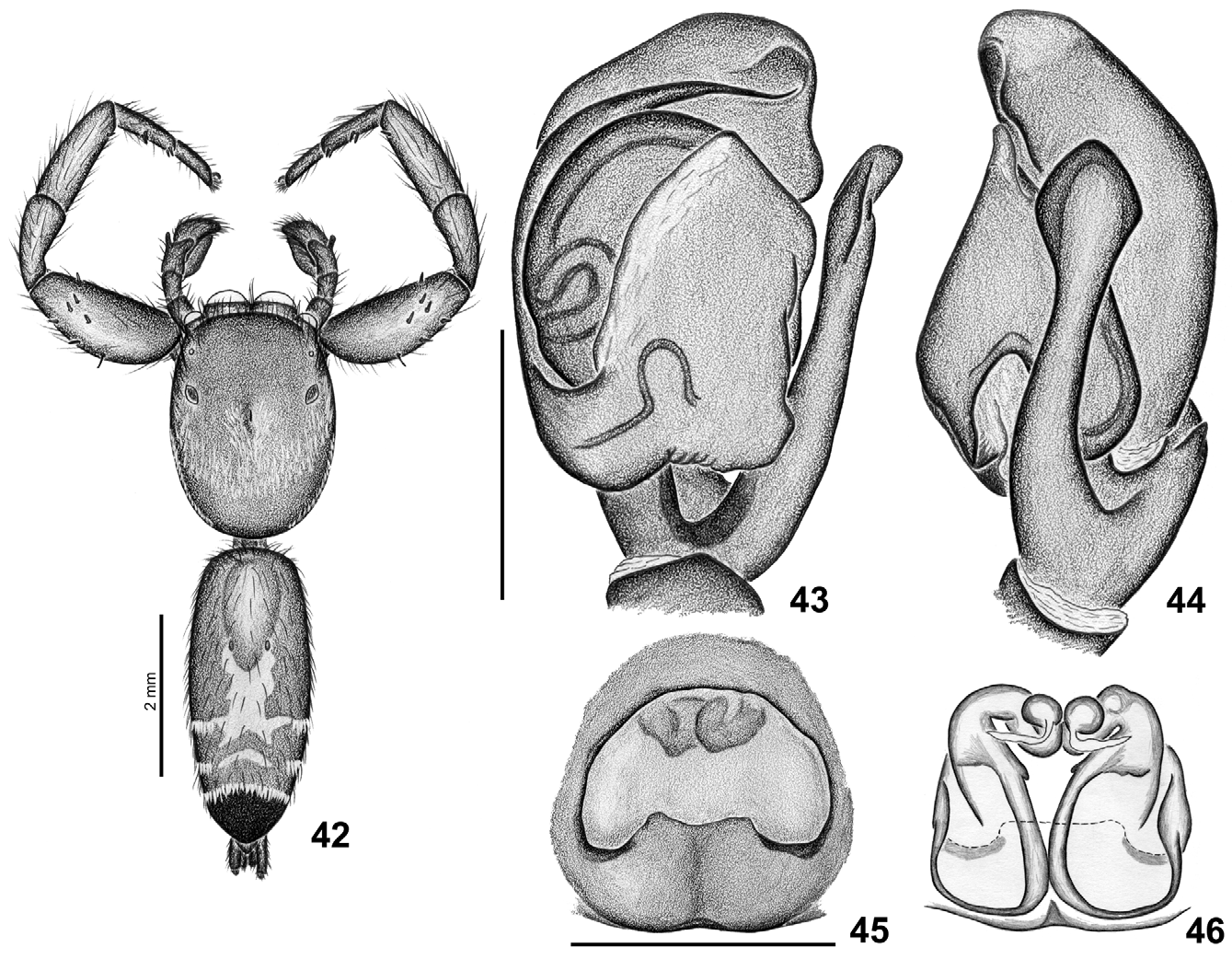
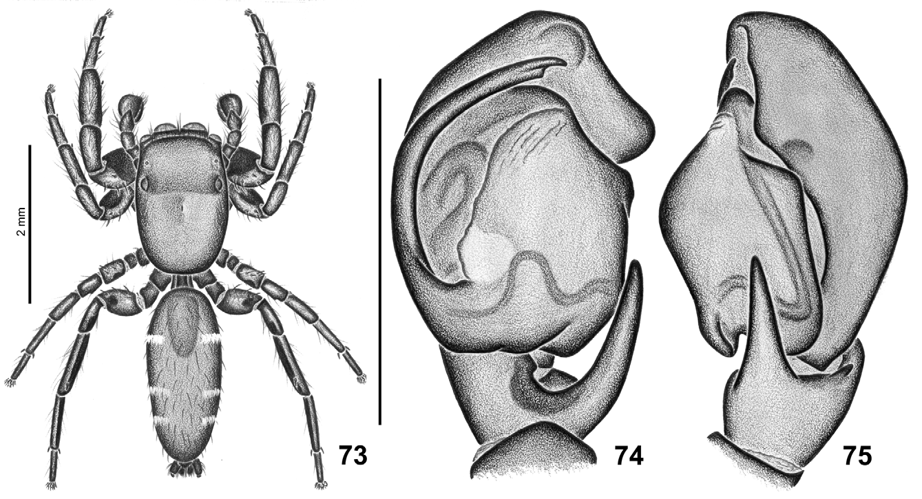
Diagnosis. Species of the genus Breda differ from those of the remaining genera of Salticidae by having the base of the embolus well developed, covering partially the bulb of the male palp in ventral view, with a retrolateral proximal depression ( Figs 10–11 View FIGURES 7 – 13 , 15–16 View FIGURES 14 – 19 , 24 View FIGURES 20 – 25. B ), and a large croissant-shaped atrium with posterior concavity on the epigynal plate of the females, through which it is possible to see the anteriorly placed spermathecae ( Figs 12–13 View FIGURES 7 – 13 ).
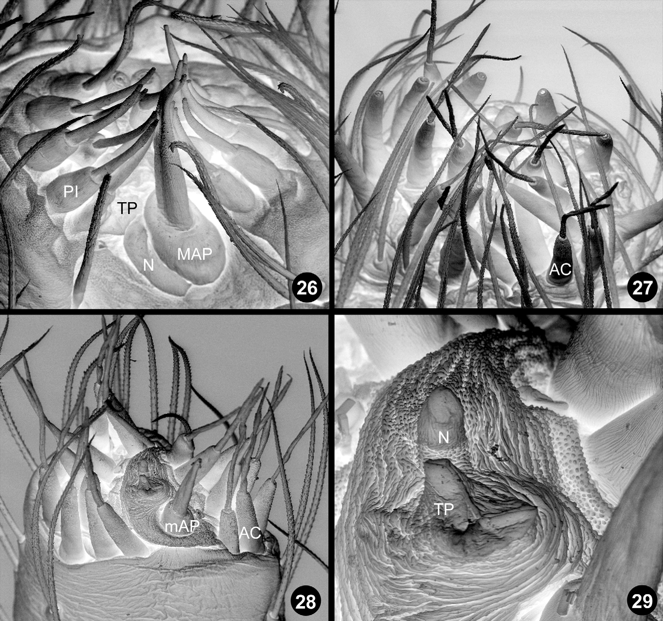
Description. Medium to large salticids (4–17mm); carapace low, always dark, covered with sparse short scales and with a well defined narrow band of white scales on the lateral borders ( Figs 20–21 View FIGURES 20 – 25. B ) (some species, such as B. milvina C.L. Koch or B. lubomirskii Taczanowski , have a longitudinal stripe of white scales on the carapace; Figs 6 View FIGURES 1 – 6 , 7 View FIGURES 7 – 13 ); chelicera short and vertical, with three to five teeth on promargin and one simple tooth on retromargin; no sexual dimorphism; endites not modified in males; leg I dilated in both sexes, mainly the tibia and femur ( Fig. 85 View FIGURES 85 – 90 ), longest in males; trochanter IV elongated; legs yellow, orange, reddish, brown or black, I always darker; ventral spines on leg I numerous and reduced in size ( Figs 8 View FIGURES 7 – 13 , 22 View FIGURES 20 – 25. B , 68 View FIGURES 68 – 72 , 92 View FIGURES 91 – 94 ); dorsal tibia IV with a longitudinal stripe of white scales ( Figs 3–4 View FIGURES 1 – 6 ); leg claws short; abdomen slightly longer than carapace, with the posterior extremity rounded; except for the two largest species, B. milvina ( Fig. 7 View FIGURES 7 – 13 ) and Breda lubomirskii ( Figs 6 View FIGURES 1 – 6 , 62 View FIGURES 62 – 67 ), which have bright orange scales on the abdomen of both sexes, the genus includes spiders with cryptic or disruptive abdominal coloration ( Figs 1–3 View FIGURES 1 – 6 ), often found living under tree bark or under rocks (e.g. B. tristis Mello-Leitão ); anterior dorsal abdominal scutum present in males ( Figs 7 View FIGURES 7 – 13 , 42 View FIGURES 42 – 46 , 73 View FIGURES 73 – 75 ); venter with a pair of rounded tufts of white (or orange) scales close to the spinnerets ( Figs 9 View FIGURES 7 – 13 , 23 View FIGURES 20 – 25. B ); anal tubercle covered with long plumose white scales ( Figs 2, 4 View FIGURES 1 – 6 ); spinnerets ( B. milvina ): The anterior lateral spinnerets have one central major ampullate (MAP) spigot and one nubbin in the male (two major ampullate spigots in the female) surrounded by several piriform spigots ( Fig. 26 View FIGURES 26 – 29 ). The posterior lateral spinnerets have only aciniform spigots ( Fig. 27 View FIGURES 26 – 29 ). The posterior median spinnerets have one central minor ampullate (mAP) spigot and a central membranous area surrounded by several aciniform spigots ( Figs 28–29 View FIGURES 26 – 29 ); male palp: femur unmodified; patella and tibia with long plumose white scales; tibia with a retroventral apophysis elongated and stout and a vestige of a dorso-retrolateral apophysis ( Figs 10–11 View FIGURES 7 – 13 , 14 View FIGURES 14 – 19 , 25 View FIGURES 20 – 25. B ); cymbium with a retrolateral bump associated with a more proximal cymbial depression close to the distal extremity of the tibial apophysis ( Figs 10– 11 View FIGURES 7 – 13 , 19 View FIGURES 14 – 19 ); a ventral depression on the distal extremity of the cymbium holds the embolus tip ( Fig. 10 View FIGURES 7 – 13 ); tegulum and subtegulum reduced, rotated at least 90° counterclockwise ( Figs 15–16 View FIGURES 14 – 19 , note proximal prolateral tegular shoulder); sperm duct forms a tegular loop just after the tegular shoulder (prolateral margin of a resting Breda tegulum, Figs 10 View FIGURES 7 – 13 , 15–16 View FIGURES 14 – 19 ); embolus elongated, with a distal origin ( Fig. 16 View FIGURES 14 – 19 ), with its base well developed, partially covering the bulb ( Figs 10 View FIGURES 7 – 13 , 15 View FIGURES 14 – 19 ); embolus base with a retrolateral proximal depression ( Figs 10–11 View FIGURES 7 – 13 , 24 View FIGURES 20 – 25. B ); distal half of the embolus thinner, extending along the prolateral border of the tegulum toward the distal extremity of the palp ( Fig. 10 View FIGURES 7 – 13 ); after penetrating the base of the embolus distally, the sperm duct goes to the posteriormost portion on the retrolateral side, deviates around the border of the embolar depression and turns toward the center of the base, where it creates a new fold (embolic loop, Fig. 10 View FIGURES 7 – 13 , 15 View FIGURES 14 – 19 ); epigyne: epigynal plate large, with a large croissant-shaped atrium ( Fig. 12 View FIGURES 7 – 13 ); posterior border of the atrium usually with a broad median projection ( Fig. 12 View FIGURES 7 – 13 ); posterior lateral openings with sclerotized border ( Fig. 12 View FIGURES 7 – 13 ); internally, the copulatory ducts begin towards the posterior border of the epigyne, converge at the center, flow anteriorly by the middle of the epigyne, where there is a pair of glandular portions associated with the copulatory ducts ( Fig. 13 View FIGURES 7 – 13 ); from this glandular area, the ducts become more slender and sclerotized and extend towards the anteriormost lateral portions of the epigyne and converge again to the center, where the spermathecae with no diameter differentiation are located ( Fig. 13 View FIGURES 7 – 13 ); fertilization ducts are easily seen in dorsal view and run laterally ( Fig. 13 View FIGURES 7 – 13 ).


No known copyright restrictions apply. See Agosti, D., Egloff, W., 2009. Taxonomic information exchange and copyright: the Plazi approach. BMC Research Notes 2009, 2:53 for further explanation.
|
Kingdom |
|
|
Phylum |
|
|
Class |
|
|
Order |
|
|
Family |
|
Kingdom |
|
|
Phylum |
|
|
Class |
|
|
Order |
|
|
Family |