
Zygophylax tizardensis Kirkpatrick, 1890
|
publication ID |
https://doi.org/10.11646/zootaxa.5214.1.1 |
|
publication LSID |
lsid:zoobank.org:pub:6E7723FD-44F7-48F0-BDB3-A5A624350ED5 |
|
DOI |
https://doi.org/10.5281/zenodo.7383655 |
|
persistent identifier |
https://treatment.plazi.org/id/03EB87C9-FFB5-4D5C-FF22-FF71FE5AFE85 |
|
treatment provided by |
Plazi (2022-11-30 12:31:04, last updated 2024-11-27 00:35:54) |
|
scientific name |
Zygophylax tizardensis Kirkpatrick, 1890 |
| status |
|
Zygophylax tizardensis Kirkpatrick, 1890 View in CoL
( Figs 22 View FIGURE 22 , 23 View FIGURE 23 , 25 View FIGURE 25 ; Tables 7 View TABLE 7 , 8 View TABLE 8 )
Zygophylax tizardensis Kirkpatrick, 1890: 12 View in CoL , pl. 3 fig. 3.— Schuchert, 2015: 337, fig. 11 (cum syn.).
Material examined. MNHN-IK-2019-2100, KANACONO Stn. DW 4764: seven sterile stems, 2.1–6 cm high; GenBank: OP724385 View Materials .—MNHN-IK-2019-2101, KANADEEP Stn. DW 4976: three sterile stems, 1.3, 2.1 and 2.9 cm high.—MNHN-IK-2019-2102, KANACONO Stn. DW 4743: a 3.6 cm high, sterile colony.—MNHN-IK-2019- 2103, KANADEEP Stn. DW 5013: a ca. 8 cm high, sterile colony with longer hydrothecae .
Description. Colonies erect, stiff, to 8 cm high, arising from dense, branched, rhizoid stolons firmly attached to various substrates; perisarc thick, from straw to brown-colored; stems unbranched or occasionally branched in a plane different to that of cladia, fascicled for most of their length, up to 1 mm wide basally; stolons and auxiliary tubes of the stem/branches bearing numerous digitiform nematothecae of varied lengths; main cauline tube undivided, slightly geniculate, composed of successive modules comprising a moderately-long proximal apophysis (together with its axillar hydrotheca atop its own apophysis) supporting a cladium, two well-developed alternate apophyses above, each carrying a hydrotheca, and a distal apophysis (and its axillar hydrotheca) on side opposite to its proximal counterpart; apophyses quite distant from one another; occasionally only one or three alternate hydrothecae intervene between two (subalternate) “pairs” of cladia, the position of the latter thus being reversed along the stem; auxiliary tubes of the stem running parallel to the main tube, occasionally branching and anastomosing. Cladia not distinctly delimited from their corresponding stem apophyses; arranged in two coplanar rows, alternate along the stem, to 1.1 cm long, monosiphonic when borne on the distal parts of the stems, fascicled basally when occurring more proximally on the caulus; usually unbranched, occasionally bearing secondary cladia (each given off from below the apophysis of a hydrotheca, the latter becoming axillar), which eventually give rise to branches through progressive lengthening and increasing polysiphony; main tube undivided, slightly geniculate, equivalents of internodes relatively long, each bearing a latero-distal, well-developed apophysis supporting a hydrotheca; up to 21 apophyses per cladium, slightly shorter than their cauline counterparts; apophyses usually coplanar, occasionally forming a wide angle between them. Hydrothecal apophyses (on both the stem and cladia) bearing a pair of spindle-shaped nematothecae (distally tapering towards the aperture), a theca confined to each side; occasionally, much longer, multiple (up to 7) renovations occur; renovated parts decidedly cylindrical (or gradually, though imperceptibly, widening distally), with slightly everted apertures, thus contrasting in shape with the original thecae. Apophyses of axillar hydrothecae devoid of nematothecae. Hydrothecae borne on indistinct pedicels, their corresponding apophyses merging imperceptibly with the thecal wall, demarcation internal, through a transversely-set diaphragm with scalloped edge, forming a raised collar for the passage of the hydranth; hydrothecae tubular, longitudinal axes distinctly S-shaped; cylindrical in proximal half, flattened “dorso-ventrally” in distal half; adaxial wall double convex, with internal, semicircular, upwardly-directed ridge projecting into lumen; abaxial wall slightly convex in its proximal half, concave in middle and curving away (at right angle) subterminally, and there with distinct, transverse notch; very distal part of hydrotheca expanding towards aperture, giving it a trumpetshaped aspect in lateral view; frontally, aperture oval, flared, rim even; up to 25 closely-set marginal renovations in many hydrothecae; apertures of stem hydrothecae facing outwards, alternately left and right with respect to the longitudinal axis of the caulus; apertures of cladial hydrothecae facing either obliquely upwards (row of thecae close to the stem) or obliquely downwards (row of thecae away from the stem). Gonosome absent in present material.
Remarks. The distinctive shape of the hydrothecae is unmistakable. The gonosome was described by Hirohito (1983, 1995) and Schuchert (2015).
Distribution. Spratly Islands ( Kirkpatrick 1890), Japan ( Hirohito 1995; Schuchert 2015), Philippines ( Nutting 1927, as Acryptolaria normani ), New Zealand ( Vervoort & Watson 2003), New Caledonia ( Vervoort & Watson 2003: 83; present study).
Hirohito, Emperor of Showa. (1983) Hydroids from Izu Oshima and Niijima. Publications of the Biological Laboratory, Imperial Household, Tokyo, 6, 1 - 83.
Hirohito, Emperor of Showa. (1995) The hydroids of Sagami Bay. II. Thecata. Publications of the Biological Laboratory, Imperial Household, Tokyo, 1 - 244.
Kirkpatrick, R. (1890) Report upon the Hydrozoa and Polyzoa collected by P. W. Bassett-Smith, Esq., Surgeon, R. N., during the Survey of the Tizard and Macclesfield Banks, in the China Sea, by H. M. S. Rambler , Commander W. U. Moore. Annals and Magazine of Natural History, (6) 5 (2), 11 - 24. https: // doi. org / 10.1080 / 00222939009460773
Nutting, C. C. (1927) Report on the Hydroida collected by the United States Fisheries steamer Albatross in the Philippine region, 1907 - 1910. In: Contributions to the biology of the Philippine archipelago and adjacent regions. Bulletin of the United States National Museum, 100 - 6 (3), 195 - 242.
Schuchert, P. (2015) On some hydroids (Cnidaria, Hydrozoa) from the Okinawa Islands, Japan. Revue Suisse de Zoologie, 122 (2), 325 - 370.
Vervoort, W. & Watson, J. E. (2003) The marine fauna of New Zealand: Leptothecata (Cnidaria: Hydrozoa) (thecate hydroids). NIWA Biodiversity Memoir, 119, 1 - 538.
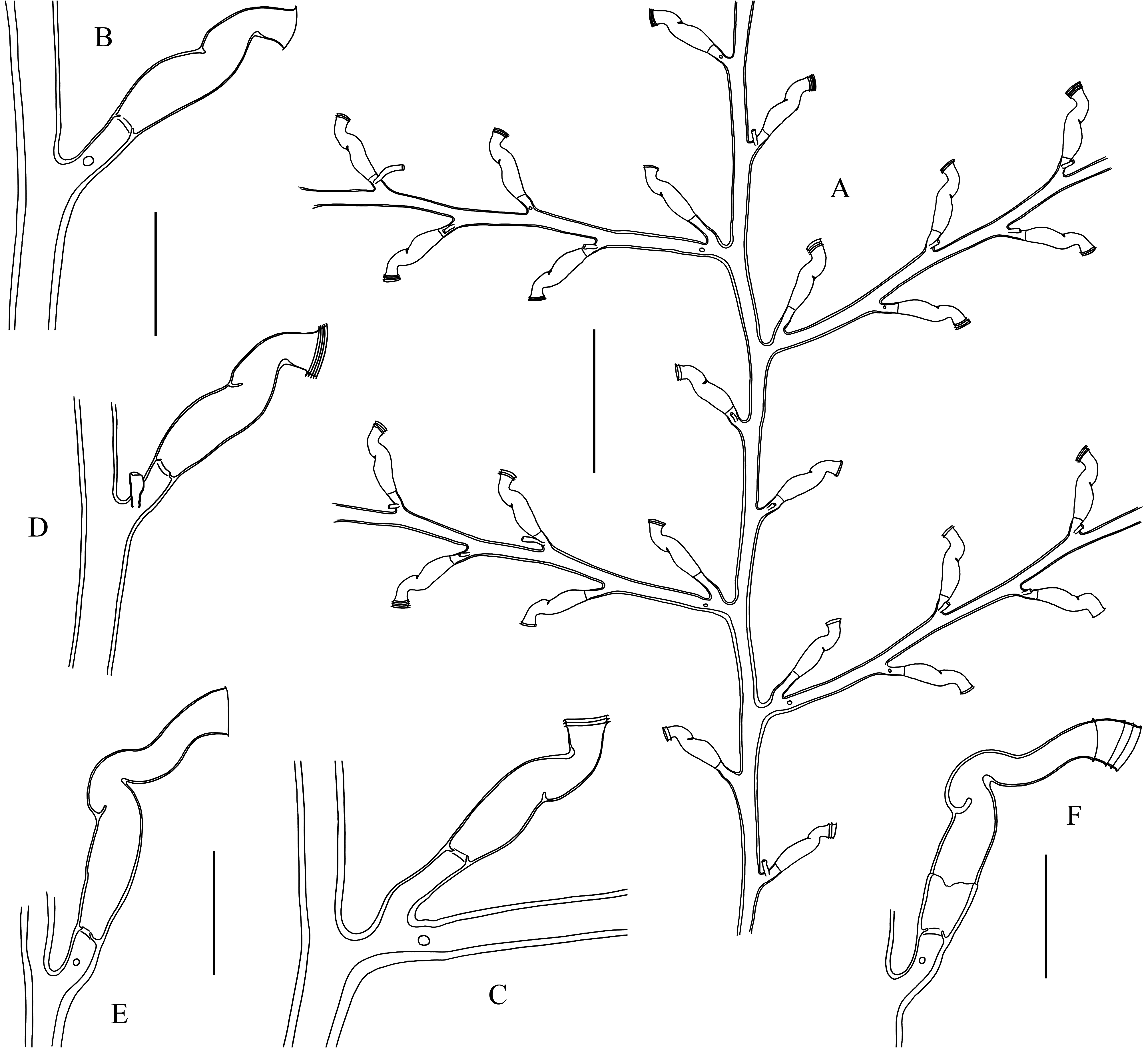
FIGURE 22. Zygophylax tizardensis Kirkpatrick, 1890 (part). A. Apical part of a colony. B–F. Cauline (B), axillar (C) and cladial (D–F) hydrothecae. From samples MNHN-IK-2019-2100 (A–D) and -2103 (E, F). Scale bars: B–F = 300 µm; A = 1 mm.
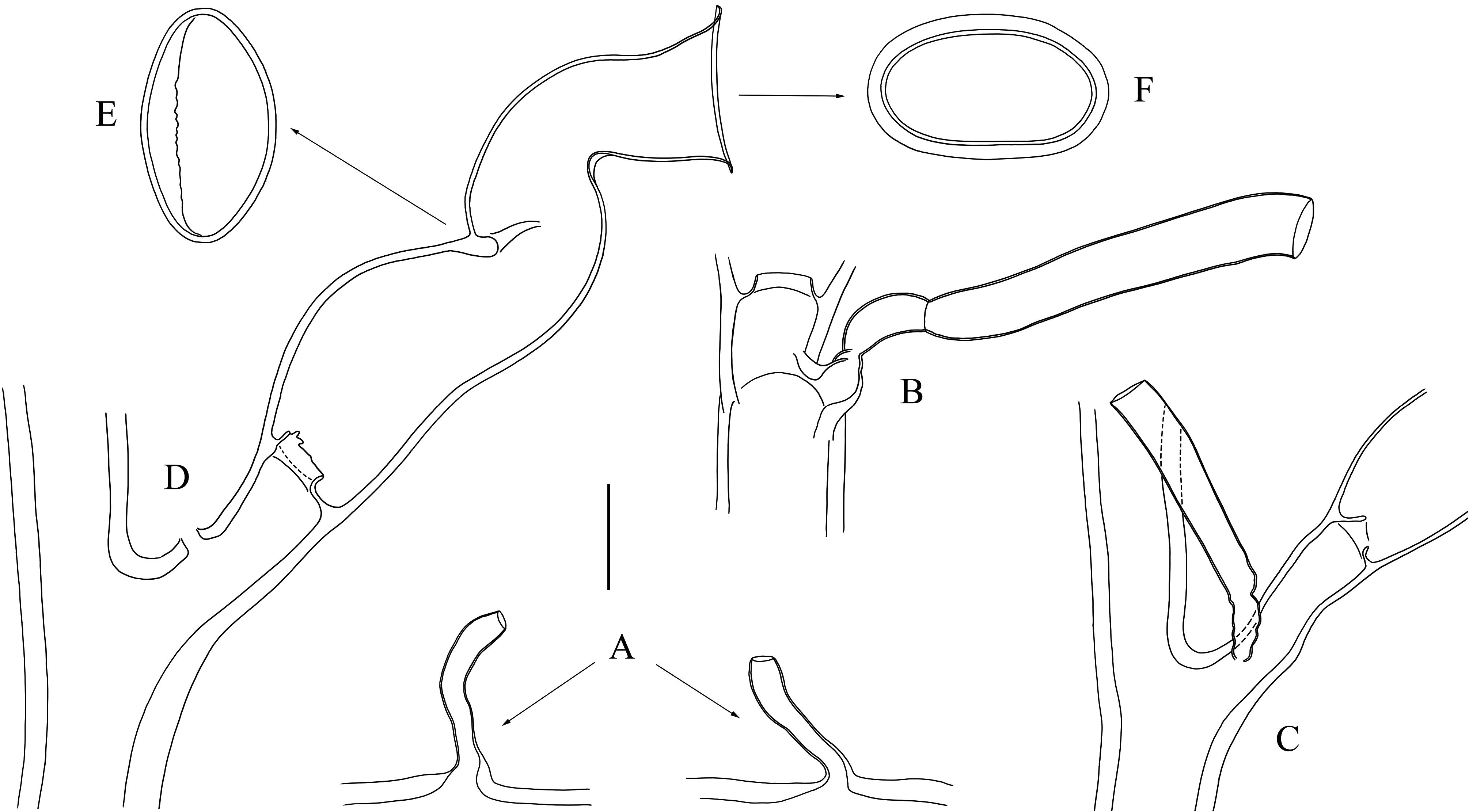
FIGURE 23. Zygophylax tizardensis Kirkpatrick, 1890 (continued). A. Nematothecae from the accessory tubes of the stem. B, C. Nematothecae borne on cladial apophyses, renovated in both cases. D–F. Details of a hydrotheca, showing the diaphragm (D, lateral view), internal septum (E, transverse section), and shape of aperture (F, apical view). All from sample MNHN-IK- 2019-2100. Scale bar: A–F = 100 µm.

FIGURE 25. Phylogenetic hypothesis for the Zygophylaciidae based on the 16S rRNA gene. Numbers at nodes refer to support values of the maximum likelihood, Bayesian inference and maximum parsimony analyses, respectively, and are shown only when> 70 and 0.7. Sequences generated in this study are followed by voucher numbers.
No known copyright restrictions apply. See Agosti, D., Egloff, W., 2009. Taxonomic information exchange and copyright: the Plazi approach. BMC Research Notes 2009, 2:53 for further explanation.
|
Kingdom |
|
|
Phylum |
|
|
Class |
|
|
SubClass |
Hydroidolina |
|
Order |
|
|
Family |
|
|
Genus |
Zygophylax tizardensis Kirkpatrick, 1890
| Galea, Horia R., Maggioni, Davide & Galli, Paolo 2022 |
Zygophylax tizardensis
| Schuchert, P. 2015: 337 |
| Kirkpatrick, R. 1890: 12 |
1 (by plazi, 2022-11-30 12:31:04)
2 (by ExternalLinkService, 2022-11-30 12:40:22)
3 (by ExternalLinkService, 2022-11-30 18:59:16)
4 (by tatiana, 2022-12-01 16:06:25)
5 (by ExternalLinkService, 2022-12-01 16:16:53)
6 (by ExternalLinkService, 2022-12-01 17:42:20)
7 (by plazi, 2023-11-07 23:12:06)