
Guimaraesiella (Cicchinella) retusa Gustafsson, Clayton
|
publication ID |
https://doi.org/10.11646/zootaxa.4543.4.1 |
|
publication LSID |
lsid:zoobank.org:pub:4F591303-AF92-4BBB-8B68-EDD27AA229DE |
|
DOI |
https://doi.org/10.5281/zenodo.5936015 |
|
persistent identifier |
https://treatment.plazi.org/id/1BE4FCA1-ABBA-4D34-B0B0-971411842624 |
|
taxon LSID |
lsid:zoobank.org:act:1BE4FCA1-ABBA-4D34-B0B0-971411842624 |
|
treatment provided by |
Plazi (2019-03-30 06:50:36, last updated 2024-11-29 11:59:54) |
|
scientific name |
Guimaraesiella (Cicchinella) retusa Gustafsson, Clayton |
| status |
new species |
Guimaraesiella (Cicchinella) retusa Gustafsson, Clayton & Bush, new species
( Figs 8–14 View FIGURES 8–9 View FIGURES 10–14 )
Type host. Trochalopteron milnei sinianum Stresemann, 1930 —red-tailed laughing-thrush ( Leiothrichidae ).
Type locality: Jingxin County, Guangxi Province , China .
Diagnosis. Guimaraesiella (C.) retusa is most similar to Guimaraesiella (C.) philiproundi n. sp., with which is shares the following characters: (1) female subgenital plate reticulated ( Figs 14 View FIGURES 10–14 , 22 View FIGURES 17–22 ); (2) anterior margin of TN broadly flattened ( Figs 12 View FIGURES 10–14 , 20 View FIGURES 17–22 ); and (3) mesosomal ventral sclerite without an anterior rugose area ( Figs 12 View FIGURES 10–14 , 20 View FIGURES 17–22 ). However, these two species can be separated by the following characters: (1) lateral margins of preantennal head straight to slightly concave in G. (C.) retusa ( Fig. 10 View FIGURES 10–14 ) but convex in G. (C.) philiproundi ( Fig. 17 View FIGURES 17–22 ); (2) antennae sexually dimorphic in G. (C.) philiproundi ( Figs 17–18 View FIGURES 17–22 ) but not in G. (C.) retusa ( Fig. 10 View FIGURES 10–14 ); (3) mesosomal ventral sclerite broad in G. (C.) retusa ( Fig. 12 View FIGURES 10–14 ) but narrow in G. (C.) philiproundi ( Fig. 20 View FIGURES 17–22 ); (4) lateral margins of mesosome more sinuous in G. (C.) retusa ( Fig. 12 View FIGURES 10–14 ) than in G. (C.) philiproundi ( Fig. 20 View FIGURES 17–22 ); (5) ames microsetae in G. (C.) retusa ( Fig. 12 View FIGURES 10–14 ) but sensilla in G. (C.) philiproundi ( Fig. 20 View FIGURES 17–22 ); and (6) reticulation of female subgenital plate more pronounced in G. (C.) philiproundi ( Fig. 22 View FIGURES 17–22 ) than in G. (C.) retusa ( Fig. 14 View FIGURES 10–14 ).
Description. Both sexes. Head pentagonal ( Fig. 10 View FIGURES 10–14 ). Lateral margins of preantennal area straight to slightly concave. Dorsal preantennal suture reaches lateral margins of head, but does not completely separate dorsal anterior plate from mean head plate. Head chaetotaxy as in Fig. 10 View FIGURES 10–14 ; pns sensilla. Coni short, not reaching distal margin of scapes. Antennae not sexually dimorphic. Gular plate triangular. Thoracic and abdominal segments as in Figs 8–9 View FIGURES 8–9 . Reentrant heads of pleurites broad and long.
Male. Thoracic and abdominal chaetotaxy as in Fig. 8 View FIGURES 8–9 . Male genitalia as in Figs 11–13 View FIGURES 10–14 . Basal apodeme broad ( Fig. 11 View FIGURES 10–14 ), roughly rectangular. Proximal mesosome rectangular ( Fig. 12 View FIGURES 10–14 ), with sinuous lateral margins. Mesosomal lobes also with sinuous lateral margins. Marginal thickening of lobes not displaced medianly in anterior end. Large trapezoidal nodi with flat anterior margins on distal mesosome. Gonopore open distally, shapes as in Fig. 12 View FIGURES 10–14 ; 2 View FIGURES 1–2 ames microsetae on each side near antero-lateral corners of mesosomal lobes; gpmes not visible in examined material; 2 lpmes microsetae on each side in concave section of lateral margins of mesosome. Parameral heads irregular ( Fig. 13 View FIGURES 10–14 ). Parameral blades slightly elongated, tapering only distally; pst1–2 close together. Measurements (n = 1): TL = 1.15; HL = 0.32; HW = 0.33; PRW = 0.19; PTW = 0.30; AW = 0.42.
Female. Thoracic and abdominal chaetotaxy as in Fig. 9 View FIGURES 8–9 ; ss of tergopleurite VIII much shorter than ss of tergopleurites II–VII, not visible in all examined females. Subgenital plate ( Fig. 14 View FIGURES 10–14 ) with central reticulation; crosspiece broad, with very broad connection to subgenital plate. Vulval margin rounded ( Fig. 14 View FIGURES 10–14 ), with 3 slender vms and 7–8 short, thorn-like vss on each side (one specimen with 12 on one side); 3–4 long, slender vos on each side; distal vos median to vss. Measurements (n = 3): TL = 1.51–1.54; HL = 0.36–0.38; HW = 0.37–0.38; PRW = 0.22– 0.23; PTW = 0.33–0.35; AW = 0.50–0.52.
Etymology. The species epithet is derived from “ retusa ”, Latin for “blunt”, referring to the broad, blunt preantennal area.
Type material. Ex Trochalopteron milnei sinianum : Holotype Ƌ, 23.122'N, 105.963'E, Jingxin County, Guangxi Province, China, 28 Sep. 2004, S.E. Bush , Bird ATP-2004-108, Louse P-314, PIPeR # 83 (NHML). Paratypes: 3♀, same data as holotype (PIPeR).
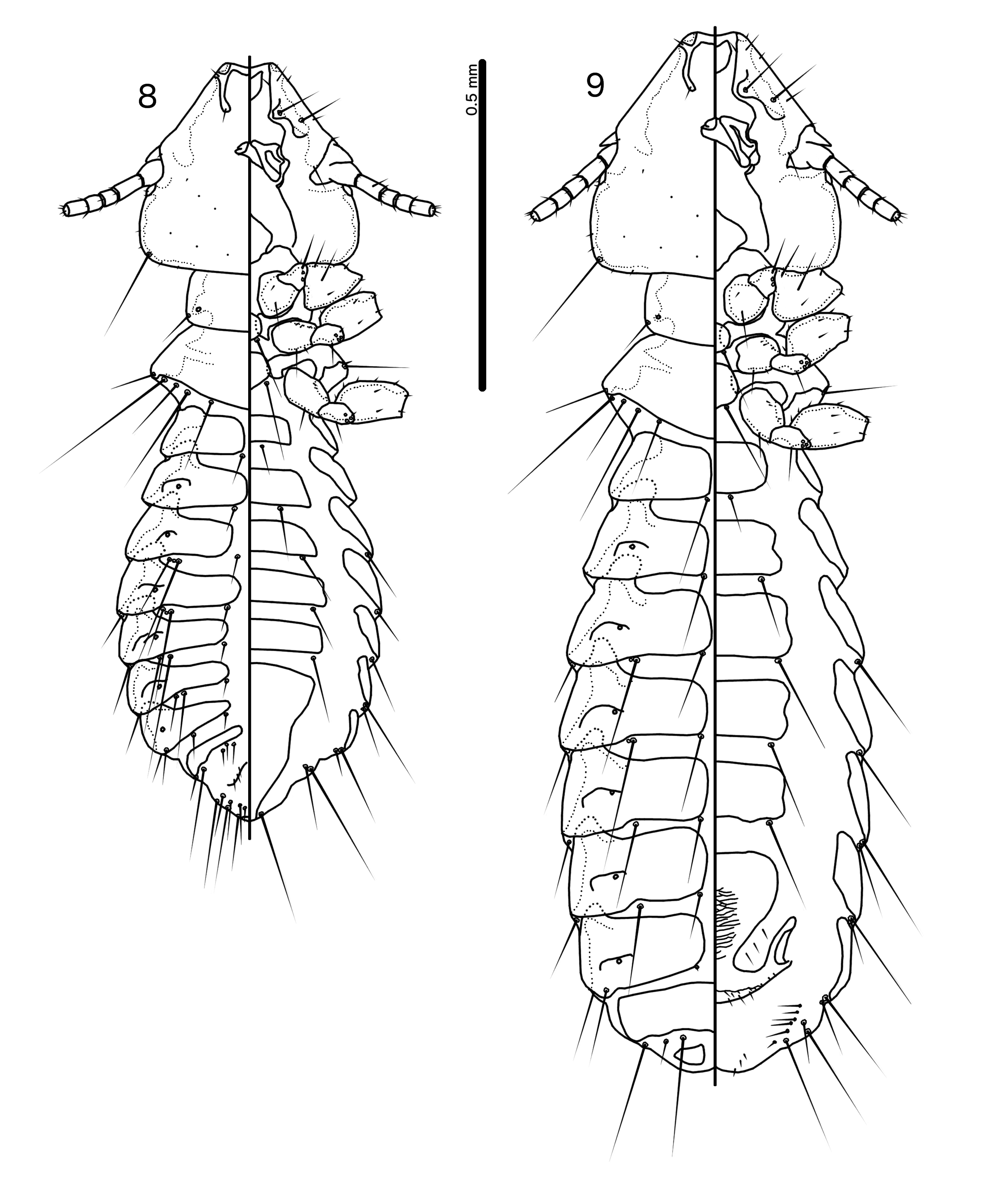
FIGURES 8–9. Guimaraesiella (Cicchinella) retusa n. sp. 8, male, whole body, dorsal and ventral views. 9, female, whole body, dorsal and ventral views.
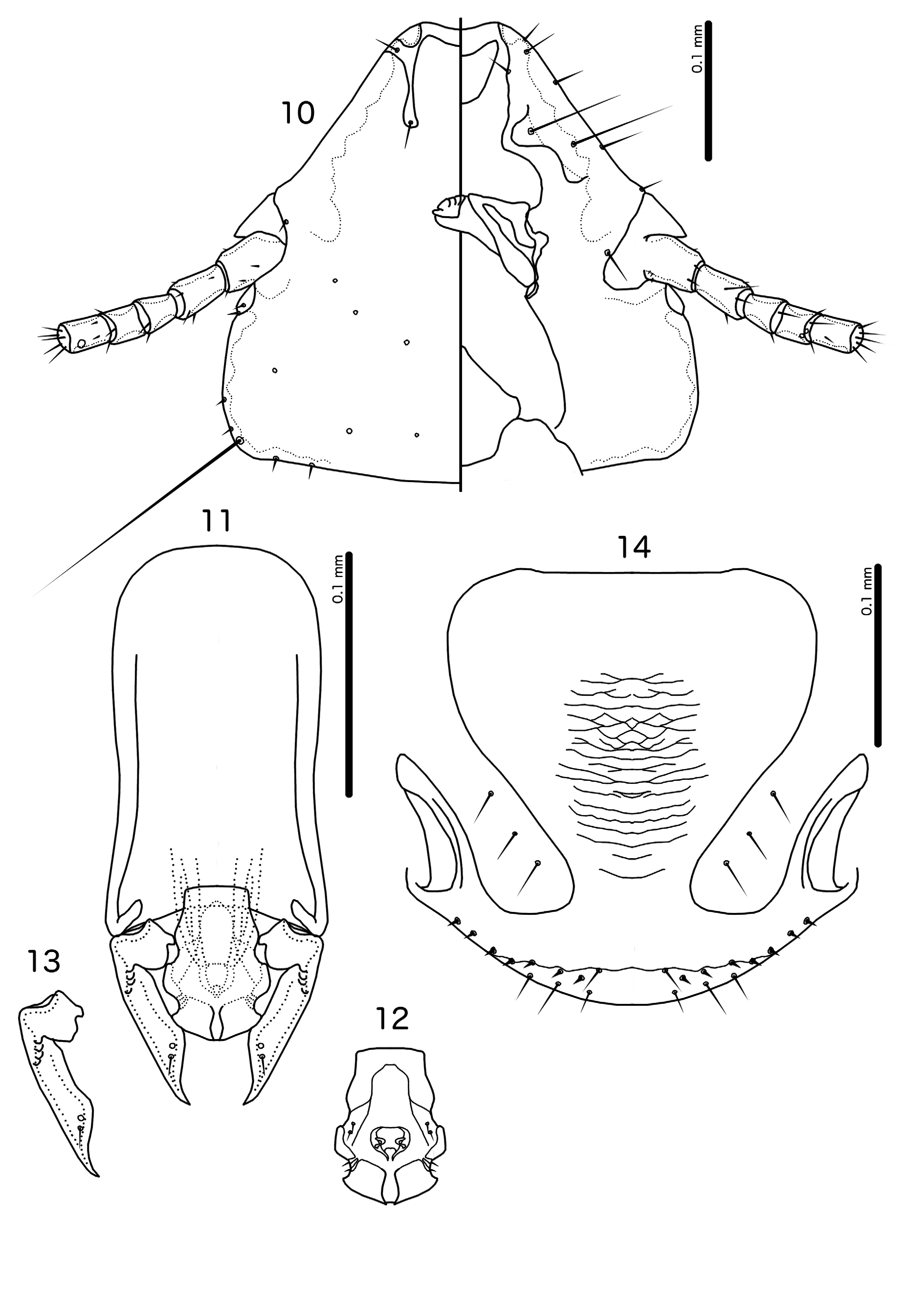
FIGURES 10–14. Guimaraesiella (Cicchinella) retusa n. sp. 10, male head, dorsal and ventral views. 11, male genitalia, dorsal view. 12, male mesosome, ventral view. 13, male paramere, dorsal view. 14, female subgenital plate and vulval margin, ventral view.
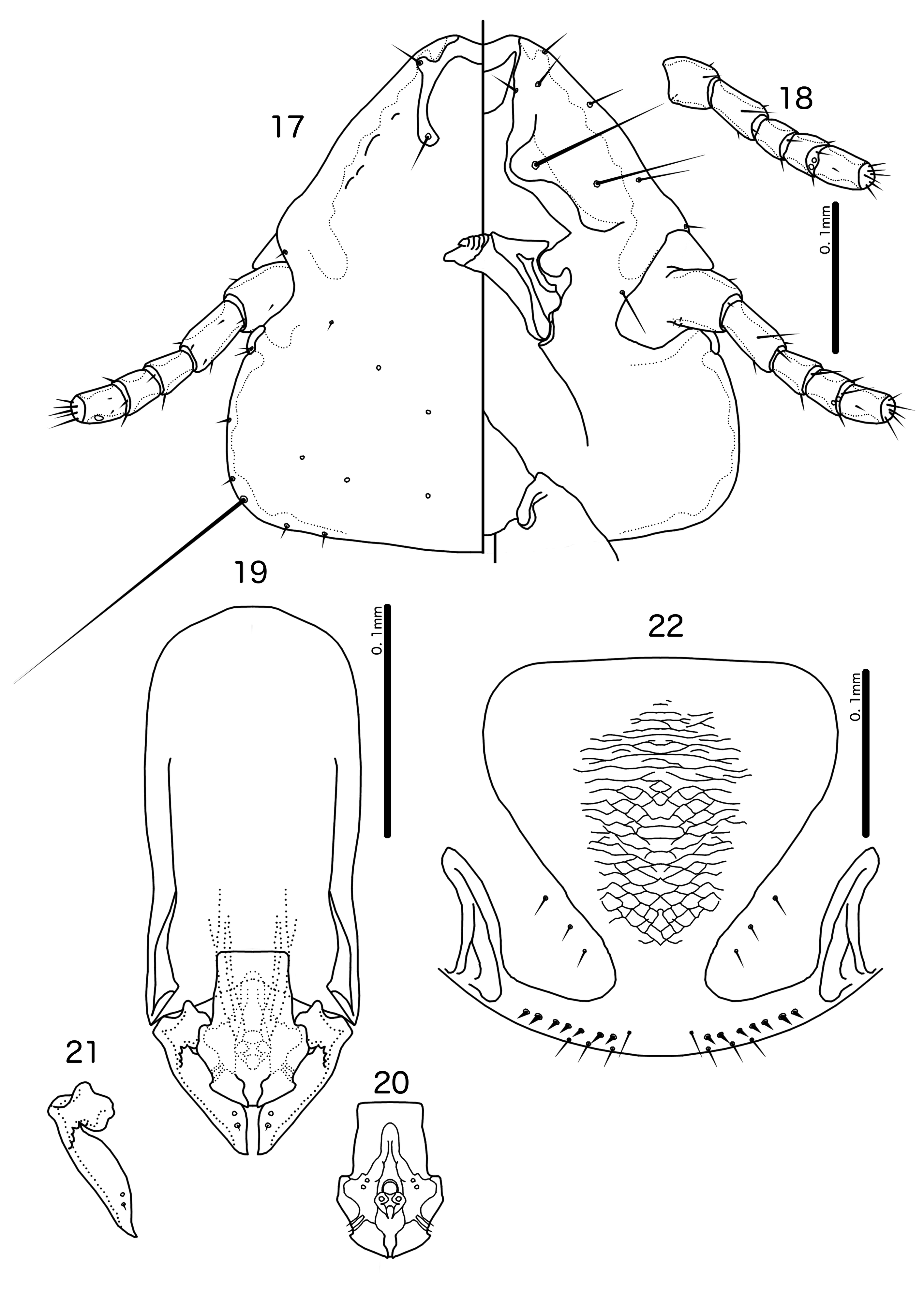
FIGURES 17–22. Guimaraesiella (Cicchinella) philiproundi n. sp. 17, male head, dorsal and ventral views. 18, female antenna, ventral view. 19, male genitalia, dorsal view. 20, male mesosome, ventral view. 21, male paramere, dorsal view. 22, female subgenital plate and vulval margin, ventral view.
No known copyright restrictions apply. See Agosti, D., Egloff, W., 2009. Taxonomic information exchange and copyright: the Plazi approach. BMC Research Notes 2009, 2:53 for further explanation.
|
Kingdom |
|
|
Phylum |
|
|
Class |
|
|
Order |
|
|
Family |
|
|
Genus |