
Sinocallipus thai, Stoev, Pavel, Enghoff, Henrik, Panha, Somsak & Fuangarworn, Marut, 2007
|
publication ID |
https://doi.org/ 10.5281/zenodo.176244 |
|
DOI |
https://doi.org/10.5281/zenodo.6243111 |
|
persistent identifier |
https://treatment.plazi.org/id/03E9386E-2659-FFF9-FF7A-FD5F78A8FE7F |
|
treatment provided by |
Plazi |
|
scientific name |
Sinocallipus thai |
| status |
sp. nov. |
Sinocallipus thai View in CoL sp. n.
Figs 1–7 View FIGURES 1 – 2 View FIGURES 3 – 5 .
Material examined. Holotype: adult male; 72 pleurotergites (PT) + telson, length ca. 56 mm, width ca. 2.0 mm; Thailand, Saraburi Province, Muang District, Sriwilai Cave Temple, 14°41’40’’N, 100°54’34’’E, 44 m a.s.l., 31.viii.2006, M. Fuangarworn leg., deposited in the collection of Animal Systematic Research Unit, Department of Biology, Faculty of Science, Chulalongkorn University, Bangkok.
Description of locality. The single specimen was collected under a rock at the base of a limestone hill. The vegetation at the site is deciduous forest which is, however, mostly destroyed by rock quarries. Other millipedes found at the site included Anurostreptus sculptus Demange, 1961 (Harpagophoridae) and two unidentified species of Pachybolidae . The pulmonate terrestrial snail Cryptozona siamensis (Pfeiffer, 1856) was dominant in the forest floor invertebrate fauna.
Etymology. The species name emphasizes the country of origin.
Diagnosis. The new species can be distinguished from S. simplipodicus by the characters summarized in Table 1.
TABLE 1. Diagnostic characters of the species of genus Sinocallipus . For S. simplipodicus , only the original data from Zhang (1993) are considered, cf. discussion.
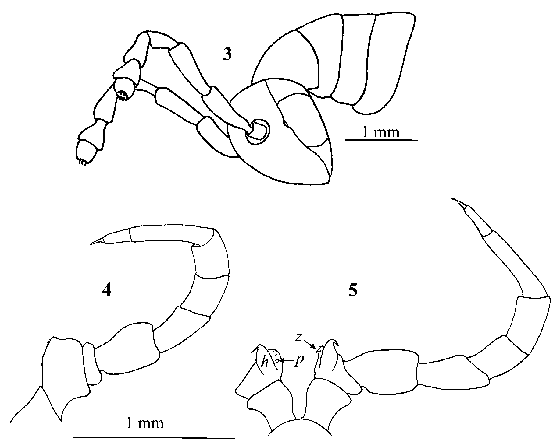
Description. Length: ca. 56 mm, width: ca. 2.0 mm, 72 PT + telson. Body colour: generally dark brown mottled with lighter spots mostly on the lateral sides of metazonites; metazonites with a posterior light brownyellowish semi-moniliform band. Dorsum with a broad snow-white medial band from PT5 to body end. PT 1- 4 snow-white, sharply contrasting to subsequent ones ( Fig. 1 View FIGURES 1 – 2 ). Head: lower 2/3rd of frontal surface, stipes and cardo white; a yellowish subtriangular spot in the middle of frons; labrum yellowish, upper 1/3 of frons and posterior side of head brown, with numerous lighter spots, especially between eyes; cephalic suture lighter. Eyes: subovoid composed of 45-50 transparent ocelli in 7 horizontal rows (ca. 12 vertical rows, RO) lying on black ground. Organ of Tömösváry: very small, inconspicuous, situated close to anteroventral side of eye. Head in male convex (without even a trace of concavity), covered with minute setae. Antennae: short, slightly extending beyond the posterior edge of PT4 ( Fig. 3 View FIGURES 3 – 5 ); length of antennomeres: 7<1=6<4=5<2=3; tip of antennomere 7 with four cones; antennomere 6 infundibular ( Fig. 2 View FIGURES 1 – 2 ); antennomere 7 short, shape intermediate between spherical and subconical. Antennomeres 6-8 snow white, 1-5 brown (basal one less so). Basal podomeres lighter, tarsi darker.
PT 1-4 slightly narrower than subsequent ones, body broadest at midlength, gently tapering towards telson. Dorsal side of collum and PT 2-3 smooth; complete crests appearing from PT 4 onwards. Each hemipleurotergum with about 6 crests on midbody PT; all crests flattened, touching anteriorly and strongly diverging posteriorly; no significant difference in the shape of crests on different PT. Ozopores situated between crests 4 and 5, visible from sixth to last but two pleurotergites. Hypoproct tripartite, median sclerite largest, subtriangular, bearing a pair of macrosetae at base, lateral sclerites with a seta each. Paraprocts divided into smaller dorsal and bigger ventral sclerites. Each dorsal sclerite with pair of macrosetae situated in vertical line on a small lobe. Spinnerets: long and slender, ending with a long seta.
First and second leg-pairs in males shorter than following legs; tarsi undivided (also probably on 3rd legpair, but hard to see under the light microscope); gonopore opening through small protuberance on posterior side of the coxa 2. Tarsal pads poorly developed; absent on the first two pairs of legs; pads thinner towards body end, almost disappearing on midbody legs. No particular modifications on coxae of pregonopodal legs. Leg-pair 7: coxa subquadrate; prefemur swollen, tarsus divided, twice as long as tibia, ending with a long claw ( Fig. 4 View FIGURES 3 – 5 ). Leg-pairs 4-6: all podomeres with generally the same shape as in leg-pair 7. Leg-pair 9: coxa subrectangular, expanded postero-laterally; trochanter with two processes: anterior one (h) higher, leafshaped, its tip very sharpened, spine-like, curved cephalad; posterior process (z) rounded with a small bulge, pointing cephalad too, at the medio-ventral side of the coxa; a small pore opening (p) under the bulge; prefemur slightly swollen ( Fig. 5 View FIGURES 3 – 5 ). Coxal sacs on leg-pairs 3–11.
Chaetotaxy (Table 2): the only traceable setae on PT are the anterior setae a and b on PT 2, 3 and 4, others either missing or broken off; it is even hard to see the pits where usually the setae are attached.
TABLE 2. Chaetotaxy of anterior PT in Sinocallipus thai sp. n.
Gonopods ( Figs 6–7): since we could not find any differences in the structure of gonopods of our specimen and the description of the Laotian Sinocallipus (see below our interpretation of its status), the following part is to a large extent a repetition of the description of gonopods made by Shear et al. (2003). Sternum (st): broad, rectangular, lying at the base of gonocoxae extending over their entire breadth. Coxae (cx): with two clavate processes extending directly ventrad on anterior side; a short, glabrous, anterior process (k) arising basally from anterior side of longer, caudal process, the latter apically setose (g); several long setae on a small bulge arising at the articulation with femoroid (d). Femoroid (= telopodite; fe): short and broad, positioned lateral to coxa and extending directly ventrad, without prostatic groove, with two slender, acicular (n), and one short and subfalcate (m) narrowly separated terminal projections directed strongly mediad; caudalmost projection longest, overlapping longer coxal process and nearly meeting corresponding projection from opposite gonopod; medial projection slightly shorter; anteriormost projection shortest, broad, extending slightly beyond telopodal margin and terminating well short of coxal processes. Cannula (ca): long and slender, not coiled, arising near midlength of caudal side of coxa at level of origin of shorter anterior process, angling dorsomediad basally and converging with opposite member, then curling and diverging, angling anteriolaterad, apices overhanging the longer femoroidal projections.
Female unknown.

No known copyright restrictions apply. See Agosti, D., Egloff, W., 2009. Taxonomic information exchange and copyright: the Plazi approach. BMC Research Notes 2009, 2:53 for further explanation.