
Mnioes Townes, 1946
|
publication ID |
https://doi.org/10.11646/zootaxa.4743.2.3 |
|
publication LSID |
lsid:zoobank.org:pub:A3EB2E6D-C484-4E24-9613-572BE7873E21 |
|
DOI |
https://doi.org/10.5281/zenodo.3688115 |
|
persistent identifier |
https://treatment.plazi.org/id/03E887DD-CC54-FFC2-FF16-FE6A37FBFA4D |
|
treatment provided by |
Plazi (2020-02-26 09:28:37, last updated 2024-11-25 12:07:16) |
|
scientific name |
Mnioes Townes, 1946 |
| status |
|
Mnioes Townes, 1946 View in CoL View at ENA
Mnioes Townes, 1946: 58 . Type-species Lampronota jacunda Cresson, 1874 , by original designation.
Diagnosis. Mnioes can be distinguished from all other genera of Atrophini by the combination of the following characters: (1) sculpture of the body, usually, granulate and matte; (2) outer surface of mandible, close to the base of upper tooth, convex and polished ( Fig. 5B View FIGURE 5 ); (3) clypeus basally convex, apically weakly sclerotized and, at least medially, concave ( Figs 1C View FIGURE 1 , 2C View FIGURE 2 ); (4) clypeal margin truncate to concave medially ( Fig. 2B View FIGURE 2 ); (5) frons weakly biconcave, granulate, without ornamentations; (6) flagellum, usually (except Mnioes iskay sp. nov.), centrally white- or yellow-banded; (7) scape apically truncated, about 60–75 o from transverse ( Fig. 2C View FIGURE 2 ); (8) submetapleural carina narrow, only slightly and evenly broadened anteriorly ( Fig. 2D View FIGURE 2 ); (9) propodeum ( Fig. 1D View FIGURE 1 ) without anterior transverse, lateral, and lateromedian longitudinal carinae, posterior transverse carina usually discernible as a vestige at middle (rarely more extensively developed), and pleural carina from absent to strong; (10) fore wing with veins 3rs-m entirely absent, with 2rs-m more than 2.0 times as long as abscissa of M between 2rs-m and 2m-cu, 2m-cu usually with two bullae, bullae separated by a short length vein which often bears a stub of a spurious vein (rarely without this abscissa of 2m-cu thus with a single long bulla); (11) hind wing with length of abscissa of Cu1 between M and Cu-a 0.7–0.9 times as long as the combined lengths this vain and cu-a; (12) tergite I without lateromedian longitudinal carinae ( Figs 1D View FIGURE 1 and 3C View FIGURE 3 ); and (13) ovipositor sheath 1.6–3.1 times as long as hind tibia.
Comments. No other South American country has records of Mnioes . The genus ranges from sea-level up to about 3000 meters, with the exception of Mnioes iskay sp. nov. recorded at about 4000 meters. The peak of diversity occurs between 700 and 1500 meters as found in Costa Rica ( Ugalde-Gómez and Gauld 2002).
Several species are widely distributed and commonly collected; although no hosts are known. Banchinae with known host associations are koinobiont endoparasitoids of Lepidoptera larvae ( Quicke 2015; Broad et al. 2018,), and this is also expected to be the case for Mnioes .
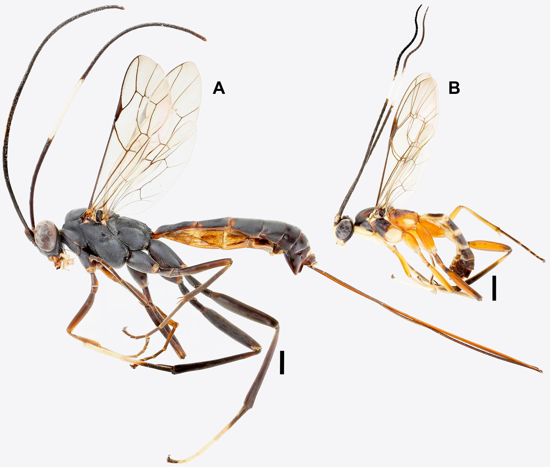
Sexual dimorphism is mostly evident in the coloration, males in several species are chromatically quite distinct from females, e.g., M. attenboroughi sp. nov. and M. tawa sp. nov. In species with similar coloration in both sexes, males generally present the white band starting more apically and covering fewer articles than in females. Additionally, males are usually much smaller than their respective females ( Figs 1 View FIGURE 1 , 7 View FIGURE 7 , 8 View FIGURE 8 ). This was also noticed for the Central American species ( Ugalde-Gómez and Gauld 2002).
Broad, G. R., Shaw, M. R. & Fitton, M. G. (2018) Ichneumonid wasps (Hymenoptera: Ichneumonidae): their classification and biology. RES Handbooks for the Identification of British Insects. Vol. 7. No. 12. Royal Entomological Society, London, 418 pp.
Quicke, D. L. (2015) The braconid and ichneumonid parasitoid wasps: biology, systematics, evolution and ecology. Wiley Blackwell, Chichester, 704 pp. https: // doi. org / 10.1002 / 9781118907085
Ugalde-Gomez, J. & Gauld, I. D. (2002) Tribe Atrophini. In: Gauld, I. D., Godoy, C., & Ugalde-Gomez, J. (Ed.), The Ichneumonidae of Costa Rica 4. Memoirs of the American Entomological Institute. Vol. 66. American Entomological Institute, Gainesville, Florida, pp. 306 - 666.

FIGURE 1. Details of Mnioes attenboroughi sp. nov. (paratype) A. Lateral habitus of female (scale bar = 1 mm), B. Lateral habitus of male (scale bar = 1 mm) C. Facial view of female D. Propodeum and first two metasomal tergites of female.

FIGURE 2. Details of Mnioes huk sp. nov. (holotype). A. Lateral habitus of female (scale bar = 1 mm) B. Frontal view of head C. Latero-frontal view of head D. Ventro-dorsal view of propodeum.

FIGURE 3. Details of Mnioes iskay sp. nov. (holotype). A. Lateral habitus of female (scale bar = 1 mm) B. Dorsal view of propodeum C. Dorsal view of metasomal tergites I–II.

FIGURE 5. Details of Mnioes pisqa sp. nov. and M. pusaq sp. nov. A–B Mnioes pisqa sp. nov. (holotype) A. Lateral habitus of female (scale bar = 1 mm) B. Lateral view of head C. Mnioes pusaq sp. nov. (holotype), lateral habitus of female (scale bar = 1 mm).

FIGURE 7. Details of Mnioes soqta sp. nov. (paratype) A. Habitus in lateral view of female B. Habitus in lateral view of male (scale bars = 1 mm).
No known copyright restrictions apply. See Agosti, D., Egloff, W., 2009. Taxonomic information exchange and copyright: the Plazi approach. BMC Research Notes 2009, 2:53 for further explanation.
|
Kingdom |
|
|
Phylum |
|
|
Class |
|
|
Order |
|
|
Family |