
Paraedessa Silva & Fernandes
|
publication ID |
https://doi.org/ 10.11646/zootaxa.3716.3.4 |
|
publication LSID |
lsid:zoobank.org:pub:798449F3-C14D-4D9D-B2E1-505EE2BC6115 |
|
DOI |
https://doi.org/10.5281/zenodo.6158036 |
|
persistent identifier |
https://treatment.plazi.org/id/03E387AB-FF88-BE1D-4589-5870FB1EF9EB |
|
treatment provided by |
Plazi |
|
scientific name |
Paraedessa Silva & Fernandes |
| status |
gen. nov. |
Paraedessa Silva & Fernandes gen. nov.
Etymology. The name makes reference to a similarity between the species of the new genus and those of Edessa . Gender: feminine.
Type species: Cimex stolidus Linneus, 1758 .
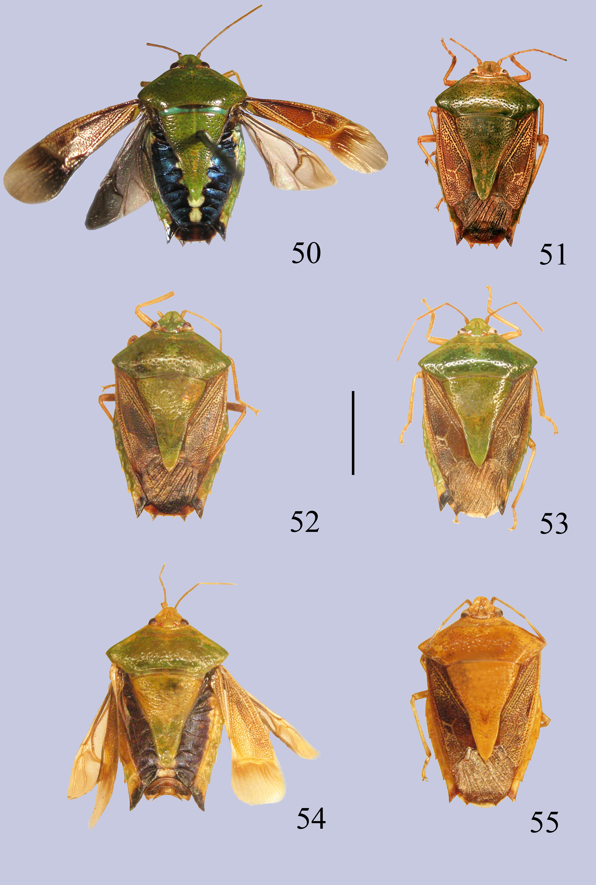
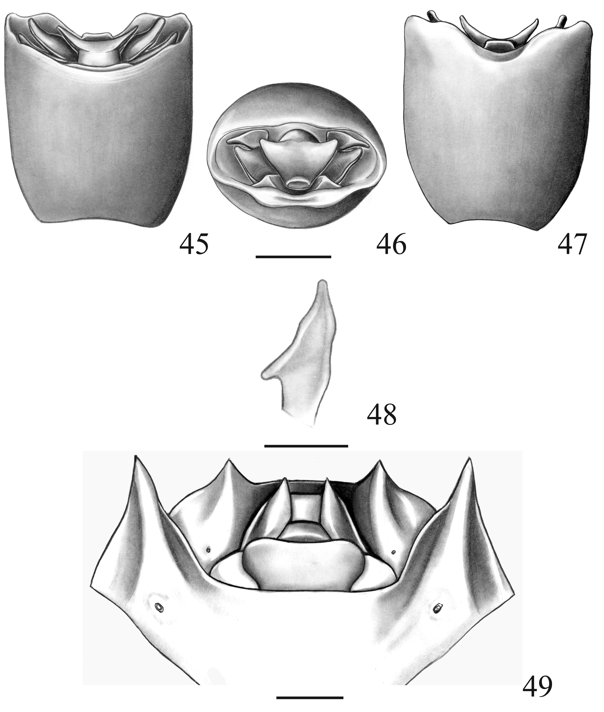
Small species (10–13 mm in length). Body green dorsally ( Figs. 50–58 View FIGURES 50 – 55 View FIGURES 56 – 58 ) and ventrally light yellow. Humeral angles not developed ( Figs. 50–58 View FIGURES 50 – 55 View FIGURES 56 – 58 ). Corium dark and densely punctured with some impunctate whitish areas. Dorsal surface of abdomen blue metallic with median white spots ( Figs. 50, 54 View FIGURES 50 – 55 , 56, 58 View FIGURES 56 – 58 ). Ventral surface with dark transverse stripes. Proctiger without a lateral dense pilose strip or tuft of hair; laterally expanded ( Figs. 2 View FIGURES 1 – 6 , 12 View FIGURES 11 – 15 , 17 View FIGURES 16 – 20 , 22 View FIGURES 21 – 25 , 27 View FIGURES 26 – 30 , 32 View FIGURES 31 – 35 , 37 View FIGURES 36 – 39 , 41 View FIGURES 40 – 44 , 46 View FIGURES 45 – 49 ). Gonocoxites 8 reduced ( Figs. 5 View FIGURES 1 – 6 , 15 View FIGURES 11 – 15 , 20 View FIGURES 16 – 20 , 25 View FIGURES 21 – 25 , 30 View FIGURES 26 – 30 , 35 View FIGURES 31 – 35 , 44 View FIGURES 40 – 44 , 49 View FIGURES 45 – 49 ) and gonapophyses 8 exposed and sclerotized
Head: Triangular, wider than long, with margins yellow, impunctate. Juga longer than tylus and contiguous in front of it, anteriorly rounded and somewhat bent ventrally. Bucculae large, evanescent and enclosing the first rostral segment. Second rostral segment longer than third and fourth together. Fourth rostral segment with a black spot on posterior portion. Antenna yellow ( Figs. 50–55 View FIGURES 50 – 55 , 57–58 View FIGURES 56 – 58 ) and pilose. Antennal segments increasing in length from I to V.
Thorax: Pronotum trapezoidal, wider than long, with yellow margin, dark deep punctures uniformly distributed throughout the length, except for the cicatrices which are calloused and impunctate. Anterolateral angle armed with small tooth or not. Anterolateral margin grooved. Scutellum with dark deep punctures concentrated mesially; apex acute, without punctures and armed with small tooth. Membrane of hemelytra brown ( Figs. 50–58 View FIGURES 50 – 55 View FIGURES 56 – 58 ). Evaporatorium concolorous with the pleura to whitish, dull and slightly rugose. Peritreme flagelliform, bright and almost reaching the lateral margin of the body. Evaporatorium with circular callosed spot on the edge. Metasternal process longer than wide, high, pilose, with a short anterior bifurcation, apex of each arm rounded and evanescent, enclosing the last rostral segment ( Fig. 6 View FIGURES 1 – 6 ). Legs yellow ( Figs. 51–53, 55 View FIGURES 50 – 55 , 57 View FIGURES 56 – 58 ) with distal margin of the femora black.
Abdomen: Connexivum exposed with sparse concolorous punctures; posterolateral angles of segments armed with small black tooth, lateral margin dark. Segment VII with dorsal black spot that occupies half of segment ( Figs. 50–58 View FIGURES 50 – 55 View FIGURES 56 – 58 ). Ventral surface with small round yellow spot close to lateral margin. Spiracles elliptical. Trichobothria aligned with spiracles. Cicatrices and intersegmental areas brown. Segment VII (VII) produced, but not reaching the level of laterotergites 8 in females ( Figs. 5 View FIGURES 1 – 6 , 15 View FIGURES 11 – 15 , 20 View FIGURES 16 – 20 , 25 View FIGURES 21 – 25 , 30 View FIGURES 26 – 30 , 35 View FIGURES 31 – 35 , 44 View FIGURES 40 – 44 , 49 View FIGURES 45 – 49 ).
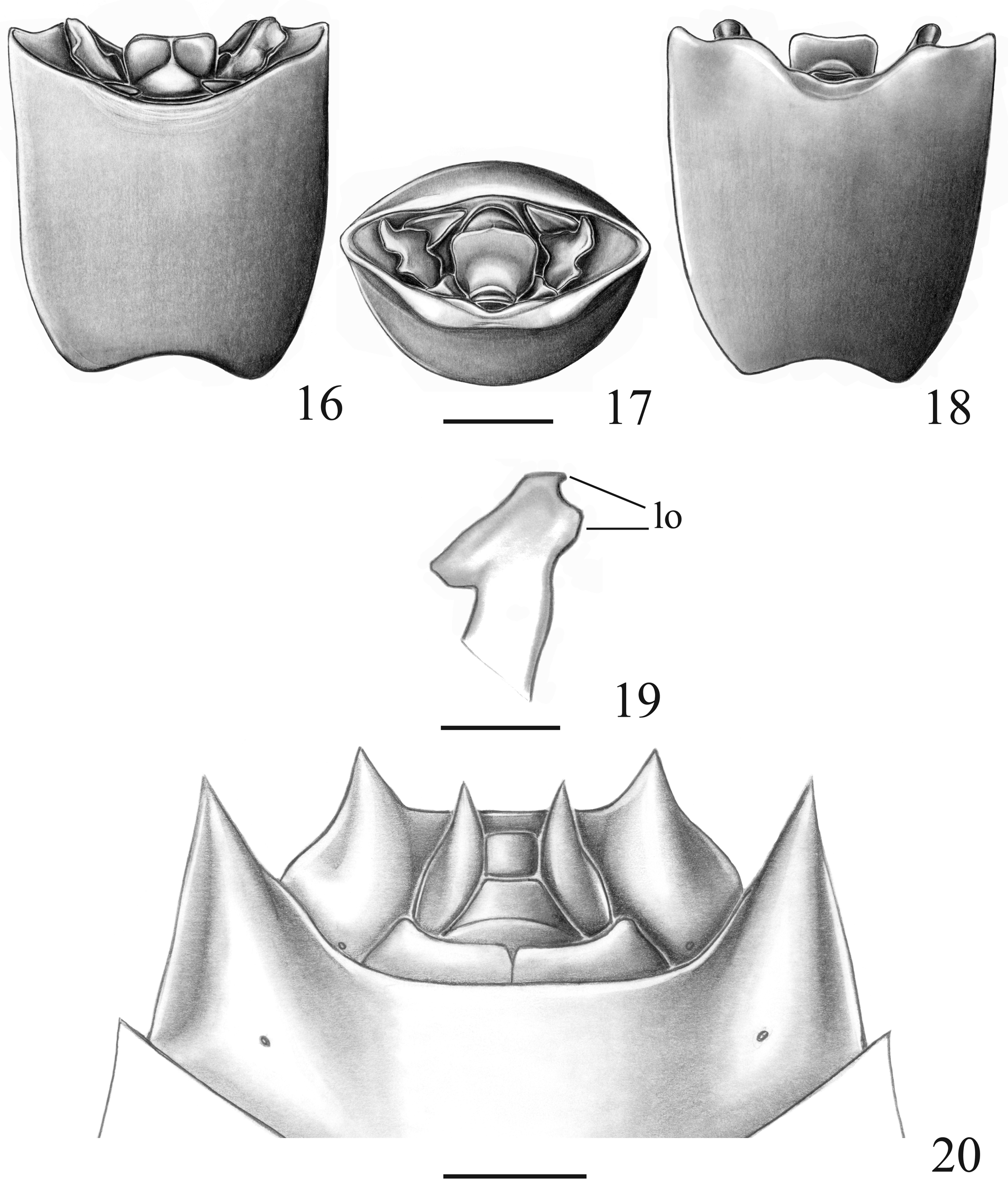
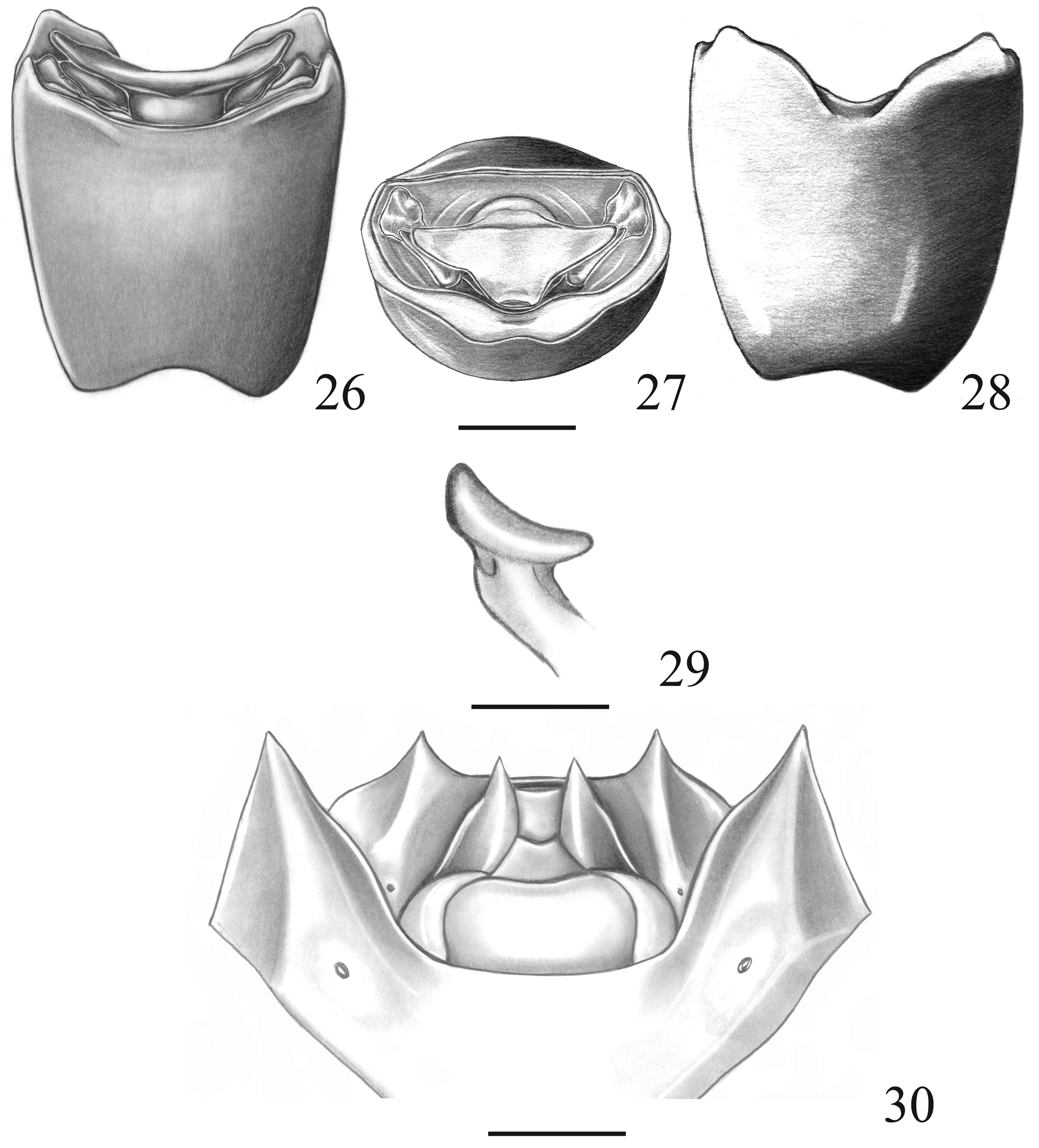
Male: Pygophore (pyg) rectangular in dorsal view, open dorsoposteriorly ( Figs. 1 View FIGURES 1 – 6 , 11 View FIGURES 11 – 15 , 16 View FIGURES 16 – 20 , 21 View FIGURES 21 – 25 , 26 View FIGURES 26 – 30 , 31 View FIGURES 31 – 35 , 36 View FIGURES 36 – 39 , 40 View FIGURES 40 – 44 , 45 View FIGURES 45 – 49 ). Dorsal rim dark or slightly dark. Surface of genital cup processes and distal part of parameres textured like lizard scales. Bottom of genital cup on each side of proctiger, forming a rough dark ridge (pgc) that partially covers base of parameres ( Figs. 2 View FIGURES 1 – 6 , 12 View FIGURES 11 – 15 , 17 View FIGURES 16 – 20 , 22 View FIGURES 21 – 25 , 27 View FIGURES 26 – 30 , 32 View FIGURES 31 – 35 , 37 View FIGURES 36 – 39 , 41 View FIGURES 40 – 44 , 46 View FIGURES 45 – 49 ), this structure continue inside pygophore as a low ridge on the side of the phallus . Genital cup processes (gp) flat ( Figs.1–2 View FIGURES 1 – 6 , 11–13 View FIGURES 11 – 15 , 17 View FIGURES 16 – 20 , 21 View FIGURES 21 – 25 , 26 View FIGURES 26 – 30 , 31 View FIGURES 31 – 35 , 36 View FIGURES 36 – 39 , 41 View FIGURES 40 – 44 , 45 View FIGURES 45 – 49 ) and entirely or partially dark. Parameres (pa) with a main distal expansion variable in shape and an anterior expansion usually subtriangular (ae) near base ( Figs. 4 View FIGURES 1 – 6 , 14 View FIGURES 11 – 15 , 19 View FIGURES 16 – 20 , 24 View FIGURES 21 – 25 , 39 View FIGURES 36 – 39 , 43 View FIGURES 40 – 44 , 48 View FIGURES 45 – 49 ); base of the paramere discoid and large (not illustrated). Proctiger (X) cylindrical, short and glossy; distal part slightly rough with a little to strong constriction just before a lateral expansion with different shapes.Ventral rim sparsely punctured and furrowed; deeply excavated medially leaving anal opening visible; margins of excavation with two lobes; vertex of excavation with a rugulose, concave and dark area ( Figs. 3 View FIGURES 1 – 6 , 13 View FIGURES 11 – 15 , 18 View FIGURES 16 – 20 , 23 View FIGURES 21 – 25 , 28 View FIGURES 26 – 30 , 33 View FIGURES 31 – 35 , 38 View FIGURES 36 – 39 , 42 View FIGURES 40 – 44 , 47 View FIGURES 45 – 49 ). Phallus : phallotheca (ph) cylindrical, elongated, curved dorsally and strongly esclerotized; vesica (v) short, about 1/10 of length of phallotheca, with a pair of convergent tooth-like dorsal processes and a single ventral membranous lobe ( Figs. 7–9 View FIGURES 7 – 10 ); ductus seminis distalis (dsd) short, not surpassing apex of vesica ( Fig. 7 View FIGURES 7 – 10 ); conjunctiva absent.
Female: Gonocoxites 8 (gc8) reduced to various extent. Gonapophyses 8 (gn8) sclerotized, and broad, forming rectangular single plate that occupies the space left by gonocoxites 8. Gonocoxite 9 (gc9) trapezoidal, anterolateral angles forming short arms that not reach apex of laterotergites 9; anterior margin medially concave. Gonapophyses 9 (gn9) with a medial thickening (mt). Thickening of vaginal intima (tvi) with a beak-like projection, visible in lateral view. Chitinellipsen (ch) present. Ductus receptaculi (dr) thin, shorter after vesicular area. Capsula seminalis (cs) with three digitiform processes, directed toward annular crests but not reaching posterior one (aac, pac); pars intermedialis (pi) slightly esclerotized ( Fig. 10 View FIGURES 7 – 10 ). Laterotergites 8 (la8) longitudinally convex and acuminate; distal margin dark; acute apex surpassing level of posterolateral angle of abdominal segment VII Spiracles present at the base of laterotergites 8. Laterotergites 9 (la9) triangular, with median depression, furrowed, and with distal margin reaching or passing band dorsally uniting laterotergites 8. Segment X (X) rectangular ( Figs. 5 View FIGURES 1 – 6 , 15 View FIGURES 11 – 15 , 20 View FIGURES 16 – 20 , 25 View FIGURES 21 – 25 , 30 View FIGURES 26 – 30 , 35 View FIGURES 31 – 35 , 44 View FIGURES 40 – 44 , 49 View FIGURES 45 – 49 ).
Comments. The proposed new genus is based on the morphology of the genitalia of both sexes. Paraedessa is easily recognizable by the proctiger presenting a remarkable lateral expansion of proctiger ( Figs. 2 View FIGURES 1 – 6 , 12 View FIGURES 11 – 15 , 17 View FIGURES 16 – 20 , 22 View FIGURES 21 – 25 , 27 View FIGURES 26 – 30 , 32 View FIGURES 31 – 35 , 37 View FIGURES 36 – 39 , 41 View FIGURES 40 – 44 , 46 View FIGURES 45 – 49 ); female genitalia also shows a unique reduction of the gonocoxites 8 and a great development and sclerotization of the gonapophyses 8 ( Figs. 5 View FIGURES 1 – 6 , 15 View FIGURES 11 – 15 , 20 View FIGURES 16 – 20 , 25 View FIGURES 21 – 25 , 30 View FIGURES 26 – 30 , 35 View FIGURES 31 – 35 , 44 View FIGURES 40 – 44 , 49 View FIGURES 45 – 49 ); less conspicuous but equally important is the rough dark ridge on the sides of proctiger and the discoid large base of the paramere. The species composing this genus have very similar facies and their identification is possible only comparing the genitalia. The general aspect of Paraedessa, except for its genitalia, is very similar to Edessa , especially for the subgenus Hypoxys. Both share a dorsal color green, but not the brown corium with whitish areas; humeral angle slightly projected, around the diameter of an eye; female with abdominal segment VII posteriorly developed but not projected beyond the genital plates.
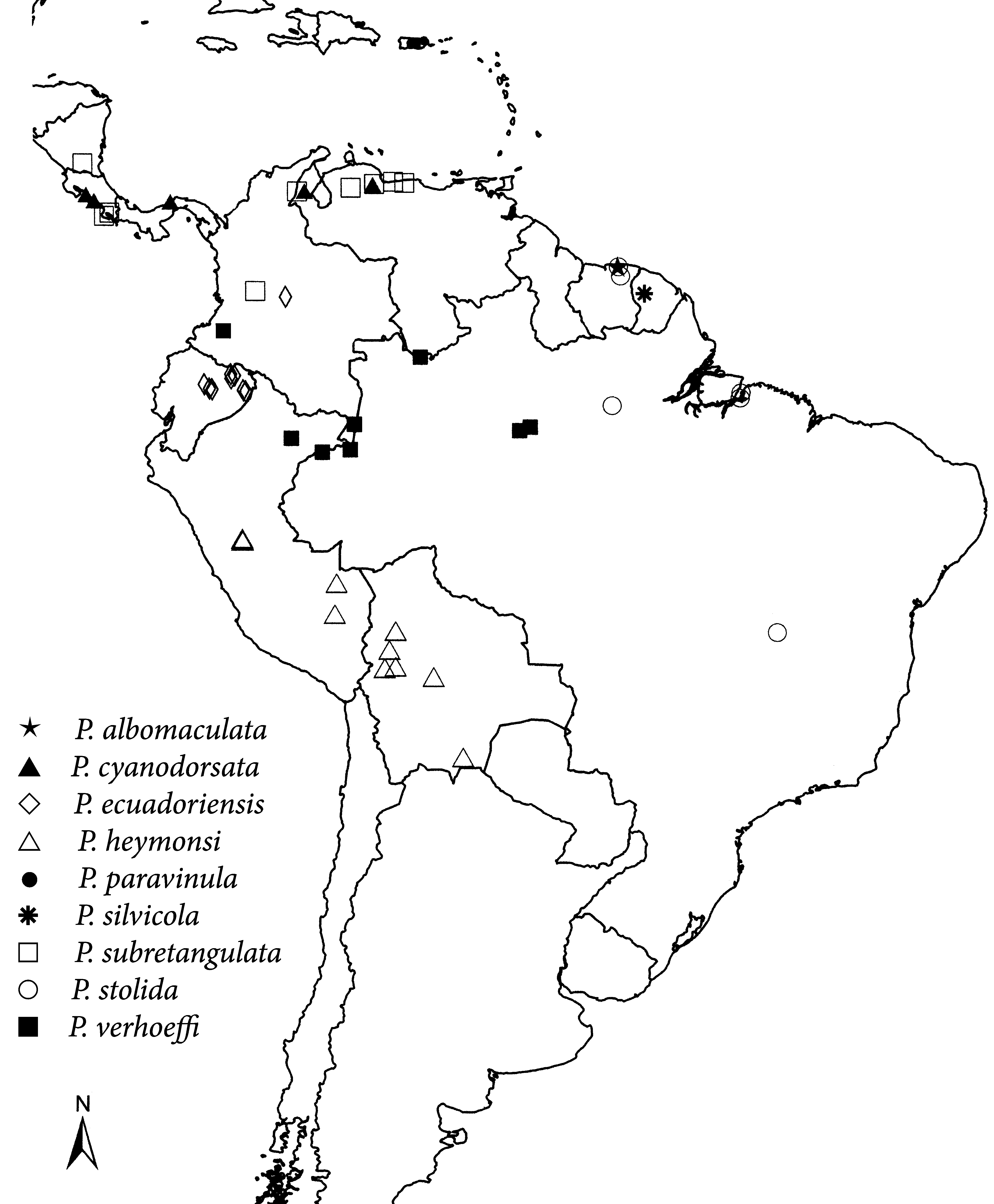
Distribution ( Fig. 59 View FIGURE 59 ): PUERTO RICO, NICARAGUA, COSTA RICA, PANAMA, VENEZUELA, COLOMBIA, SURINAME, FRENCH GUIANA, ECUADOR, PERU, BRAZIL, BOLIVIA.


No known copyright restrictions apply. See Agosti, D., Egloff, W., 2009. Taxonomic information exchange and copyright: the Plazi approach. BMC Research Notes 2009, 2:53 for further explanation.