
Progomphus, Selys, 1854
|
publication ID |
https://doi.org/10.11646/zootaxa.5297.2.4 |
|
publication LSID |
lsid:zoobank.org:pub:454B72E3-2659-4F3D-9C9A-503DBABEE9CE |
|
DOI |
https://doi.org/10.5281/zenodo.7999765 |
|
persistent identifier |
https://treatment.plazi.org/id/03E22F20-A771-2220-FF1C-F25AFA46FCBC |
|
treatment provided by |
Plazi (2023-06-01 06:56:03, last updated 2024-11-27 08:15:40) |
|
scientific name |
Progomphus |
| status |
|
Key to the larvae of Progomphus View in CoL from Argentina
(four species remain unknown in this stage: P. auropictus , P. australis , P. basistictus and P. costalis ).
1 Last antennomere (the fourth) short, spherical ( Figs. 6a View FIGURE 6 , 11b View FIGURE 11 , 13b View FIGURE 13 ), pro- and mesotibiae with distal spur ( Figs. 6a View FIGURE 6 , 13a View FIGURE 13 ), abdomen with posterolateral spines on S6–9........................................................................ 2
- Last antennomere long (at least longer than antennomere1-2 combined) and slender, cylindrical ( Figs. 2b View FIGURE 2 , 4b View FIGURE 4 , 9b View FIGURE 9 ), pro- and mesotibiae without distal spur ( Figs. 2c View FIGURE 2 , 4c View FIGURE 4 ), abdomen with posterolateral spines at least on S5–9 ( Figs. 1i View FIGURE 1 , 4d View FIGURE 4 , 9a View FIGURE 9 )........ 4
2 Sternum of S8 formed by 3 plates ( Fig. 6c View FIGURE 6 )....................................................... P. joergenseni View in CoL
- Sternum of S8 formed by 5 plates ( Figs. 11d View FIGURE 11 , 13h View FIGURE 13 )........................................................... 3
3 Third antennomere flat and wide, subcircular in shape ( Fig. 11b View FIGURE 11 ), ligula with 14 marginal tubercles ( Fig. 10f View FIGURE 10 )..... P. lepidus View in CoL
- Third antennomer subcylindrical, elongated ( Fig. 13b View FIGURE 13 ), ligula with only 2 median marginal tubercles ( Fig. 12e View FIGURE 12 ).................................................................................................. P. phyllochromus View in CoL
4 Anal pyramid short (epiproct subequal in length to S10, Figs. 4d–e View FIGURE 4 ), posterolateral spine absent on S4 ( Fig. 4d View FIGURE 4 )................................................................................................... P. complicatus View in CoL
- Anal pyramid long and slender (epiproct as long as or longer than S9 and 10 combined, Figs. 2d, 2g –i View FIGURE 2 , 9d–g View FIGURE 9 ), posterolateral spine present on S4 ( Figs. 1i View FIGURE 1 , 8c View FIGURE 8 )......................................................................... 5
5 Small abdominal dorsal tubercles present on S1–9, sometimes very reduced on S5 to 8 ( Figs. 1h View FIGURE 1 , 2d–e View FIGURE 2 ), posterolateral spines on S2–9 ( Figs. 1i View FIGURE 1 , 2d View FIGURE 2 ).......................................................................... P. aberrans View in CoL
- Small abdominal dorsal tubercles present on S1–3 ( Figs. 8a–b View FIGURE 8 , 9c View FIGURE 9 ), posterolateral spines on S4–9 ( Figs. 8c View FIGURE 8 , 9a View FIGURE 9 )... P. kimminsi View in CoL
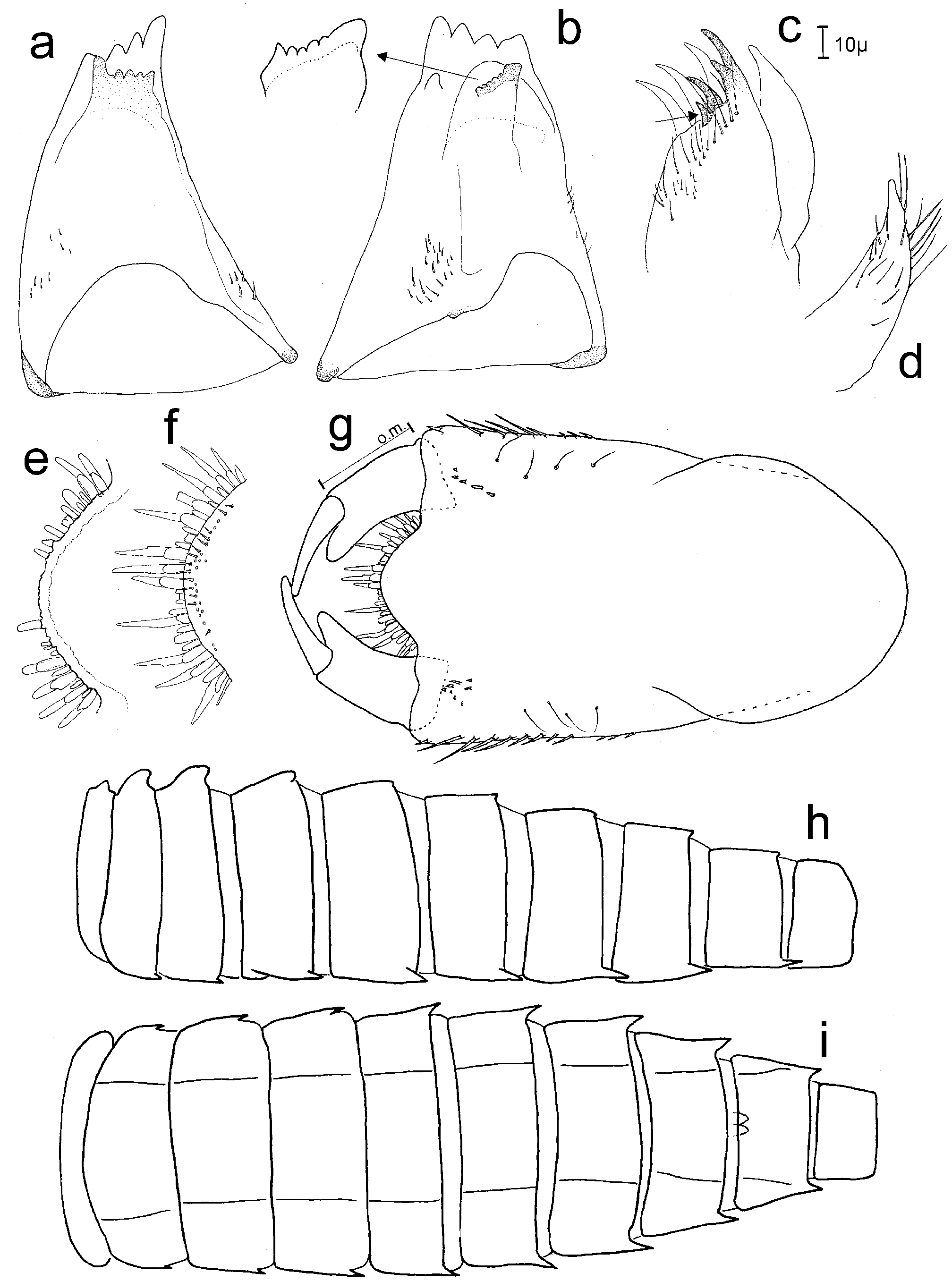
FIGURE 1. P. aberrans Belle, last instar larval exuvia: a, left mandible, occlusal view; b, right mandible with detail, occlusal view; c, maxilla, ventral view (arrow indicates first tooth); d, maxilar palp, dorsal view; e, detail of ligula; f, same (other specimen); g, prementum, dorsal view (om= outer margin of palpal segment); h, abdominal lateral outline (sterna omitted); i, abdominal ventral outline.
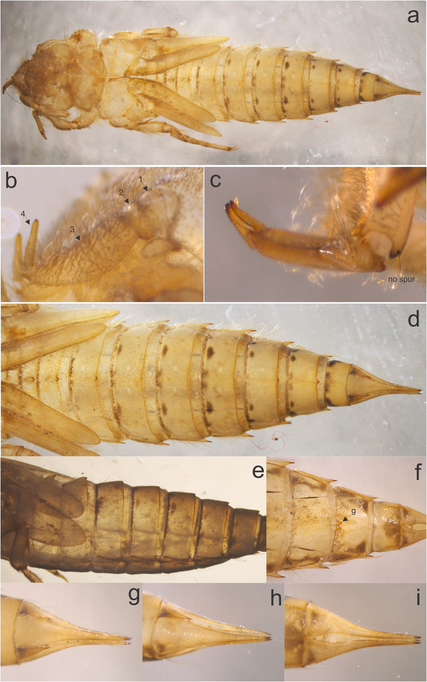
FIGURE 2. P. aberrans Belle, female last instar larval exuvia: a, dorsal habitus; b, antennae, lateral (numbers indicate antennomeres); c, fore leg with cheliform claws; d, abdomen, dorsal; e, abdomen, lateral; f, abdominal S8–10, ventral (g= gonapophyses); g, anal pyramid, dorsal; h, same, lateral; i, same, ventral.
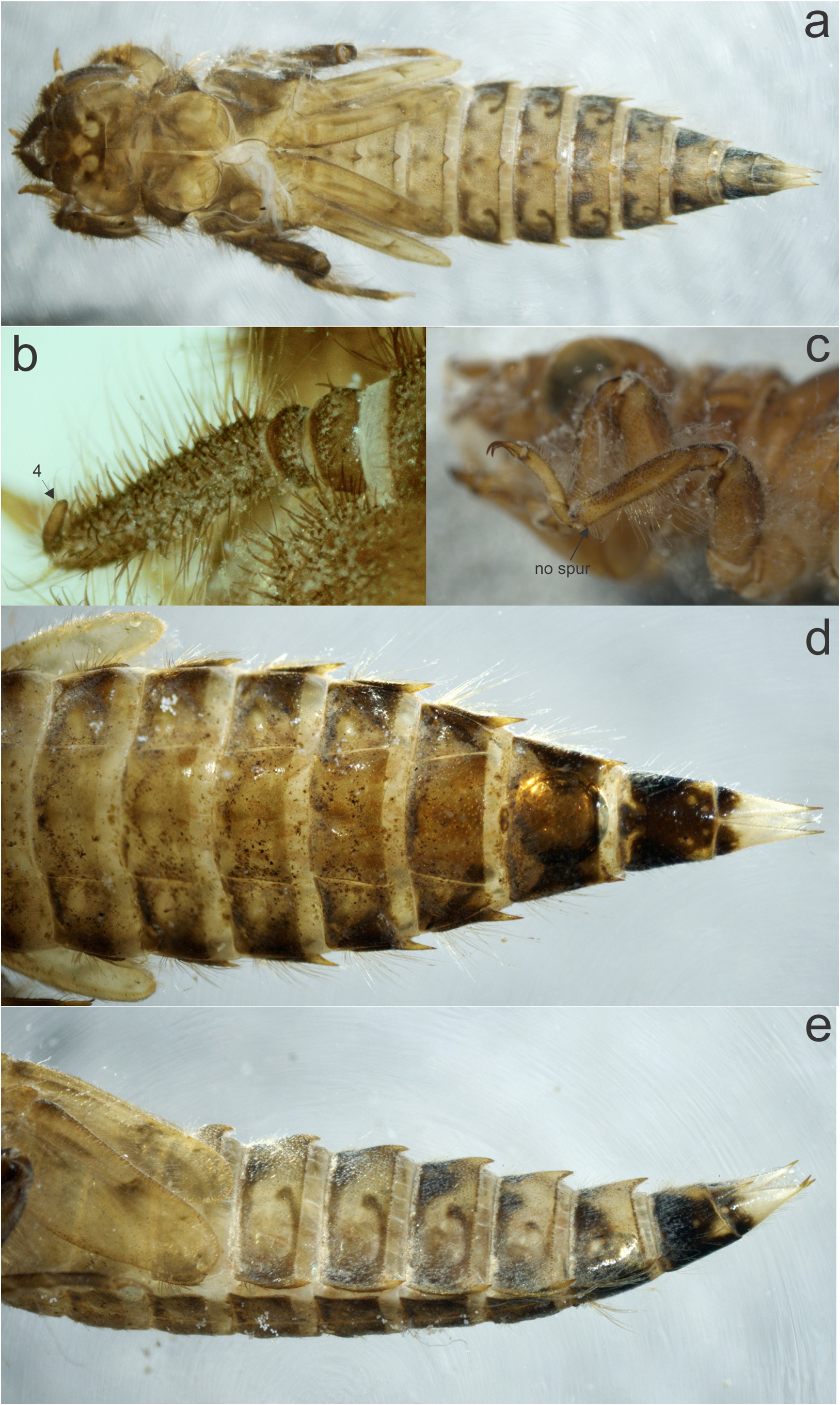
FIGURE 4. P. complicatus Selys, female last instar larval exuvia: a, dorsal habitus; b, antennae, dorsal (number indicate 4th antennomere); c, “normal” fore tarsal claws, lateral; d, abdomen, ventral; e, abdomen, lateral.
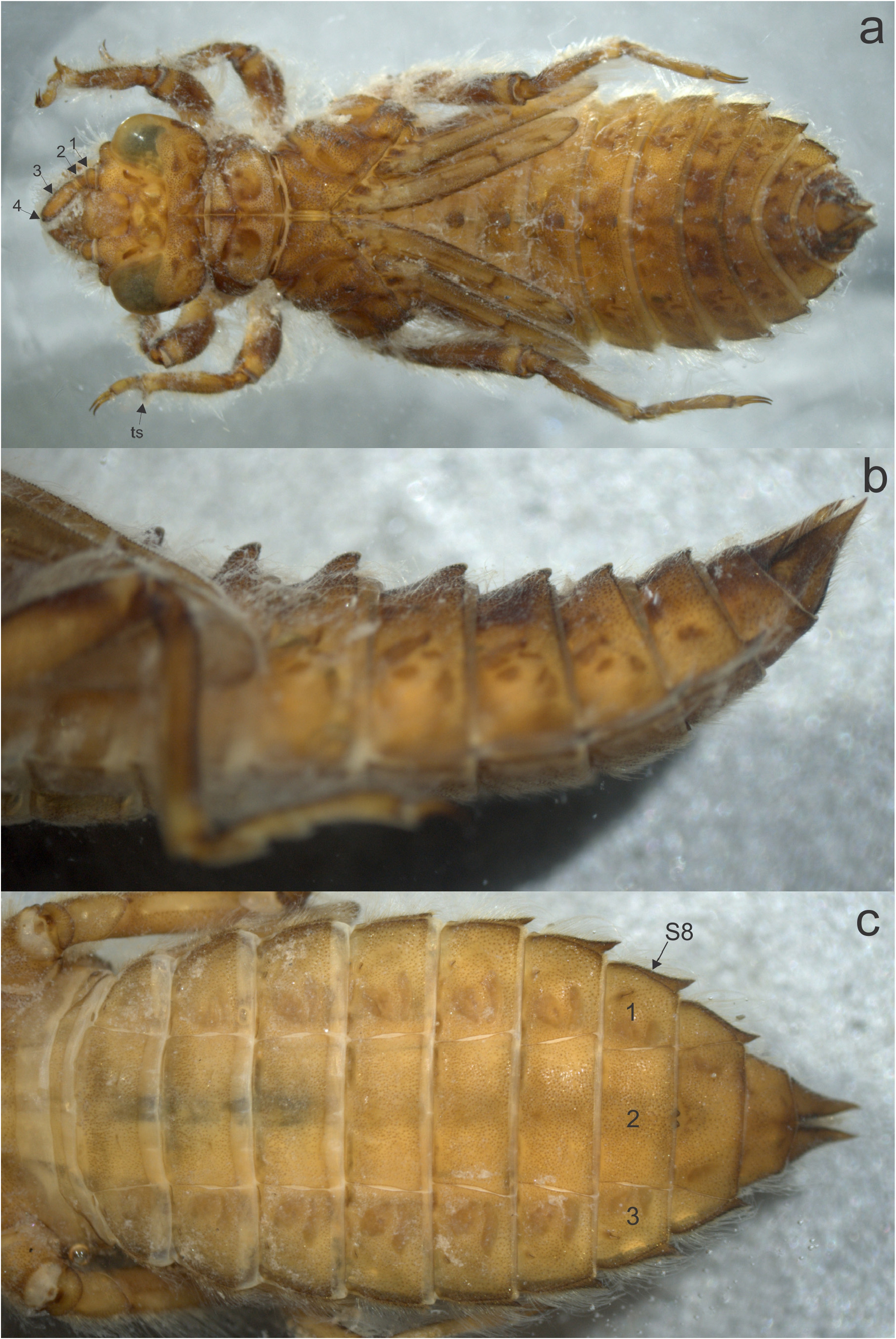
FIGURE 6. P. joergenseni Ris, last instar larval exuvia: a, dorsal habitus (antennomeres are numbered, ts=tibial spur); b, abdomen, lateral; c, abdomen, ventral.
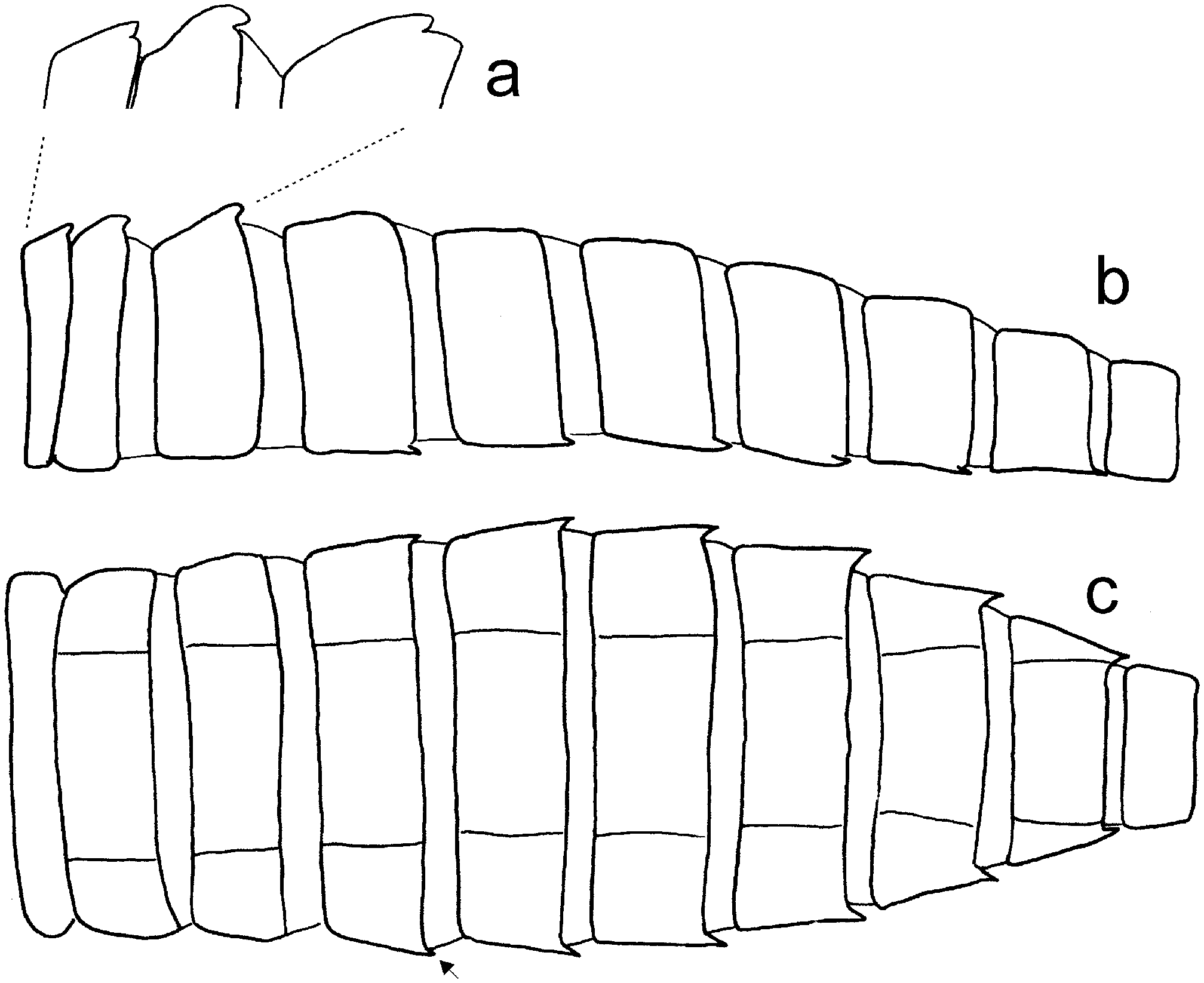
FIGURE 8. P. kimminsi Belle, last instar larval exuvia: a, lateral abdominal outline, detail of tubercles on segments 1–3; b, lateral abdominal outline; c, ventral abdominal outline (arrow indicate spine present on S4).
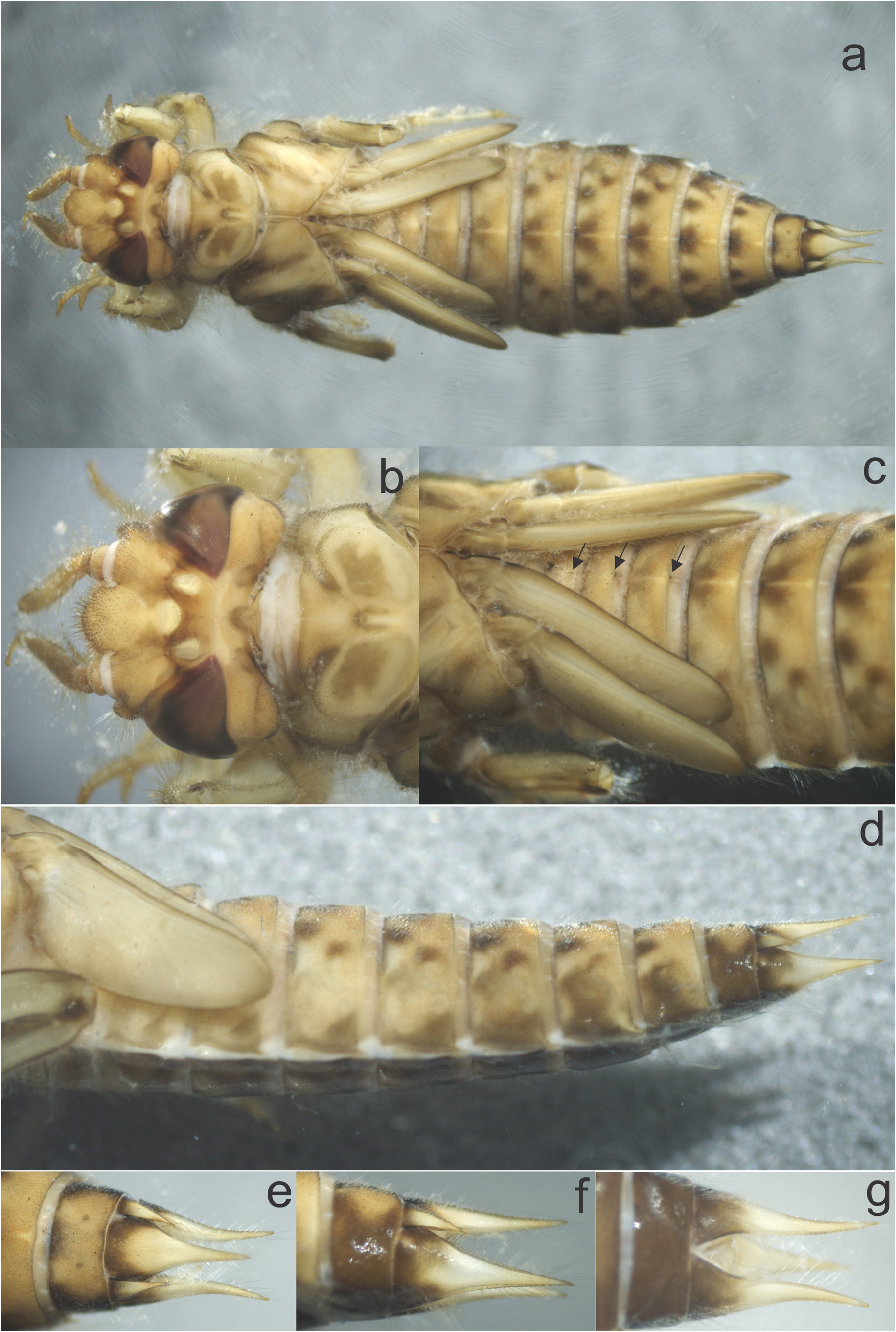
FIGURE 9. P. kimminsi Belle, last instar larval exuvia: a, dorsal habitus; b, detail of head, dorsal; c, detail of abdominal dorsal hooks (arrows); d, abdomen, lateral; e, anal pyramid, dorsal; f, same, lateral; g, same, ventral.

FIGURE 10. P. lepidus Ris, last instar female larva: a, left mandible, occlusal view; b, right mandible, occlusal view; c, same, dorso-occlusal view; d, maxilla, dorsdal view; e, maxila, ventral view (arrow indicates first tooth); f, detail of ligula; g, prementum, ventral view with detail of marginal microspines; h, subgenital lobe from pharate female.
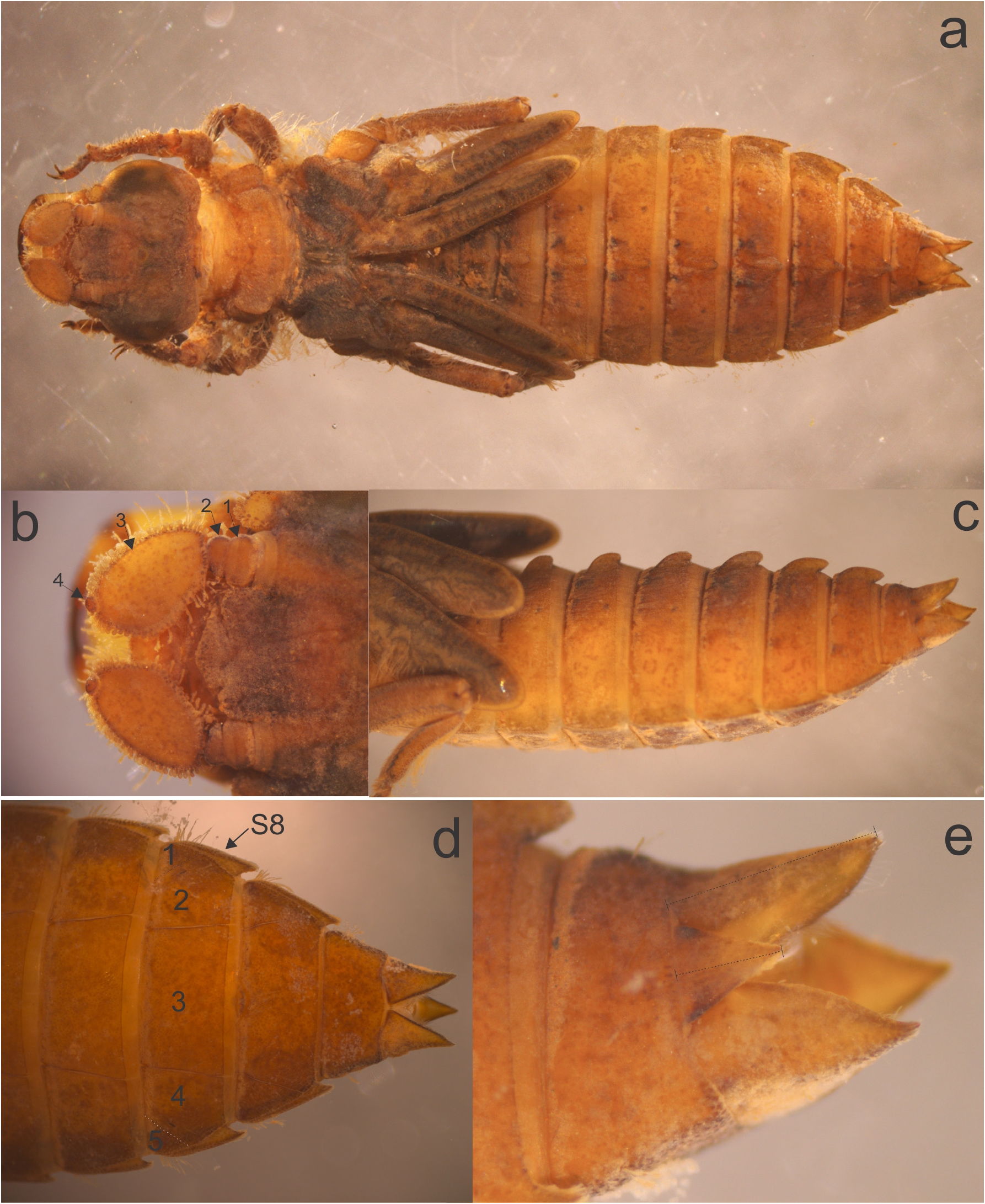
FIGURE 11. P. lepidus Ris, mature larva, female: a, dorsal habitus; b, antennae (antennomeres are numbered); c, abdomen lateral; d, abdominal sterna S7–10 (numbers indicate plates on S8, dotted lines indicate lateral sutures); e, anal pyramid, lateral.

FIGURE 12. P. phyllochromus Ris, last instar larval exuvia: a, left mandible, occlusal view; b, right mandible, occlusal view; c, maxilla, ventral view (arrow indicates first tooth); d, same, dorsal view; e, ligula. detail of flat marginal setae; f, prementum, ventral view; g, detail of labial palp, ventral view; h, prementum with detail, dorsal view.

FIGURE 13. P. phyllochromus Ris, last instar male larva: a, dorsal habitus; b, head and pronotum, dorsal (antennomeres are numbered); c, detail of tibial spurs (ts), ventral; d, abdomen, lateral; e, same, ventral; f, anal pyramid, dorsal; g, same, lateral; h, abdominal sterna S8–10 (numbers indicate plates on S8).
No known copyright restrictions apply. See Agosti, D., Egloff, W., 2009. Taxonomic information exchange and copyright: the Plazi approach. BMC Research Notes 2009, 2:53 for further explanation.