
Macedomeris ivoi Antić, 2021
|
publication ID |
https://doi.org/10.11646/zootaxa.4908.3.4 |
|
publication LSID |
lsid:zoobank.org:pub:29916FEE-7462-4AAE-8C54-DC5E0B10B83D |
|
DOI |
https://doi.org/10.5281/zenodo.4441974 |
|
persistent identifier |
https://treatment.plazi.org/id/03DE87BF-FF95-FFF6-FF3D-F8A8FB1AFE88 |
|
treatment provided by |
Plazi (2021-01-15 09:11:07, last updated 2024-11-29 18:46:26) |
|
scientific name |
Macedomeris ivoi Antić |
| status |
sp. nov. |
Macedomeris ivoi Antić View in CoL , new species
Figs 1–9 View FIGURE 1 View FIGURE 2 View FIGURE 3 View FIGURE 4 View FIGURE 5 View FIGURE 6 View FIGURE 7 View FIGURE 8 View FIGURE 9
Diagnosis. As for the genus.
Etymology. The new species is dedicated to our friend and colleague Ivo Karaman, a specialist of Opiliones and Isopoda, and biospeleologist who was the first to discover this beautiful creature. Noun in the genitive case.
Material examined. Holotype ♁ (IZB), NORTH MACEDONIA, Municipality of Struga, Tašmaruništa village, Mlečnik Cave (41.27° N, 20.65° E), 27.07.2019, leg. D. Antić. GoogleMaps Paratypes (IZB): 1 ♀ (paratype female I), 1 subadult ♁, same data as holotype; 1 GoogleMaps ♁, 1 ♀ (paratype female II), same cave but 21.04.2006, leg. I. Karaman.

FIGURE 1. Macedomeris ivoi gen. et sp. nov., holotype male (IZB), habitus. A. Lateral view. B. Dosral view. C. Ventral view. Scale bars: 0.5 mm.
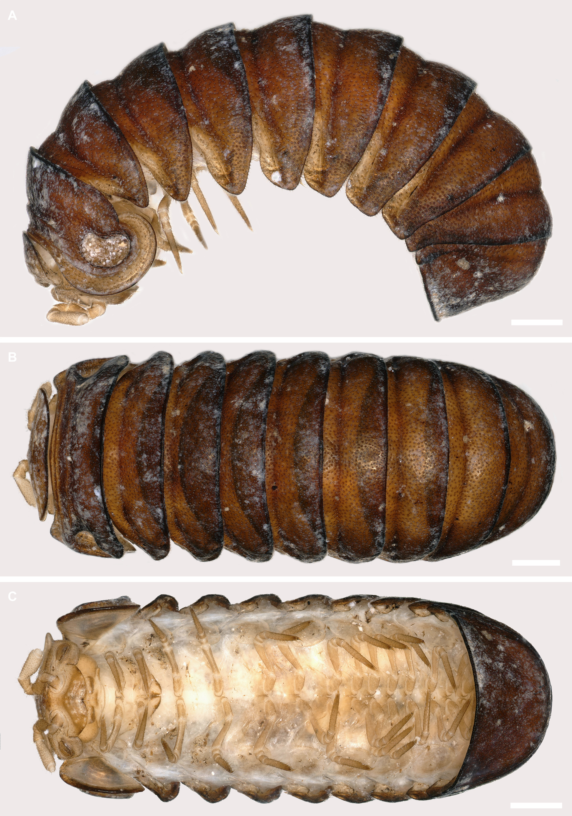
FIGURE 2. Macedomeris ivoi gen. et sp. nov., paratype female I (IZB), habitus. A. Lateral view. B. Dosral view. C. Ventral view. Scale bars: 0.5 mm.

FIGURE 3. Macedomeris ivoi gen. et sp. nov., paratype female I (IZB), coiled habitus. A. Right lateral view. B. Left lateral view. C. Midbody tergites, dorsal view. D. Thoracic schield, tergite 3 and anal shield. Abbreviations: r1: marginal thoracic ridge; r2: second thoracic ridge; r3: shortest thoracic ridge. Scale bars: 0.5 mm.
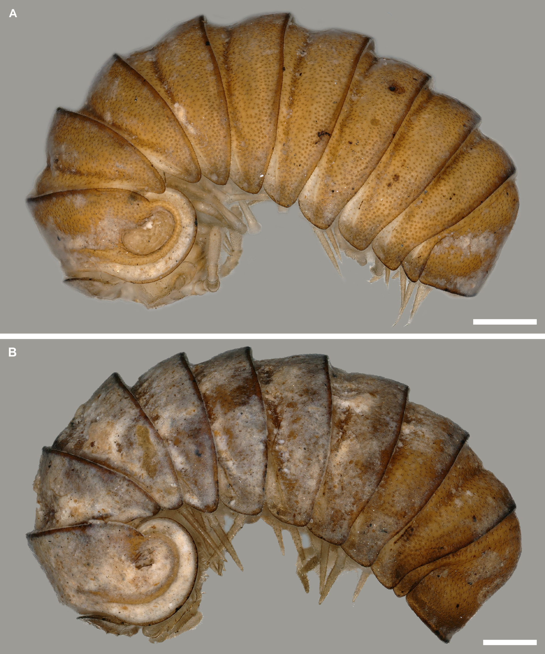
FIGURE 4. Macedomeris ivoi gen. et sp. nov., habitus, lateral view. A. Paratype female II (IZB). B. Paratype male (IZB). Scale bars: 0.5 mm.
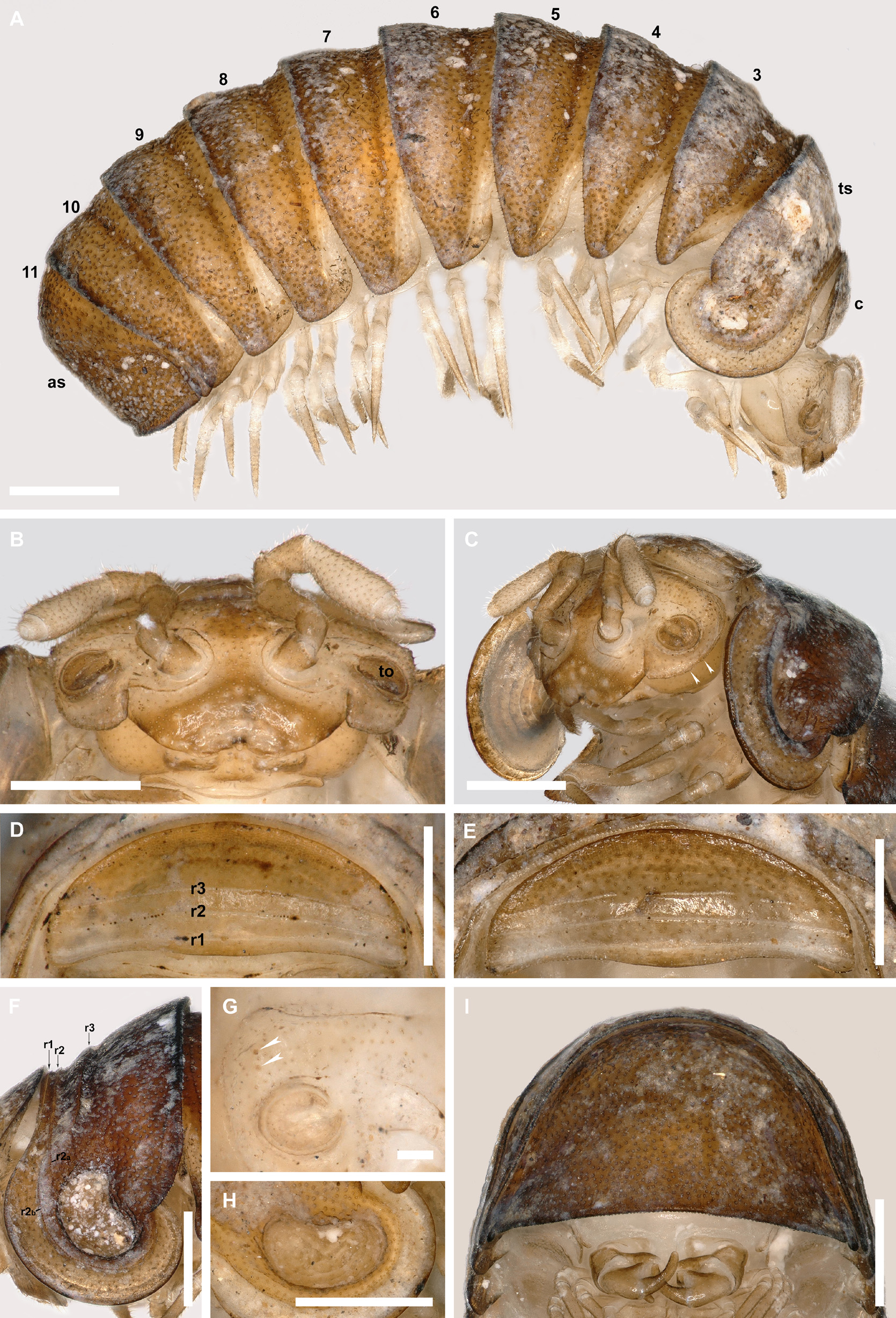
FIGURE 5. Macedomeris ivoi gen. et sp. nov. (IZB) A. Paratype subadult male, habitus, lateral view. B. Paratype female I, head, anterior view. C. Paratype female I, head and thoracic shield, anterolateral view. D. Paratype male, collum, dorsal view. E. Holotype male, collum, dorsal view. F. Paratype female I, thoracic shield, left lateral view. G. Paratype female II, right side of the head, anterior view. H. Paratype female II, left thoracic pit, lateral view. I. Holotype male, anal shield, posterior view. Abbreviations: as: anal shield; c: collum; to: organ of Tömösváry; ts: thoracic shield; r1, r2, r3: ridges on collum and thoracic shield; r2a, r2b: parts of thoracic r2; the numbers indicate tergites. White arrows indicate sclerotized nodules in C or vestigial ommatidia in G. Scale bars: 0.5 mm (A–F, H, I), 0.1 mm (G).
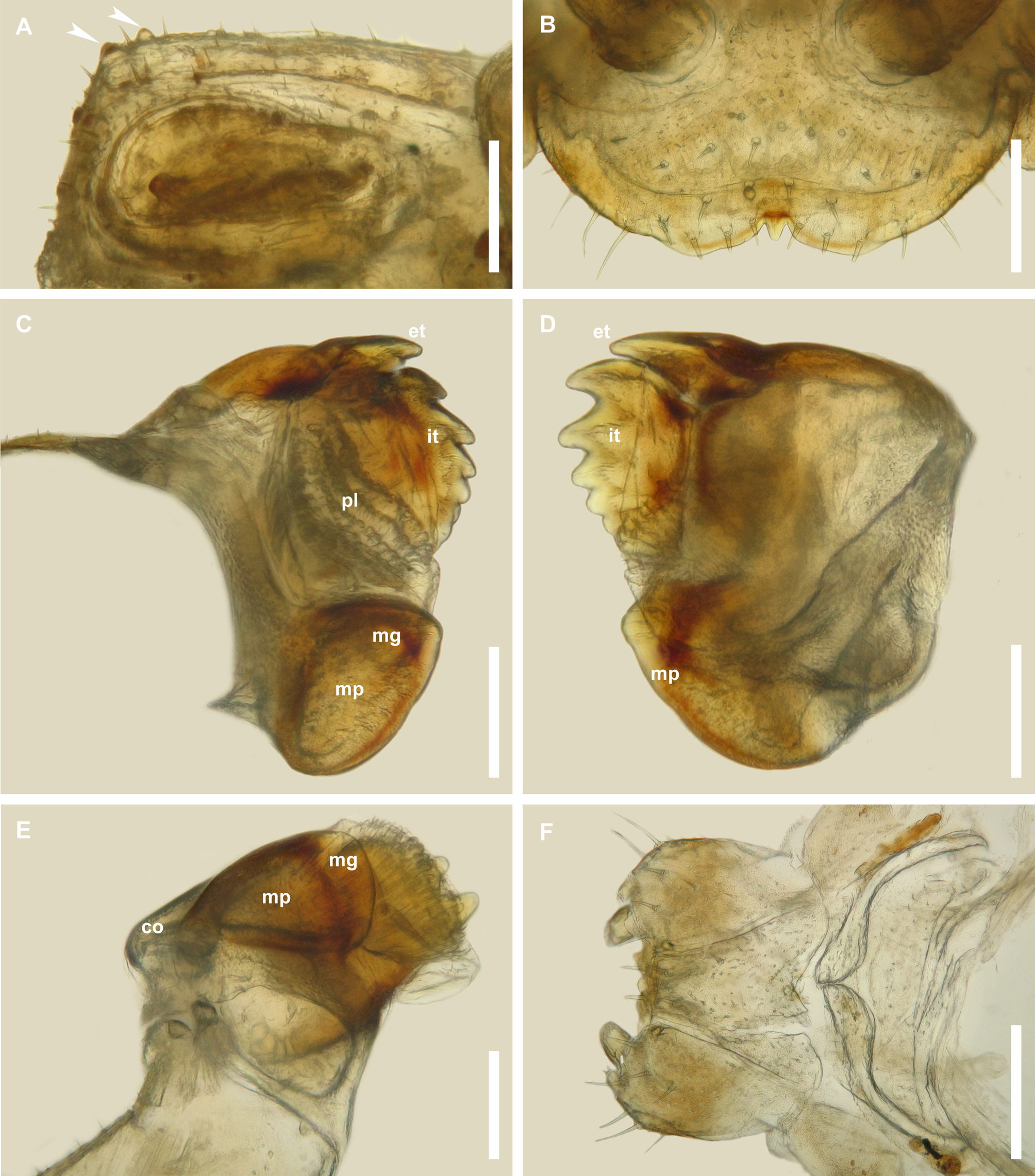
FIGURE 6. Macedomeris ivoi gen. et sp. nov., paratype male (IZB). A. Right side of the head, anterior view. B. Labrum, dorsal view. C. Right mandible, ventral view. D. Right mandible, dorsal view. E. Left mandible, mesal view. F. Gnathochilarium, ventral view. Abbreviations: co: condylus; et: extrenal tooth; it: inner tooth; mg: groove; mp: molar plate; pl: pectinate lamellae. White arrows indicate vestigial ommatidia. Scale bars: 0.5 mm (A, B), 0.2 mm (F), 0.1 mm (C–E).
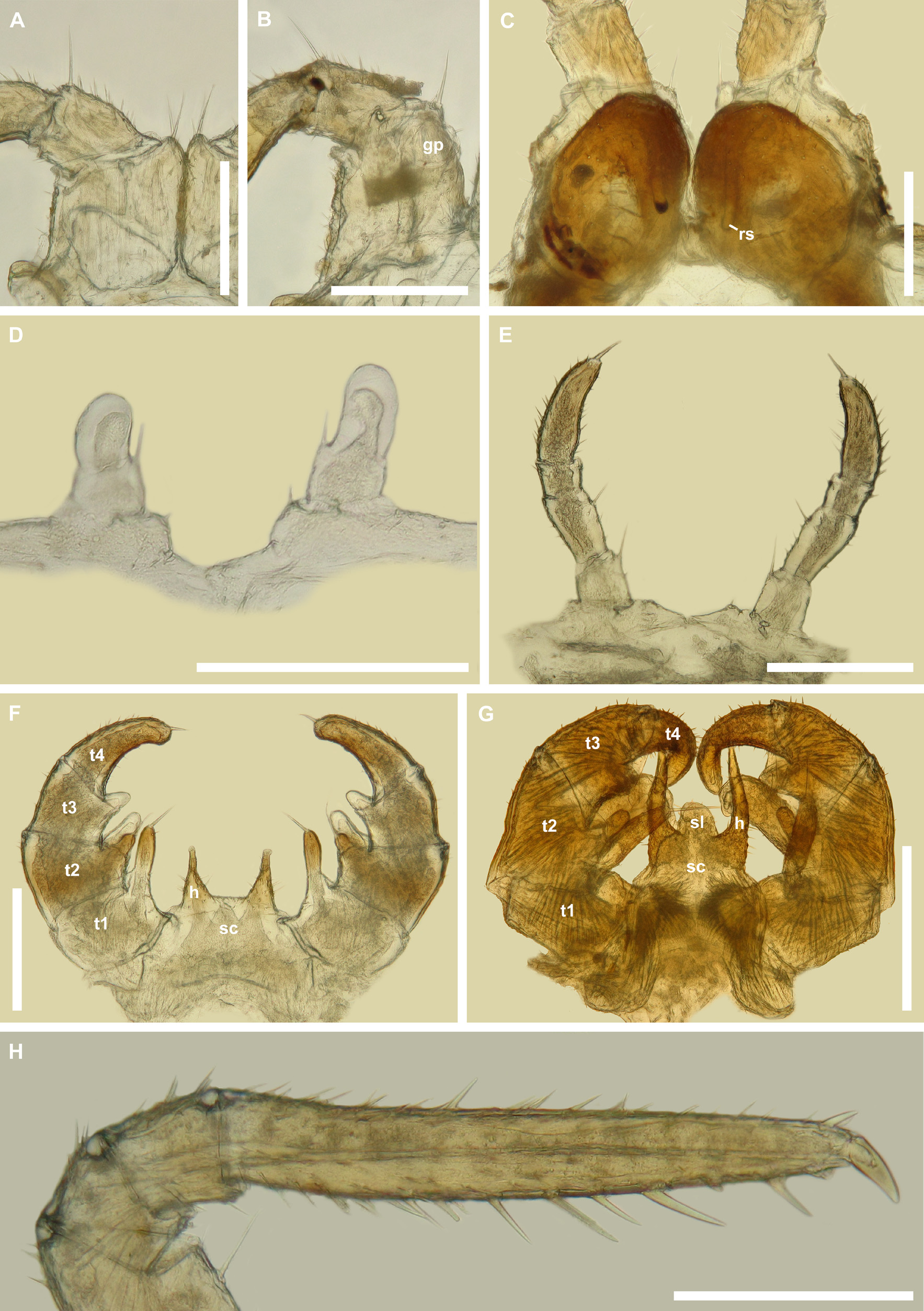
FIGURE 7. Macedomeris ivoi gen. et sp. nov. (IZB). A. Paratype male, right leg 1, posterior view. B. Paratype male, right leg 2, posterior view. C. Paratype female I, vulvae, posterior view. D. Paratype subadult male, leg-pair 17, anterior view. E. Paratype subadult male, leg-pair 18, anterior view. F. Paratype subadult male, telopods, anterior view. G. Paratype male, telopods, anterior view. H. Paratype male, left leg 9, anterior view. Abbreviations: gp: gonopore; h: syncoxital horn; rs: receptaculum seminis; sc: syncoxite; sl: syncoxital median lobe; t1: telopoditomere 1 (prefemur); t2: telopoditomere 2 (femur); t3: telopoditomere 3 (tibia); t4: telopoditomere 4 (tarsus). Scale bars: 0.3 mm (G), 0.2 mm (A–F, H).
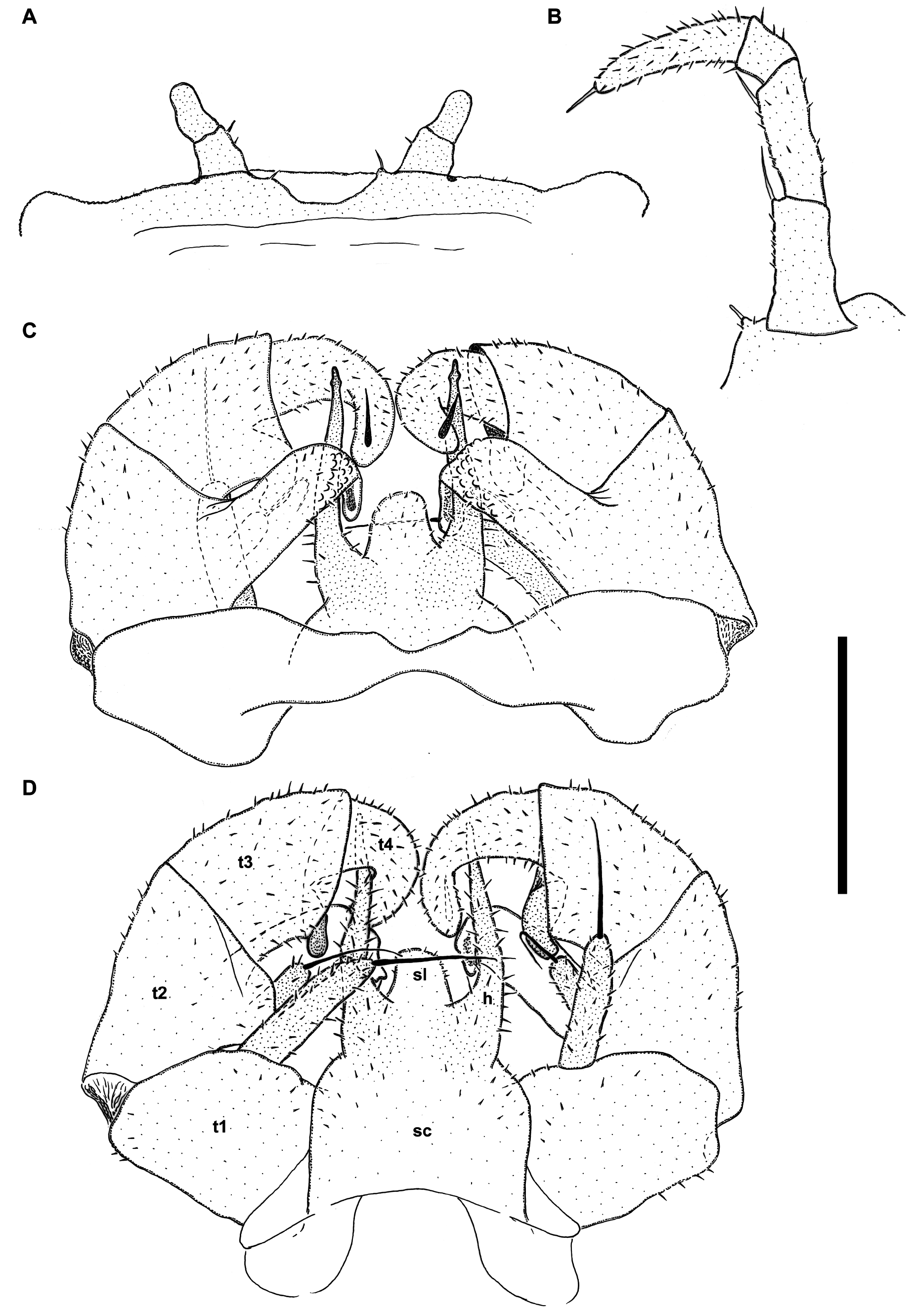
FIGURE 8. Macedomeris ivoi gen. et sp. nov., paratype male (IZB). A. Leg-pair 17, anterior view. B. Left leg 18, anterior view (flipped). C. Telopods, posterior view. D. Telopods, anterior view. Abbreviations: h: syncoxital horn; sc: syncoxite; sl: syncoxital median lobe; t1: telopoditomere 1 (prefemur); t2: telopoditomere 2 (femur); t3: telopoditomere 3 (tibia); t4: telopoditomere 4 (tarsus). Scale bar: 0.3 mm.
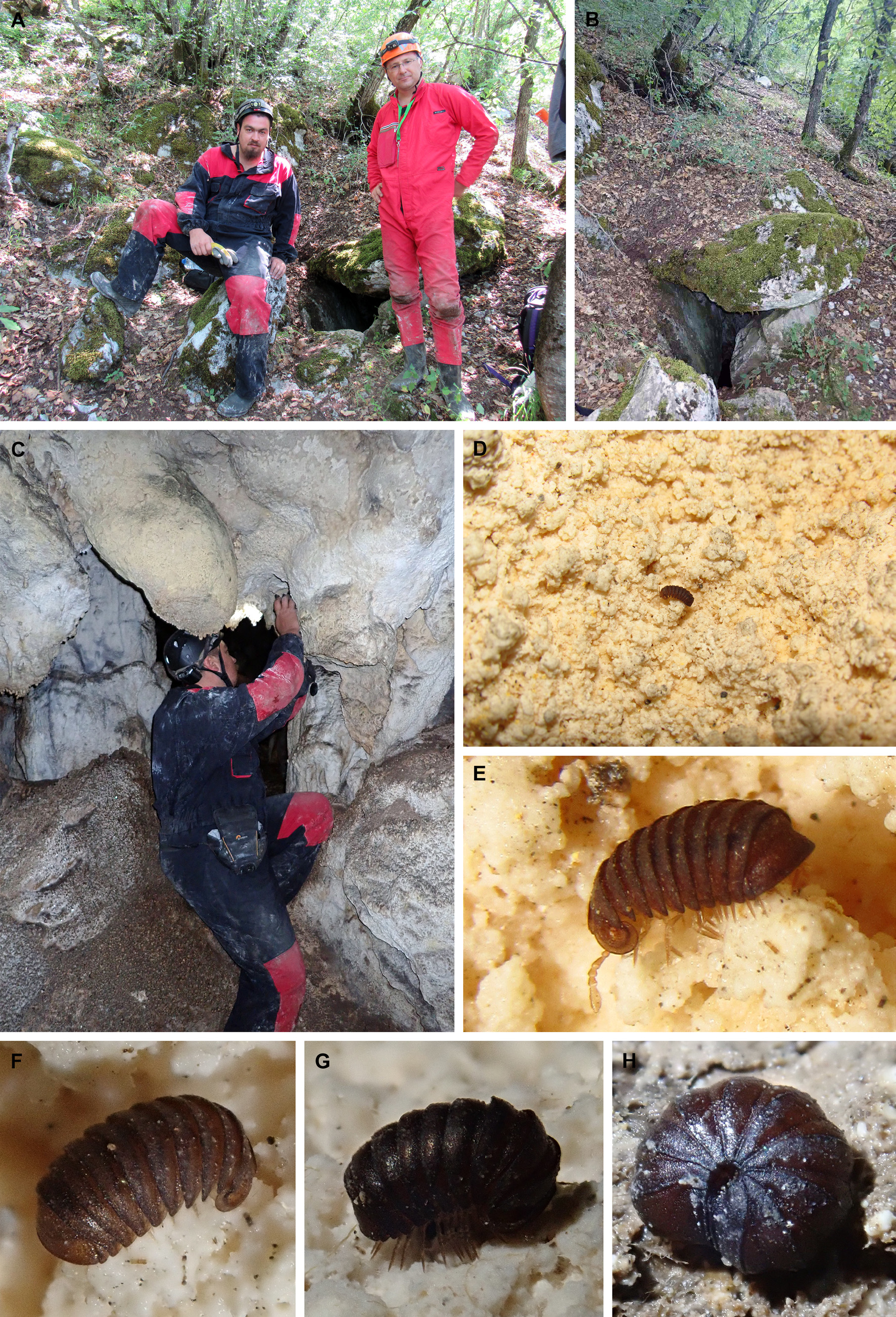
FIGURE 9. Type locality and habitus in situ of Macedomeris ivoi gen. et sp. nov. A. In front of the Mlečnik Cave. B. Entrance of the Mlečnik Cave. C. Small chamber where specimens were found. D–F. Future holotype male on cave popcorn. G. Future paratype female I on cave popcorn. H. Future paratype female I, disturbed and coiled. Photo credit: A, D, E. Dragan Antić, B, C, F–H. Marjan Komnenov.
No known copyright restrictions apply. See Agosti, D., Egloff, W., 2009. Taxonomic information exchange and copyright: the Plazi approach. BMC Research Notes 2009, 2:53 for further explanation.
|
Kingdom |
|
|
Phylum |
|
|
Class |
|
|
Order |
|
|
Family |
|
|
SubFamily |
Glomerinae |
|
Tribe |
Doderiini |
|
Genus |