
Eulophophyllum kirki Ingrisch & Riede
|
publication ID |
https://doi.org/ 10.1665/034.025.0205 |
|
DOI |
https://doi.org/10.5281/zenodo.6073579 |
|
persistent identifier |
https://treatment.plazi.org/id/03D987D4-A63A-FF93-0DC1-FEC485F02B77 |
|
treatment provided by |
Plazi |
|
scientific name |
Eulophophyllum kirki Ingrisch & Riede |
| status |
sp. nov. |
Eulophophyllum kirki Ingrisch & Riede sp. n.
urn:lsid: Orthoptera .speciesfile.org:TaxonName:493861
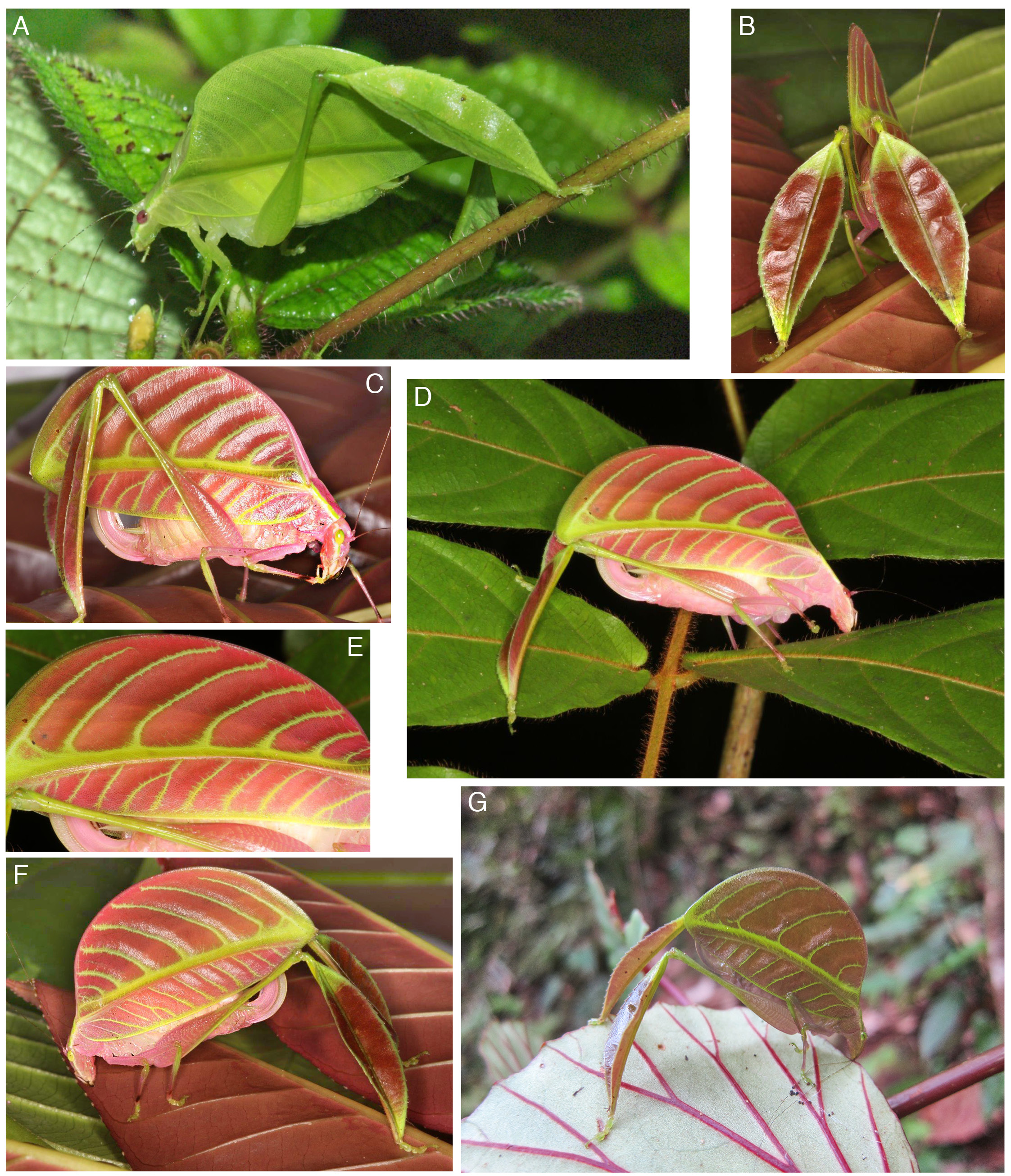
Holotype (female): East Malaysia: Sabah, Danum Valley (N 4° 57’ 55’’; E 117° 41’ 25’’, ca 170 m), 6. vi.2013, photographed by Peter Kirk ( Fig. 3 View Fig. 3 B-F). GoogleMaps
Other material (photograph of a male, Fig. 3 View Fig. 3 A): East Malaysia: Sabah, Danum Valley field centre, night walk, 24.i.2009, photographed by Paul Bertner (https://www.flickr.com/photos/rainforests/3430798861).
Diagnosis.— This species is very similar to E. lobulatum sp. n. with regard to the strongly widened tegmina, lobate hind tibiae and green and pink color polymorphism. It differs by the more semioval and relatively longer tegmina with seven or eight instead of five transverse veins in the medial field. The apical area of the male cerci is narrower than in E. lobulatum .
Description (female holotype).— Pronotum with concave anterior and convex posterior margin; disc flat, lateral margins straight and subangular; paranota a little longer than high, posterior area covered by a projecting flap of tegmen.Tegmen strongly widened;anterior margin moderately convex near both ends, nearly substraight in middle; posterior margin strongly convex, nearly semi-circular ( Fig. 3 View Fig. 3 C-F).
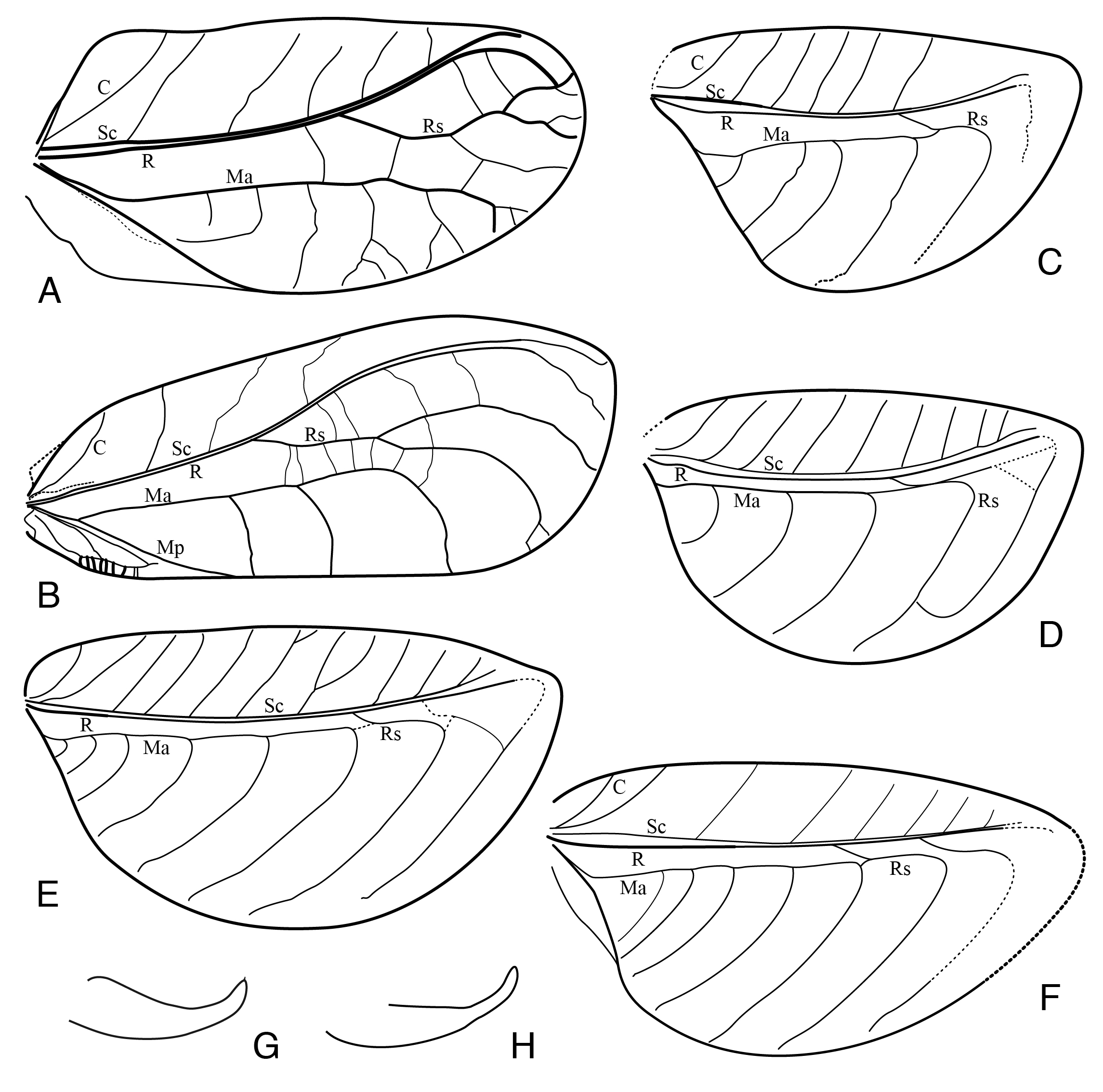
Tegminal venation: costa normal, costal field widened with oblique cross veins; subcosta and radius parallel and closely approaching each other, little diverging before apex of tegmen; radius without distinct branches but connected by numerous weak veinlets to media anterior, two of those veinlets in posterior half of tegmen that are slightly stronger and run more obliquely than the other veinlets in this area, can be regarded as branches of radius that are fused with media anterior; media forked shortly behind base; media anterior running parallel with and close to radius, both veins connected by numerous faint cross veinlets; media posterior fused with cubitus and the fused veins forming the hind margin of tegmen; media field (area between media anterior and media posterior) extremely widened with all cross veins within this field curved (in situ) dorso-craniad, the last of those cross veins with a twofold base; dorsal area of tegmen narrow, triangular and short ( Fig. 1 View Fig. 1 E).
Legs: Fore tibia normal (quadrangular), tibial tympana conchate on anterior (internal), open on posterior (external) side; mid tibia with dorsal margins little widened in basal half; hind femur widened in basal half, narrow in apical half; hind tibia with both dorsal margins strongly expanded conferring a leaf-like appearance ( Fig. 3 View Fig. 3 B).
Coloration (living female, pink color variant, Fig. 3 View Fig. 3 B-F). Head, pronotum, thorax, abdomen, and ovipositor light pink. Antennae pink at base, otherwise dark brown with white and black spaced annulation. Head with a white band from base of mandibles to compound eyes, running along and continued behind eyes; compound eyes light green,ocelli white; tips of mandibles and maxillary palpi black. Pronotum with pale green lateral angles. Tegmen pink at very base, later red with anterior margin, veins and main cross veins and area between radius and media anterior pastel green;
subcosta, radius and fused media - cubitus posterior for the greatest part pink or red. Fore and mid legs pink; fore tibia at base and mid tibia in basal half green, both with a black subapical mark; tarsi dirty green. Hind femur pink at base, getting darker posteriorally, apical half and ventral margin except at base green; posterior tibia green at base and apex, the larger expanded central area dark red on ventral, brown on dorsal side, margins green; posterior tarsus green.
Coloration (living male, green color variant, Fig. 3 View Fig. 3 A). Rather uniformly green; thorax, fore and mid legs pale green; abdomen for the greater part yellowish green. Head green with a white band bordering compound eyes on posterior side and running down to clypeus; antennal flagellum blackish, narrowly annulated and with spaced white rings; maxillary palpi with black tips. Pronotum green; disc with white lateral bands. Tegmen green; anterior margin, most veins and main cross-veins are a little lighter yellowish green; along radius in the area between radius and media there is a dark greyish green band. Fore tibiae with a black spot near ventral end. The male cerci have the apical area distinctly narrowed setose before tip.
Etymology.— Named after the photographer of the holotype, Peter Kirk.
No known copyright restrictions apply. See Agosti, D., Egloff, W., 2009. Taxonomic information exchange and copyright: the Plazi approach. BMC Research Notes 2009, 2:53 for further explanation.
|
Kingdom |
|
|
Phylum |
|
|
Class |
|
|
Order |
|
|
Family |
|
|
Genus |