
Moloha tumida, Kumar, 2015
|
publication ID |
https://doi.org/10.5852/ejt.2015.166 |
|
publication LSID |
lsid:zoobank.org:pub:FE328725-7017-417D-B140-71D830B89316 |
|
DOI |
https://doi.org/10.5281/zenodo.3815851 |
|
persistent identifier |
https://treatment.plazi.org/id/25B20CDD-06DA-4A14-BB61-1F7954ED6357 |
|
taxon LSID |
lsid:zoobank.org:act:25B20CDD-06DA-4A14-BB61-1F7954ED6357 |
|
treatment provided by |
Carolina (2020-05-06 18:00:21, last updated by Juliana 2025-02-27 14:03:36) |
|
scientific name |
Moloha tumida |
| status |
sp. nov. |
Moloha tumida View in CoL sp. nov.
urn:lsid:zoobank.org:act:25B20CDD-06DA-4A14-BB61-1F7954ED6357
Figs 1B View Fig , 3B View Fig , 4C View Fig , 5B View Fig , 9 View Fig , 13 View Fig , 14B View Fig , 15 View Fig D–G, 17B
Diagnosis
Carapace with pseudorostral and supraocular spines long, subequal; supraocular spine with distinct submedian accessory spine; gastric region with many sharp granules in addition to 3 major spines; branchial regions distinctly inflated; subhepatic region swollen, with 2 large dorsal and 3 small ventral spines; protogastric region with 2 major spines; basal antennal spine acute; P2–P4 long, slender, subcylindrical, merus with 8–10 spines on dorsal margin, outer surface with 1–13 small spines, ventral margin with 19–28 spines; P5 with 2 spines on dorsal margin, 2 small spines on outer surface, 4 spines on ventral margin, subchelate structure stout, propodus with 3 large basal spines, rest of margin with distinct, closely arranged, similarly sized spines. G1 stout, short, groove on ventral surface submedian, dorso-median surface flat, distal part less rounded, opening large, auriculiform, directed towards median part of sternum.
Etymology
The species is named after the relatively swollen carapace.
Material examined
Holotype
INDIA: ♂ (tcl 64.4 mm, cl 55.7 mm, tcw 56.1 mm, cw 50.6 mm), Kerala, 300–350 m, 3 Dec. 2014 ( DABFUK).
Description
Medium-sized species, spiny on carapace, appendages, chelipeds and ambulatory legs ( Figs 1B View Fig , 3B View Fig , 4C View Fig , 5B View Fig , 13 View Fig , 14B View Fig ). Dorsal surface of carapace with deep grooves, well marked regions ( Figs 1B View Fig , 3B View Fig , 4C View Fig , 5B View Fig , 14B View Fig ). Pseudorostral spine simple, as long as or slightly longer than supraorbital spines ( Figs 1B View Fig , 3B View Fig , 14B View Fig ). Proepistome with sharp spine. Supraocular spine long, straight, with prominent laterally directed accessory spine on anterior third ( Figs 1B View Fig , 3B View Fig , 4C View Fig , 5B View Fig , 14B View Fig ); infraorbital spine long, gently curved outwards, about ⅔ length of supraorbital spine, visible in dorsal view ( Figs 4C View Fig , 5B View Fig ); buccal spine prominent, just visible in dorsal view ( Figs 4C View Fig , 5B View Fig ); basal antennal spine sharp, acute ( Figs 4C View Fig , 5B View Fig ); protogastric region with 2 major spines and smaller spinules posterior and lateral to them ( Figs 1B View Fig , 2B View Fig , 5B View Fig , 14B View Fig ); mesogastric region with 1 large median spine ( Figs 1B View Fig , 2B View Fig , 5B View Fig , 14B View Fig ); mesobranchial region with 5 or 6 large, laterally directed spines and numerous spinules behind them ( Figs 1B View Fig , 2B View Fig , 5B View Fig , 14B View Fig ); subhepatic region swollen, with 2 large dorsal spines, 1 median spine and 2 small ventral spines ( Figs 1B View Fig , 2B View Fig , 4C View Fig , 5B View Fig , 14B View Fig ); anterolateral spine distinct, pointing obliquely anteriorly, with smaller spine below it ( Figs 1B View Fig , 2B View Fig , 5B View Fig , 14B View Fig ); posterolateral spines distinct, 3 largest laterally directed, first largest, with many spinules between them ( Figs 1B View Fig , 2B View Fig , 5B View Fig , 14B View Fig ); cardiac region small, with 1 short spine ( Figs 1B View Fig , 2B View Fig , 4C View Fig , 5B View Fig , 14B View Fig ). Cervical groove shallow, but clearly visible ( Figs 1B View Fig , 2B View Fig , 14B View Fig ); transverse cardio-intestinal groove shallow ( Figs 1B View Fig , 2B View Fig , 14B View Fig ). Posterior carapace margin strongly concave; lateral margins of branchiostegite almost smooth ( Figs 1B View Fig , 2B View Fig , 14B View Fig ). Eyes short; podophthalmite short, stout; basophthalmite slender, elongate; cornea bulbous ( Figs 1B View Fig , 2B View Fig , 4C View Fig , 5B View Fig , 14B View Fig ). Antennules with a swollen basal article, other articles long, slender, with elongate flagellum ( Fig. 4C View Fig ). Antennae short, first article with large urinary article ( Figs 4C View Fig , 5B View Fig ). Epistome truncate; posterior margin gently sinuous, with median part weakly triangular, lateral parts gently concave ( Fig. 4C View Fig ). Third maxilliped subpediform, elongated; inner margins lined with dense, long setae; basis-ischium with 4 submedian tubercles; merus with 1 large subproximal tubercle, anterolateral margin dentate; carpus short, unarmed; propodus and dactylus elongated, unarmed ( Fig. 13A View Fig ).
Cheliped relatively long, slender, spiny ( Figs 1B View Fig ); coxa with 2 short spines; ischium subtrigonal in crosssection, with 3–7 short spines; merus with 3 rows of short or long spines: dorsal row with 17–19 spines, outer surface with 12 or 13 spines, ventral margin with 14–18 spines ( Figs 1B View Fig , 9D View Fig ); carpus elongate, outer surface granulated, not spinose ( Figs 1B View Fig , 9 View Fig D–E). Palm slender, covered with small granules and setae, those on inner surface denser and longer; fingers shorter than palm, with hooked tips, pigmented throughout most of length except near base, that of pollex not extending substantially into palm; cutting edges blade-like; dactylus with 1 low subproximal tubercle ( Figs 1B View Fig , 9 View Fig E–F).
Ambulatory legs long, slender; meri subcylindrical; P3 longest ( Figs 1B View Fig , 13 View Fig B–H). P2 coxa with 3 spines; ischium with 4 short spines; merus with 3 rows of short or long spines: dorsal row with 9 or 10 spines, outer surface with 1–13 spines, ventral margin with 19–21 spines ( Fig. 13B, E View Fig ). P3 coxa with 3 spines; ischium with 4–7 short spines; merus with 3 rows of short or long spines: dorsal row with 9 spines, outer surface with 10–13 spines, ventral margin with 20–25 spines ( Fig. 13C, F View Fig ). P4 coxa with 2 or 3 spines; ischium with 3–6 short spines; merus with 3 rows of short or long spines: dorsal row with 8 spines, outer surface with 11 spines, ventral margin with 26–28 spines ( Fig. 13D, G View Fig ). P5 coxa with 1 spine; ischium with 2 short spines; merus with 3 rows of short or long spines: dorsal row with 2 spines, outer surface with 2 spines, ventral margin with 4 spines; carpus elongate, unarmed; propodus and dactylus forming subchelate structure; propodus curved, relatively shorter, flexor margin with 3 large, curved spines and 4 or 5 smaller spines anterior to it in a row, dactylus curved with 6–9 small spines on flexor margin ( Fig. 13 View Fig H–I).
Male pleon ovate, completely covering thoracic sternal surface; telson pentagonal, with distal half triangular, with sharp tip, basal part quadrate, lateral margin convex to distinctly convex ( Fig. 9 View Fig A–B); somite 6 trapezoidal, with deeply concave lateral margins, distal median margin with prominent spine ( Fig. 9 View Fig A–B); somites 4 and 5 subquadrate, with lateral margins expanded, triangular, somite 5 distal median margin with distinct tubercle, somite 4 with median tubercle ( Fig. 9B View Fig ); somites 2 and 3 trapezoidal, with long median spine; somite 1 short, with sharp median tubercle ( Fig. 9B View Fig ). Sternopleonal cavity deep, smooth; thoracic sternites 1–5 without median longitudinal line, suture between sternites 5 and 6 shallow, complete; pair of partially flattened prominences (homolid button, cf. Guinot & Bouchard 1998: 635, fig. 9c) on sternite 4 at margin of sternopleonal cavity, fits into pair of sockets on internal marginal surface of somite 6 ( Fig. 9C View Fig ).
G1 short, stout, distal part rounded; opening directed towards median part of thoracic sternum; groove submedian; dorso-median surface flat; distal part less rounded; opening auriculiform ( Figs 9C View Fig , 15 View Fig D–F); marginal and submarginal surfaces along distal part with long setae ( Fig. 15 View Fig D–F). G2 stout, as long as G1, basal part dilated, with long setae; distal part cup-like ( Fig. 15G View Fig ).
Colour
In life, the carapace and chelipeds are red; the ambulatory legs are red with patches of white and the ventral surfaces are dirty white ( Fig. 17B View Fig ).
Distribution
The species is known only from southwestern India.
Remarks
See the Discussion section.
Guinot D. & Richer De Forges B. 1995. Crustacea Decapoda Brachyura: Revision de la famille des Homolidae de Haan, 1839. In: Crosnier A. (ed.) Resultats des Campagnes MUSORSTOM, Vol. 13. Memoires du Museum national d'Histoire naturelle 163: 283 - 517. Museum national d'Histoire naturelle, Paris.
Guinot D. & Bouchard J. M. 1998. Evolution of the abdominal holding systems of brachyuran crabs (Crustacea, Decapoda, Brachyura). Zoosystema 20 (4): 613 - 694.
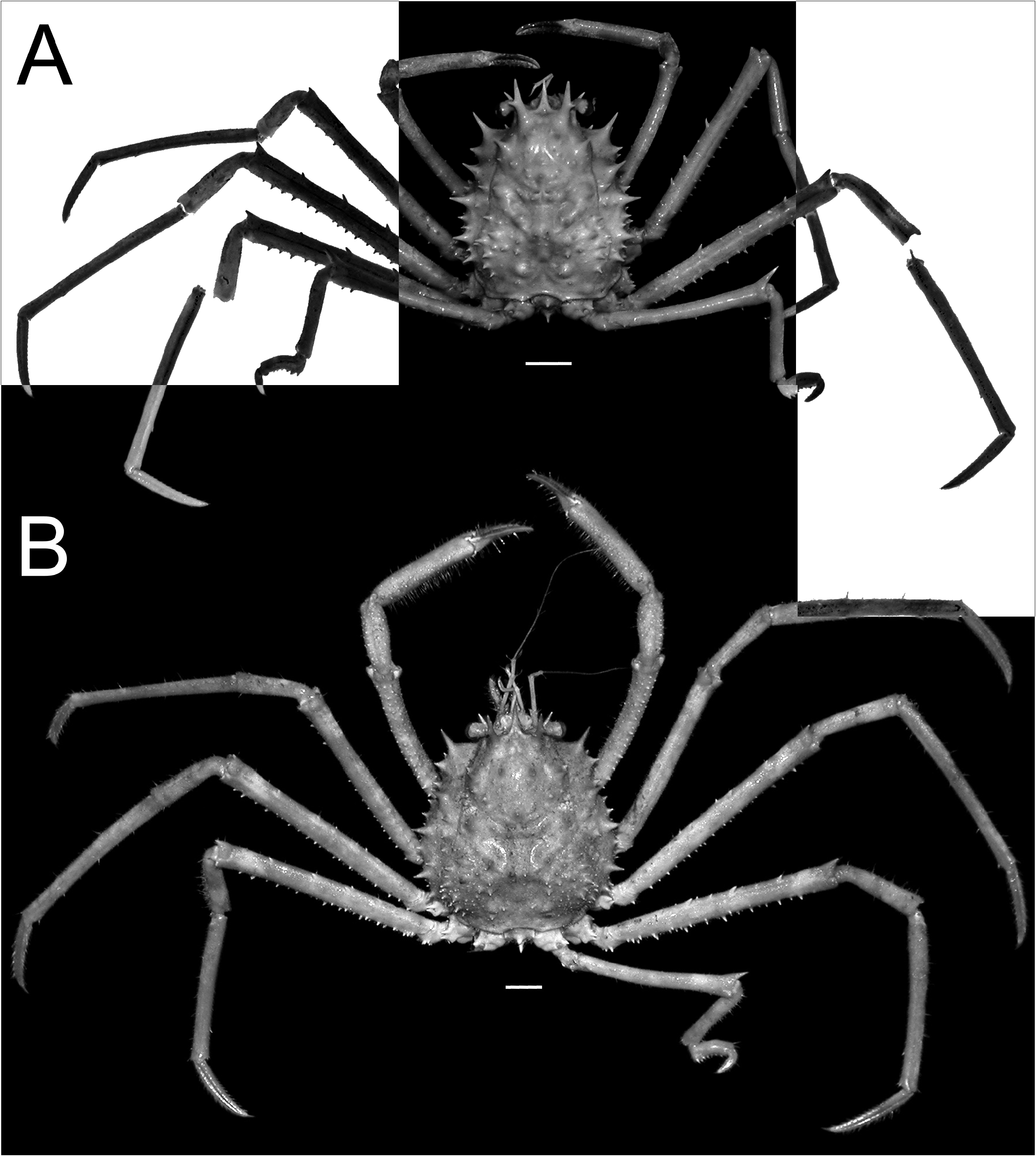
Fig. 1. Overall habitus. A. Moloha grandperrini Guinot & Richer de Forges, 1995. Holotype, ♂ (cl 46.5 mm, cw 39.0 mm) (NHM 1948.9.7.27), Maldives. B. Moloha tumida sp. nov. Holotype, ♂ (cl 55.7 mm, cw 50.6 mm) (DABFUK), Kerala, India. Scale bars = 10 mm.
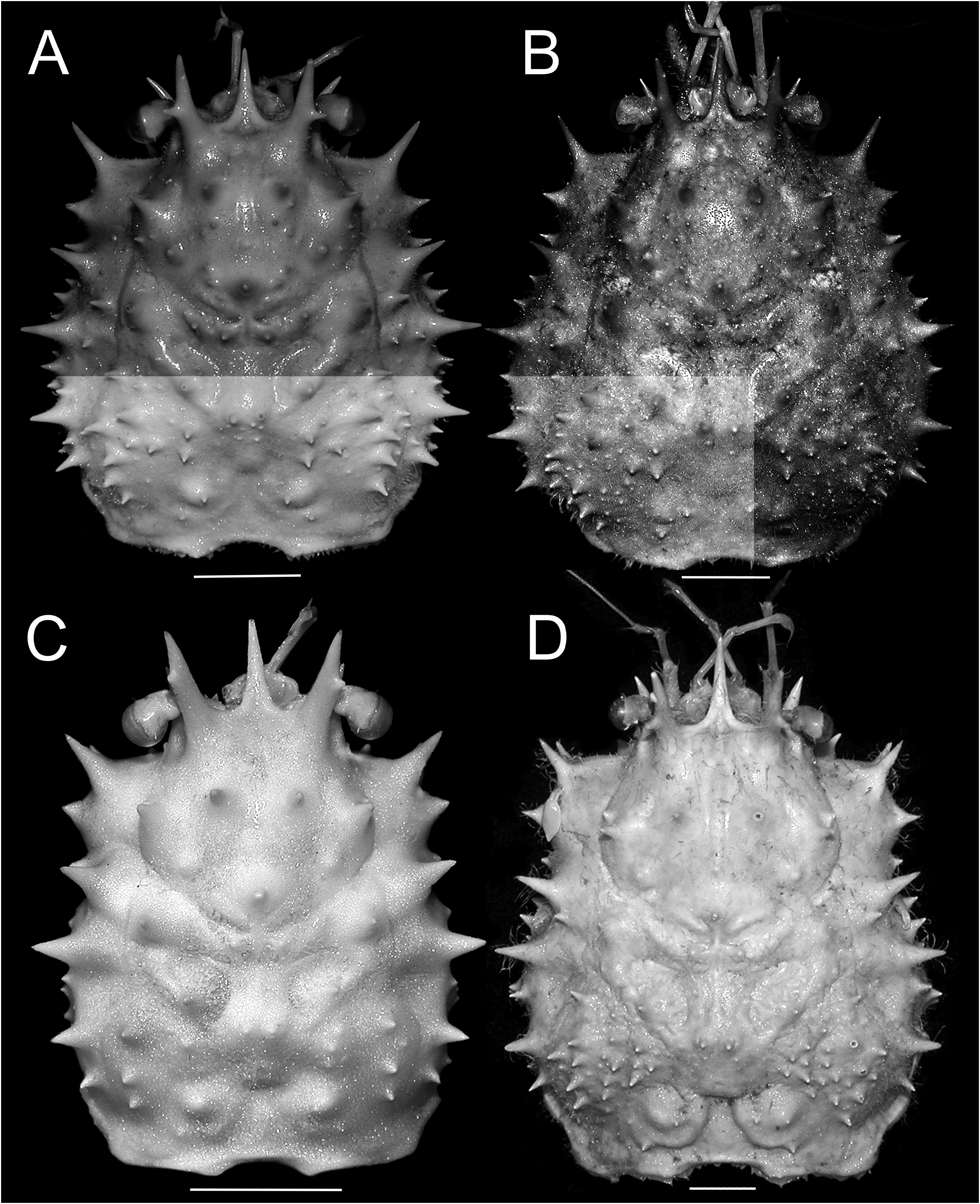
Fig. 3. Dorsal view of carapace. A. Moloha grandperrini Guinot & Richer de Forges, 1995. Holotype, ♂ (cl 46.5 mm, cw 39.0 mm) (NHM 1948.9.7.27), Maldives. B. Moloha tumida sp. nov. Holotype, ♂ (cl 55.7 mm, cw 50.6 mm) (DABFUK), Kerala, India. C. Moloha alisae Guinot & Richer de Forges, 1995. Holotype, ♂ (cl 36.1 mm, cw 29.7 mm) (MNHN-IU-2008-11077), Seychelles. D. Moloha alisae Guinot & Richer de Forges, 1995. ♂ (cl 67.7 mm, cw 56.2 mm) (ZRC 2008.1250a), South Africa. Scale bars = 10 mm.
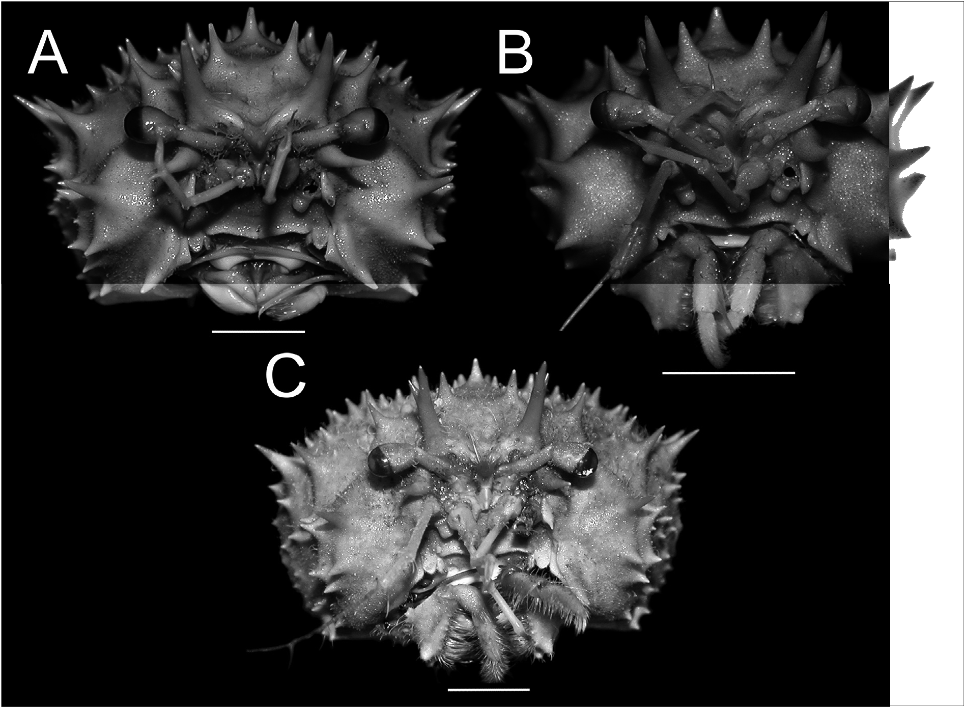
Fig. 4. Frontal view of cephalothorax. A. Moloha grandperrini Guinot & Richer de Forges, 1995. Holotype, ♂ (cl 46.5 mm, cw 39.0 mm) (NHM 1948.9.7.27), Maldives. B. Moloha alisae Guinot & Richer de Forges, 1995. Holotype, ♂ (cl 36.1 mm, cw 29.7 mm) (MNHN-IU-2008-11077), Seychelles. C. Moloha tumida sp. nov. Holotype, ♂ (cl 55.7 mm, cw 50.6 mm) (DABFUK), Kerala, India. Scale bars = 10 mm.
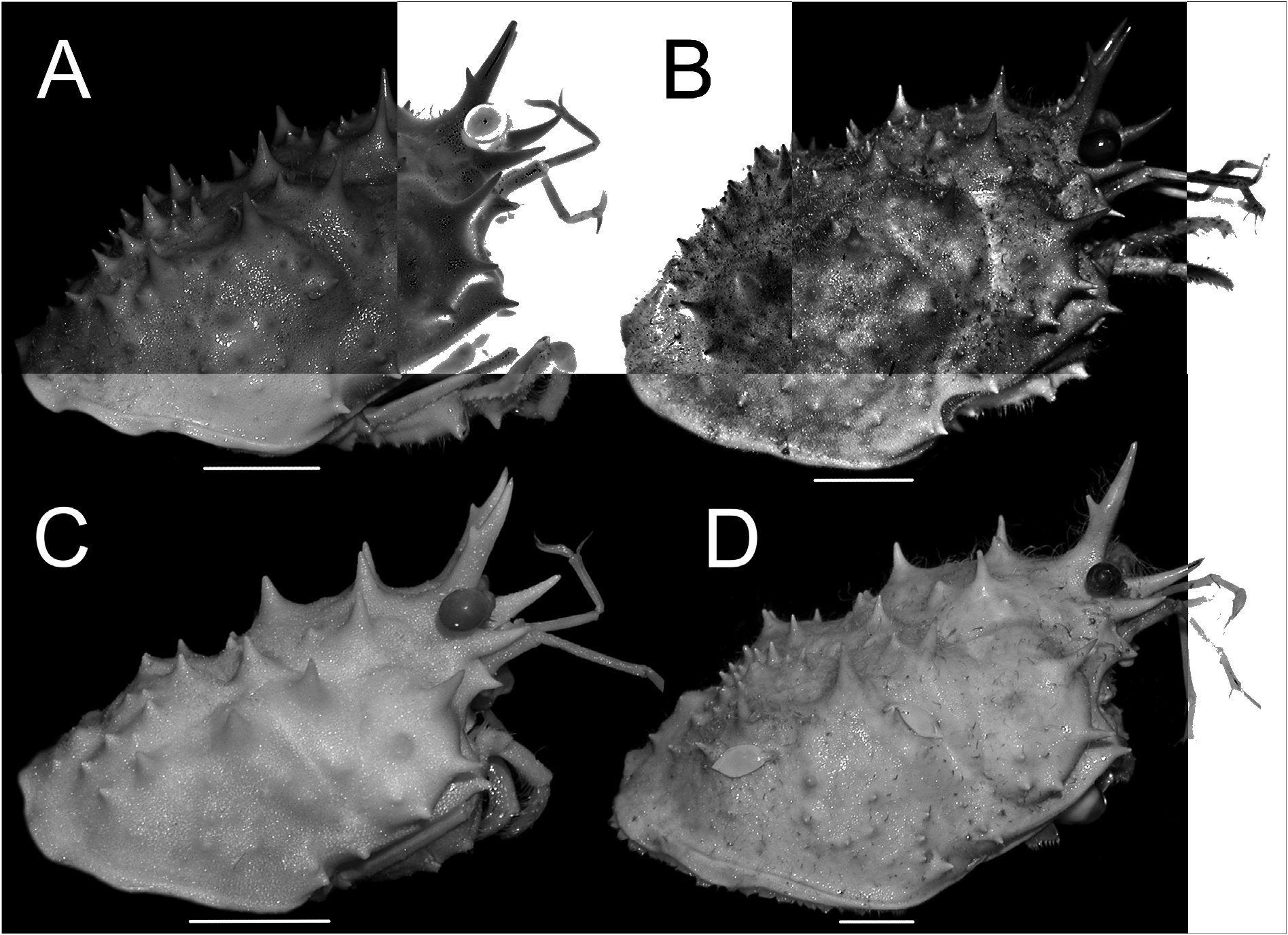
Fig. 5. Lateral view of cephalothorax. A. Moloha grandperrini Guinot & Richer de Forges, 1995. Holotype, ♂ (cl 46.5 mm, cw 39.0 mm) (NHM 1948.9.7.27), Maldives. B. Moloha tumida sp. nov. Holotype, ♂ (cl 55.7 mm, cw 50.6 mm) (DABFUK), Kerala, India. C. Moloha alisae Guinot & Richer de Forges, 1995. Holotype, ♂ (cl 36.1 mm, cw 29.7 mm) (MNHN-IU-2008-11077), Seychelles. D. Moloha alisae Guinot & Richer de Forges, 1995. ♂ (cl 67.7 mm, cw 56.2 mm) (ZRC 2008.1250a), South Africa. Scale bars = 10 mm.
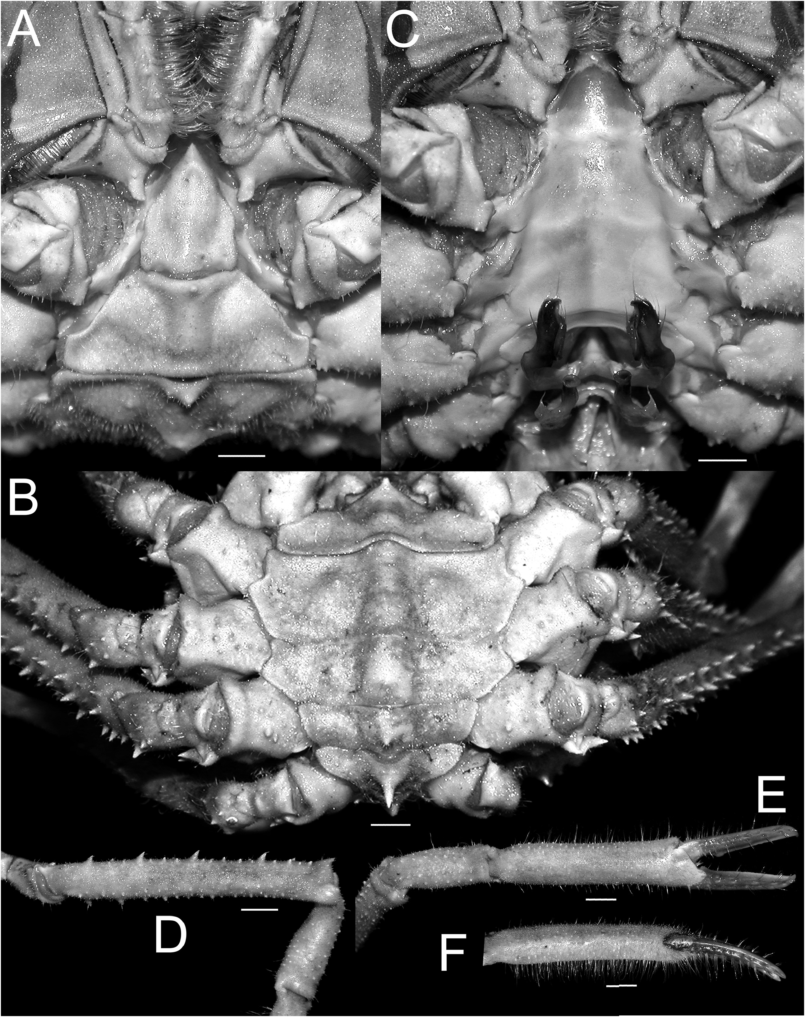
Fig. 9. Moloha tumida sp. nov. Holotype, ♂ (cl 55.7 mm, cw 50.6 mm) (DABFUK), Kerala, India. A. Telson and pleonal somites 5 and 6. B. Pleonal somites 2–6. C. Sternopleonal cavity. D. Merus and carpus of right cheliped. E. Outer view of right chela. F. Ventro-marginal view of right chela. Scale bars = 5 mm.

Fig. 13. Moloha tumida sp. nov. Holotype, ♂ (cl 55.7 mm, cw 50.6 mm) (DABFUK), Kerala, India. A. Right third maxilliped. B–D. Left P2–P4, respectively. E–H. Right P2–P5, respectively. I. Right P5 subchelate dactylus and propodus. Scale bars = 10 mm.

Fig. 14. Dorsal view of carapace showing position of folded right P5. A. Moloha grandperrini Guinot & Richer de Forges, 1995. Holotype, ♂ (cl 46.5 mm, cw 39.0 mm) (NHM 1948.9.7.27), Maldives. B. Moloha tumida sp. nov. Holotype, ♂ (cl 55.7 mm, cw 50.6 mm) (DABFUK), Kerala, India. C. Moloha alisae Guinot & Richer de Forges, 1995. Holotype, ♂ (cl 36.1 mm, cw 29.7 mm) (MNHN- IU-2008-11077), Seychelles. D. Moloha alisae Guinot & Richer de Forges, 1995. ♂ (cl 67.7 mm, cw 56.2 mm) (ZRC 2008.1250a), South Africa. Scale bars = 10 mm.
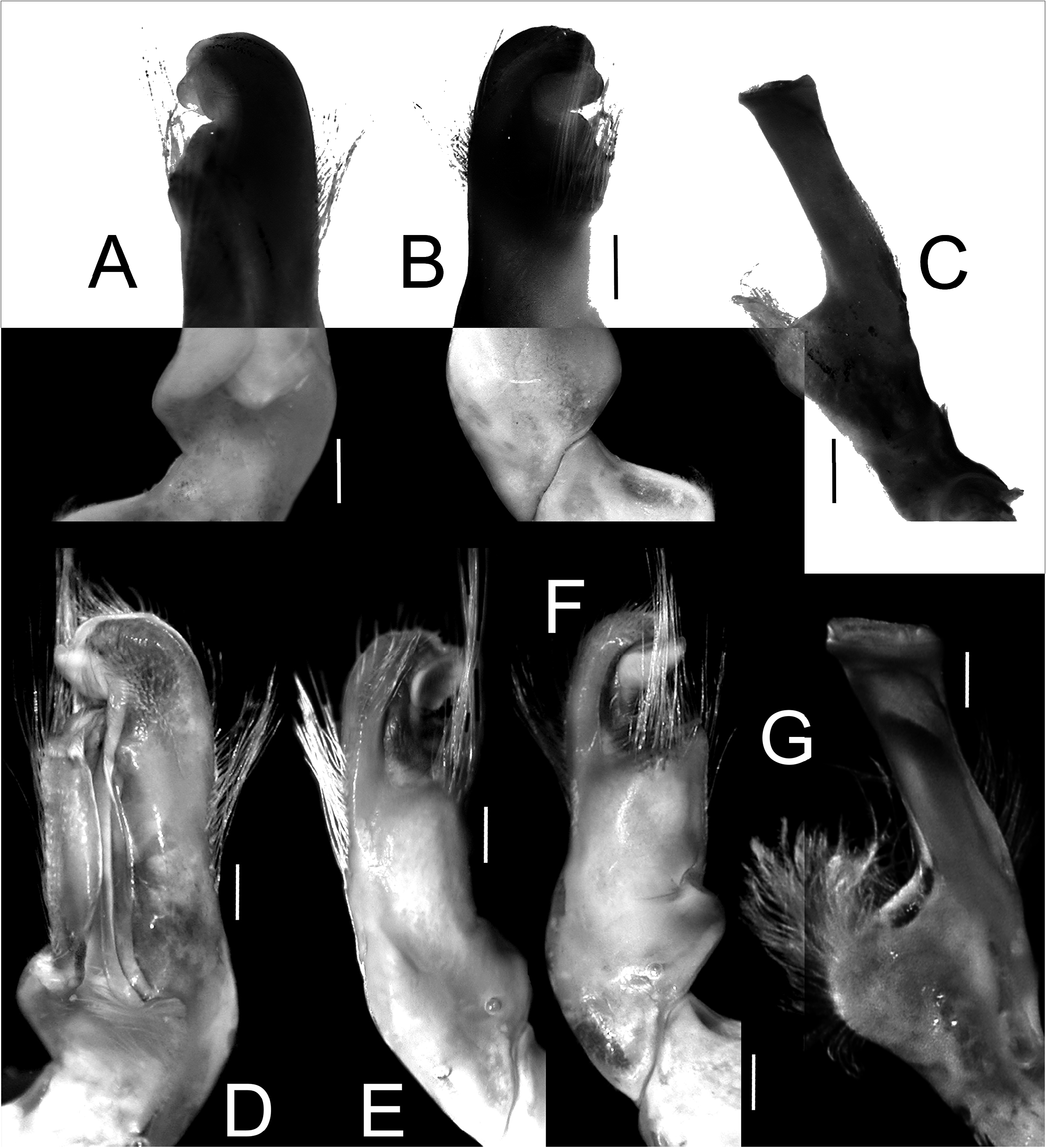
Fig. 15. Gonopods. A–C. Moloha grandperrini Guinot & Richer de Forges, 1995. Holotype, ♂ (cl 46.5 mm, cw 39.0 mm) (NHM 1948.9.7.27), Maldives. D–G. Moloha tumida sp. nov. Holotype, ♂ (cl 55.7 mm, cw 50.6 mm) (DABFUK), Kerala, India. — A, D. Ventral view of left G1. B, F. Dorsal view of left G1. E. Outer-lateral view of left G1. C, G. Left G2. Scale bars = 1 mm.
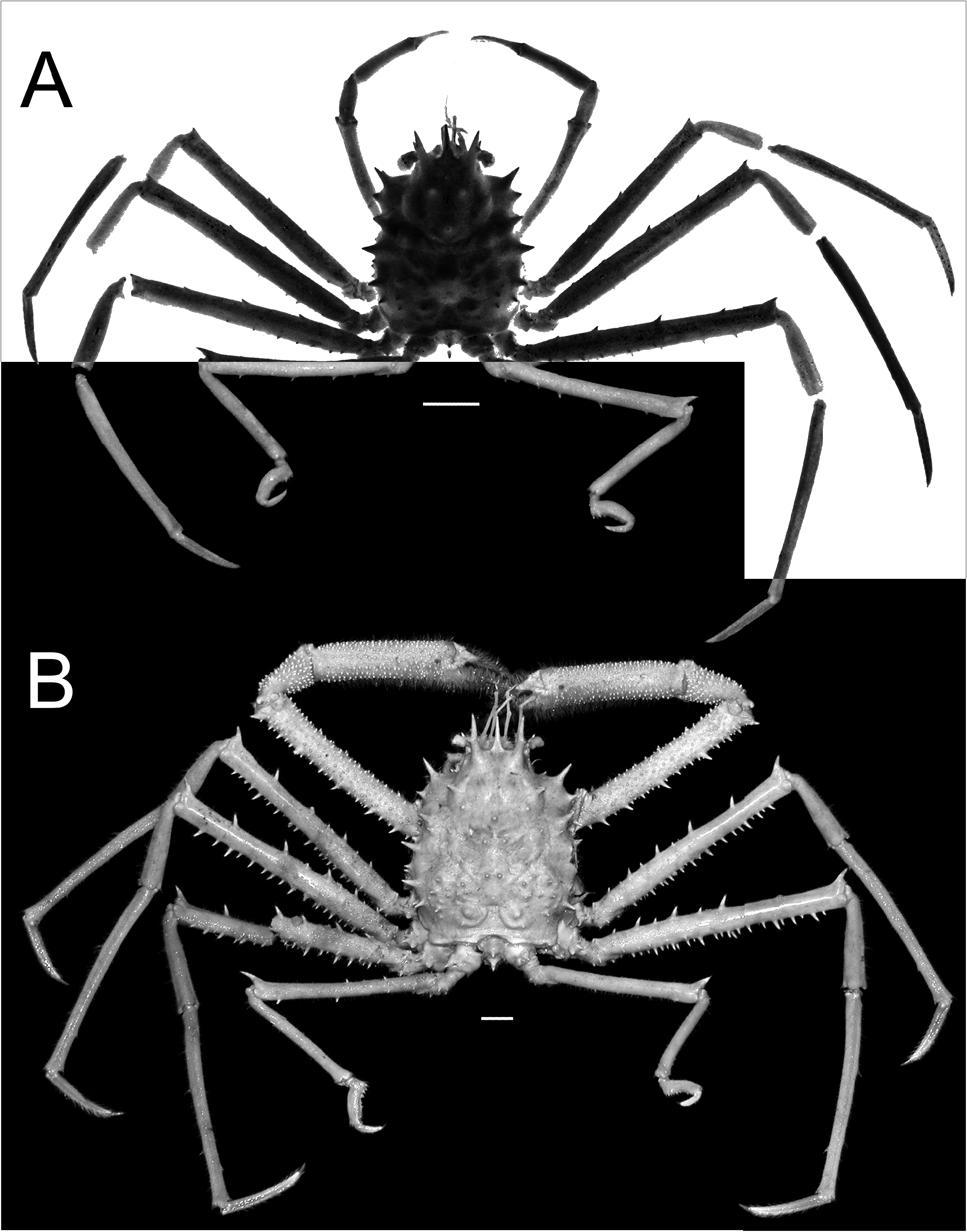
Fig. 2. Overall habitus. Moloha alisae Guinot & Richer de Forges, 1995. A. Holotype, ♂ (cl 36.1 mm, cw 29.7 mm) (MNHN-IU-2008-11077), Seychelles. B. ♂ (cl 67.5 mm, cw 56.5 mm) (ZRC 2008.1250b), South Africa. Scale bars = 10 mm.

Fig. 17. Colour in life, dorsal view. A. Moloha alisae Guinot & Richer de Forges, 1995. ♂ (cl 67.7 mm, cw 56.2 mm) (ZRC 2008.1250a), South Africa [photograph by Sean Fennessy]. B. Moloha tumida sp. nov. Holotype, ♂ (cl 55.7 mm, cw 50.6 mm) (DABFUK), India [photograph by Biju Kumar]. Scale bars = 20 mm.
No known copyright restrictions apply. See Agosti, D., Egloff, W., 2009. Taxonomic information exchange and copyright: the Plazi approach. BMC Research Notes 2009, 2:53 for further explanation.
|
Kingdom |
|
|
Phylum |
|
|
Class |
|
|
Order |
|
|
InfraOrder |
Brachyura |
|
SuperFamily |
Homoloidea |
|
Family |
|
|
Genus |
1 (by carolina, 2020-05-06 18:00:21)
2 (by ExternalLinkService, 2020-05-06 18:05:25)
3 (by ExternalLinkService, 2020-05-06 18:20:10)
4 (by ExternalLinkService, 2020-05-06 20:18:49)
5 (by ExternalLinkService, 2020-05-08 04:19:42)
6 (by ExternalLinkService, 2021-11-09 19:35:04)
7 (by ExternalLinkService, 2021-11-10 11:43:08)
8 (by plazi, 2023-10-31 12:37:19)
9 (by ExternalLinkService, 2023-11-01 11:38:37)