
Acanthochitona sp.
|
publication ID |
https://doi.org/10.11646/zootaxa.2673.1.1 |
|
persistent identifier |
https://treatment.plazi.org/id/03D7EE78-FFC0-FF9E-FF7F-FEBEFBF7B3FE |
|
treatment provided by |
Felipe (2021-08-23 07:23:52, last updated by Plazi 2023-11-04 11:46:51) |
|
scientific name |
Acanthochitona sp. |
| status |
|
( Figures 7E–G View FIGURE 7 )
Material examined: LB05: 2 intermediate valves.
Description: Intermediate valves twice as wide as long, front margin slightly convex at both sides of concave jugal part, hind margin concave at both sides of slightly protruding apex, jugal area large and smooth, latero-pleural area sculptured with neatly separated, roundish to oval, raised granules, convex at the top, with many aesthetes (from 6 to 14, not differentiated in megalaesthetes and micraesthetes) irregularly arranged on the whole surface and the periphery of the granules.
Remarks: The two intermediate valves from LB05 cannot be attributed with certainty to the other North Indian Ocean Acanthochitona , mainly for the lack of informations on the granules structures of these species. Based on the available characters (the shape of intermediate valves and the granules structure), this species is different from the other Maldive Acanthochitona species , A. penicillata (see the preceding species).
A. mahensis Winckworth, 1927 known from Red Sea, India (Madras), Seychelles and Andaman Islands ( Anseeuw & Terryn 2004) and A. penetrans Winckworth, 1933 known from Andaman Islands have a different structure of aesthetes ( Leloup 1941: pl. 3, Fig. 1B View FIGURE 1 ; pl. 3, Fig. 2B View FIGURE 2 ).
Notoplax curvisetosa (Leloup, 1960) known from Red Sea, Madagascar and Glorieuses Islands (Dell’Angelo et al. 2004) has rather conspicuous polygonal granules in the latero-pleural areas.
A. jugotenuis Kaas, 1979 known from Mozambique has a different shape of intermediate valves, the jugal area is finely longitudinally ribbed, and latero-pleural areas are densely granulose ( Kaas 1979).
A. quincunx Leloup, 1981 known from Madagascar has a particular granules structure, with small hemispherical granules quincuncially arranged ( Leloup 1981).
A. limbata Kaas, 1986 known from Madagascar has a different granules structure (Dell’Angelo et al. 2004: pl. 6, figs 3–4).
A. woodwardi Kaas & Van Belle, 1988 known from Kuwait and Qatar has the jugal area sculptured with about five weak, flat longitudinal ribs ( Kaas & Van Belle 1988). Unfortunately the structure of the granules is unknown.
Notoplax arabica Kaas & Van Belle, 1988 known from Kuwait and Qatar, has a peculiar sculpture of large, elongate, convex, widely spaced granules ( Kaas & Van Belle 1988).
Acanthochitona mastalleri Leloup MS, Strack, 1989 known from Red Sea, Oman and Kenya ( Anseeuw & Terryn 2004), is a very different species, with the tegmentum reduced in lateropleural areas and the overlapping dorsal girdle.
Hong et al. (1990) report Acanthochitona defilippii (Tapparone-Canefri, 1874) for the Maldive Islands, basing on a preceding mention of Is. Taki (1962: 35 “Sylvia Bay, Maldive Archipelago, Indian Ocean”). A. defilippii is a different species, characterized by intermediate valves of cup-like form, beaked, not comparable with the material examined. The report of this species for the Maldive Islands remains probably a misinterpretation, Sylvia Bay is not present at our knowledge on the Maldive Islands maps, and there is not a word corresponding to “Bay” in the Maldivian vocabulary.
Distribution: Acanthochitona sp. is known only from the Maldive Islands.
Anseeuw, B. & Terryn, Y. (2004) Intertidal chitons (Mollusca: Polyplacophora) from the coast of Jordan, Red Sea, with the description of a new species of Parachiton Thiele, 1909. Bollettino Malacologico, suppl. 5 (2003), 1 - 24.
Kaas, P. (1979) The Chitons (Mollusca: Polyplacophora) of Mozambique. Annals of the Natal Museum, 23, 855 - 879.
Kaas, P. (1986) Revision of the chitons (Mollusca: Polyplacophora) from the coral-reefs of Tulear, SW Madagascar, and of the Mascarene Islands. Mesogee, 46, 9 - 23.
Kaas, P. & Van Belle, R. A. (1988) Chitons (Mollusca: Polyplacophora) from the coasts of Oman and the Arabian Gulf. American Malacological Bulletin, 6, 115 - 130.
Leloup, E. (1941) A propos de quelques Acanthochitons peu connus ou nouveaux, 1. - Region Indo-Pacifique. Bulletin du Musee royal d'Histoire naturelle de Belgique, 17 (24), 1 - 19, 3 pls.
Leloup, E. (1981) Chitons de Tulear, Reunion, Maurice et Tahiti. Bulletin Institut Royale des Sciences Naturelles de Belgique, 53 (3), 1 - 46, pls 1 - 4.
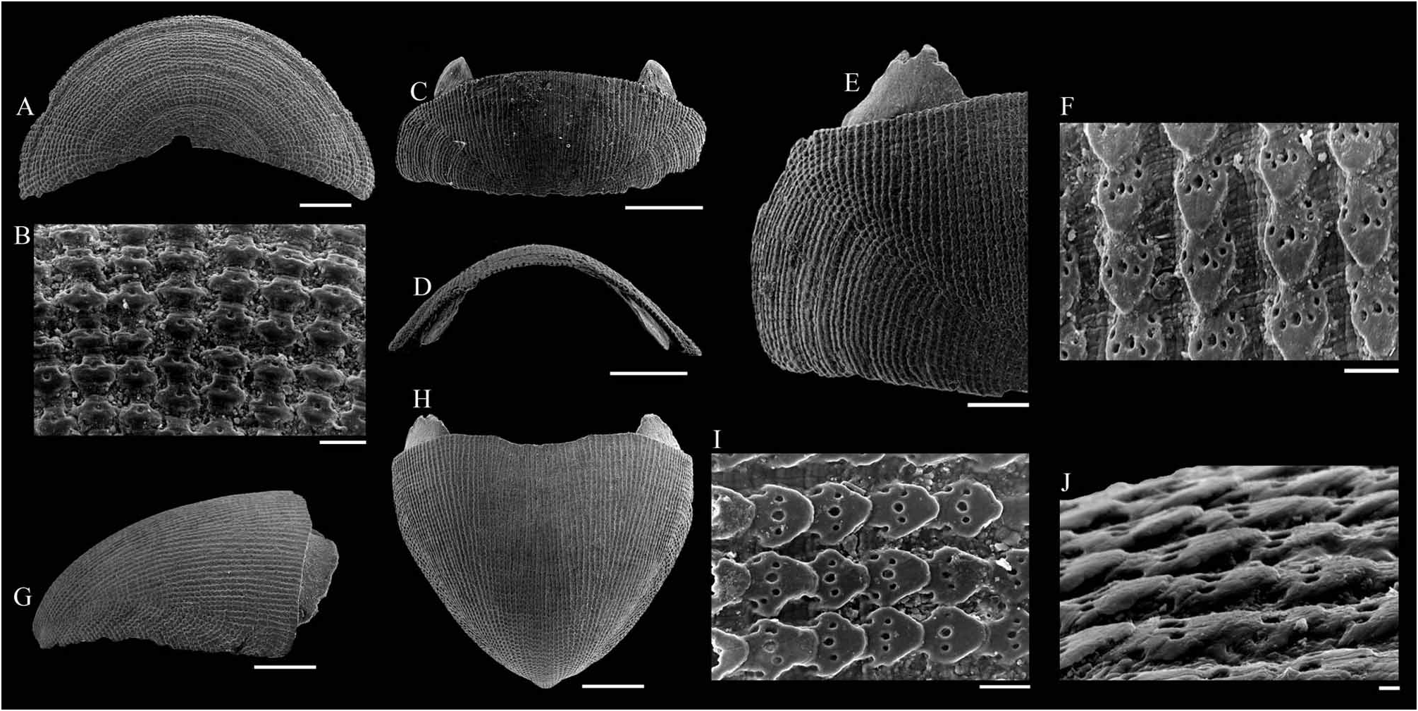
FIGURE 1A–J, Parachiton hylkiae mauricejayi Schwabe, 2002, Maldive. A–B, head valve detached from a living specimen, MLD02A. A, dorsal view. B, detail of the sculpture. C–D, I–J, intermediate valve detached from a living specimen, MLD02A. C, dorsal view. D, frontal view. I, detail of the sculpture of the central area, granules in the longitudinal chains. J, the same, by a lateral view. E–F, intermediate valve, M65. E, dorsal view of the half side. F, detail of the sculpture of the central area. G–H, tail valve, M65. G, lateral view. H, dorsal view. Scale bars 1 mm: C, D, G, H; 500 µm: A, E; 50 µm: B, F, I; 10 µm: J.
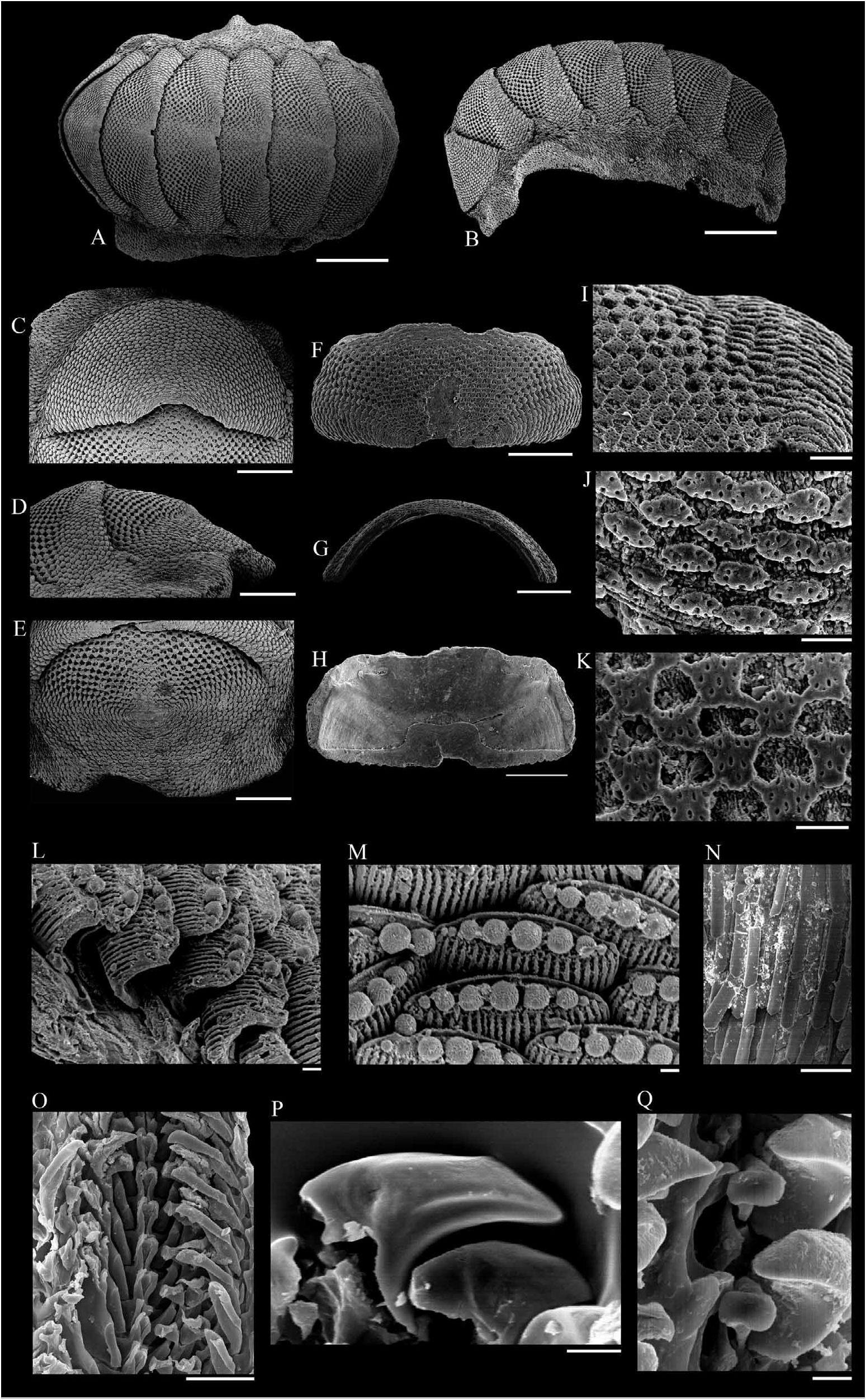
FIGURE 2A–Q, Ischnochiton albinus Thiele, 1911, Maldive, M13. A–E, L–Q, living specimen. A–B, dorsal and lateral view (respectively) of a complete specimen. C, close-up of the head valve. D, lateral view of valves vii-viii. E, close-up of the tail valve. L, perinotum elements in situ. M, detail of the perinotum elements, showing the sphaerules along the distal margin in situ. N, hyponotum elements in situ. O, radula. P, an isolated blade of the second lateral tooth. Q, detail of the central teeth. F–K, intermediate valve. F, dorsal view. G, frontal view. H, ventral view. I, detail of the sculpture, showing the contact between the right lateral and central areas. J, lateral view of elongate granules of the lateral area. K, the same, by a dorsal view. Scale bars 1 mm: A, B; 500 µm: C–H; 100 µm: I; 50 µm: J, K, N, O; 10 µm: L, M, P, Q.
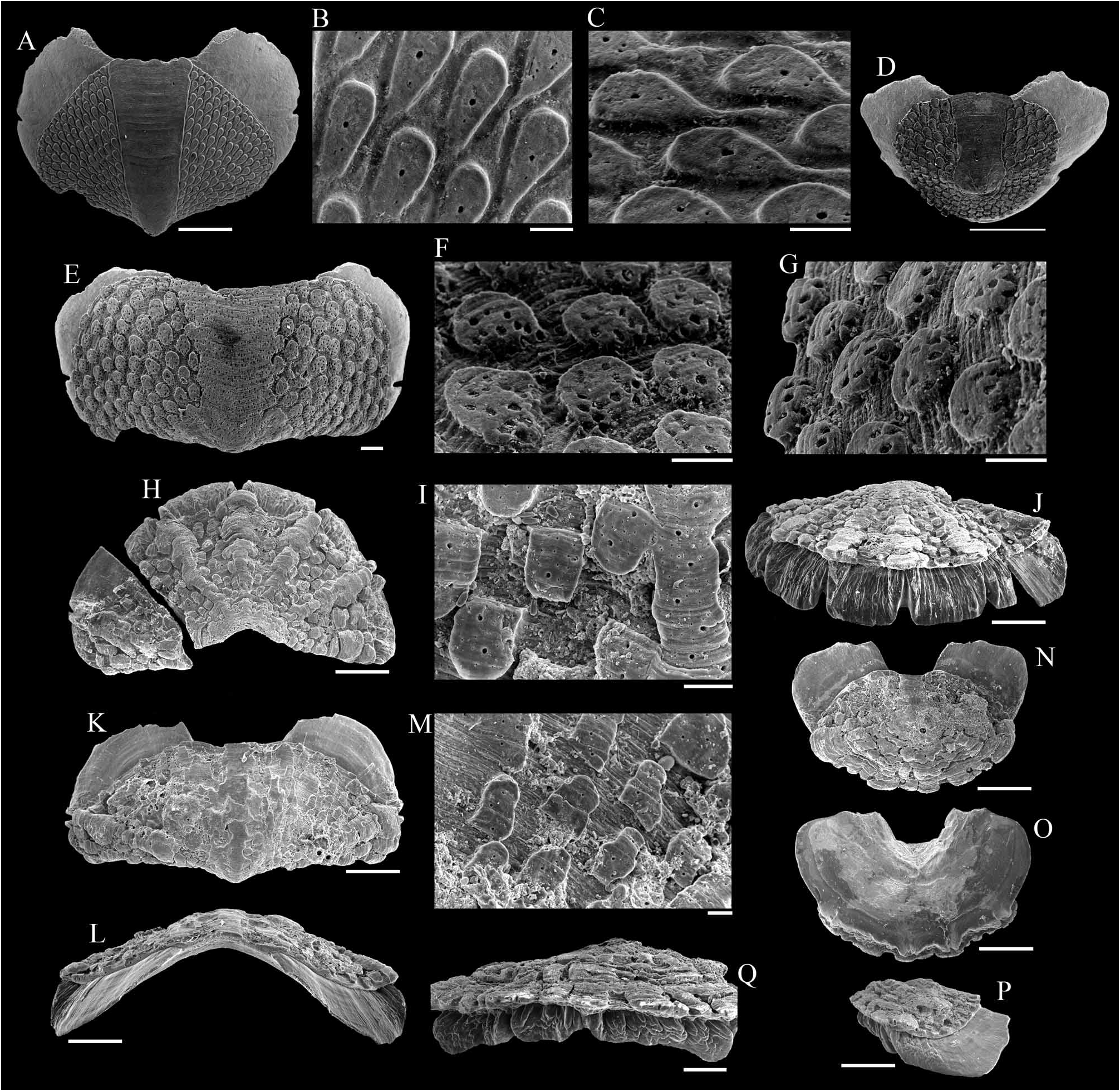
FIGURE 7A–D, Acanthochitona penicillata (Deshayes, 1863), Maldive. A–C, intermediate valve, MLD02A. A, dorsal view. B–C, granules, dorsal and lateral views, respectively. D, dorsal view of tail valve, LB05,. E–G, Acanthochitona sp., LB05, intermediate valve. E, dorsal view. F–G, granules, dorsal and lateral views. H–Q, Craspedochiton cf. laqueatus (Sowerby, 1842), Maldive, valves dismounted from living specimen, M59. H–J, head valve. H, dorsal view. I, detail of the sculpture. J, frontal view. K–M, intermediate valve. K, dorsal view. L, frontal view. M, detail of the sculpture. N–Q, tail valve. N, dorsal view. O, ventral view. P, lateral view. Q, posterior view. Scale bars 1 mm: H, J–L, N–P; 500 µm: A, D, Q; 100 µm: E, I, M; 50 µm: B, C, F, G.
No known copyright restrictions apply. See Agosti, D., Egloff, W., 2009. Taxonomic information exchange and copyright: the Plazi approach. BMC Research Notes 2009, 2:53 for further explanation.
|
Kingdom |
|
|
Phylum |
|
|
Class |
|
|
Order |
|
|
Family |
|
|
Genus |