
Matinta tatianae, Matos & Ruiz, 2023
|
publication ID |
https://doi.org/10.11646/zootaxa.5343.2.2 |
|
publication LSID |
lsid:zoobank.org:pub:63876BA9-D973-40E1-BB4B-03F60CD088A7 |
|
DOI |
https://doi.org/10.5281/zenodo.8324691 |
|
persistent identifier |
https://treatment.plazi.org/id/03D587F3-F154-FFDF-FF79-A2F2FC0CE8FA |
|
treatment provided by |
Plazi (2023-09-07 07:04:19, last updated 2024-11-27 01:47:46) |
|
scientific name |
Matinta tatianae |
| status |
sp. nov. |
Matinta tatianae sp. nov.
Figs 62–68 View FIGURES 60–65 View FIGURES 66–68 , 81 View FIGURES 75–81 , 92 View FIGURES 82–92
Type material. Holotype: ♁ from Reserva Extrativista de Catuaba [10.067⁰S 67.633⁰W], Senador Guimard, Acre, Brazil, 2003, E. Morato leg. ( IBSP 161431 View Materials ) . Paratype: 1♁ from Platô Miltônia 3, Hydro-Alunorte extraction area (3.222⁰S 47.754⁰W), Paragominas, Pará, Brazil, unknown date, A. Viana-Junior leg. ( MPEG 37347 View Materials ) .
Etymology. Species named in honor of Tatiana de Carvalho, a Greenpeace activist who died in 2012. She was honored by Greenpeace in 2020, upon the creation of the Tatiana de Carvalho Program for Research and Conservation of the Amazon, which supported the first author of this work during the development of her master’s Dissertation.
Diagnosis. This species is similar to the three species previously included in the saperda species-group. They all have no tegular slit, acute embolus and exposed subtegulum on the retrolateral side on the palpal bulb. Matinta procax , M. pardo and M. saperda have simple RTA and poorly developed RvTA. Matinta tatianae can be distinguished from them for having a dorsally directed RTA and a developed RvTA bearing a ventrally directed apophysis ( Fig. 65 View FIGURES 60–65 ). Also, M. tatianae has the largest tegulum in the species-group ( Fig. 64 View FIGURES 60–65 ).
Description. Male ( Figs 66–67 View FIGURES 66–68 ). Total length: 7.71. Carapace dark brown, 3.84 long, 2.92 wide and 1.77 high. Ocular area 2.15 long, with white scales on sides. Anterior eye row 2.48 and posterior 2.19 wide. Chelicera dark brown, strongly divergent, with paracondylic projection ( Figs 68 View FIGURES 66–68 , 81 View FIGURES 75–81 ); PMT: 2, RMT: 5, PIMT: 4, RIMT: 4 ( Fig. 92 View FIGURES 82–92 ). Palp ( Figs 62–65 View FIGURES 60–65 ) light brown, with filiform embolus emerging prolaterally from tegulum. Sternum light brown. Legs 1342; I: with proximal femur yellow, rest light brown; II–IV: light brown. Length: Leg I 9.98 (femur: 2.77; patella: 1.67; tibia: 2.83; metatarsus: 1.76 and tarsus: 0.95); II 7.64 (2.51; 1.22; 2.01; 1.15; 0.75); III 8.70 (2.97; 1.43; 1.90; 1.45; 0.95); IV 7.69 (2.69; 0.88; 1.75; 1.65; 0.72). Leg spination: femur I–IV d1-1-1, p0-0-2, r0-0-1; patella I–II p0-1-0, r0; III–IV p0-1-0, r0-1-0; tibia I v2-2-2, p1-0-0, r0; II v1r-2-2, p1-1-1, r0; III v1p-0-2, p1-1-1, r1-1-1; IV v1p-0-2; p0-1-1, r1-1-1; metatarsus I–II v2-2; III v2-0-2, p1-0-2, r2-0-2; IV v1p-0-2, p2-0-2, r1-1-2. Abdomen dorsally and ventrally cream-colored ( Fig. 66 View FIGURES 66–68 ), with poorly conspicuous ventral dark stripe ( Fig. 67 View FIGURES 66–68 ). Spinnerets light brown.
Female. Unknown.
Distribution. Known from the states of Acre and Pará ( Brazil).
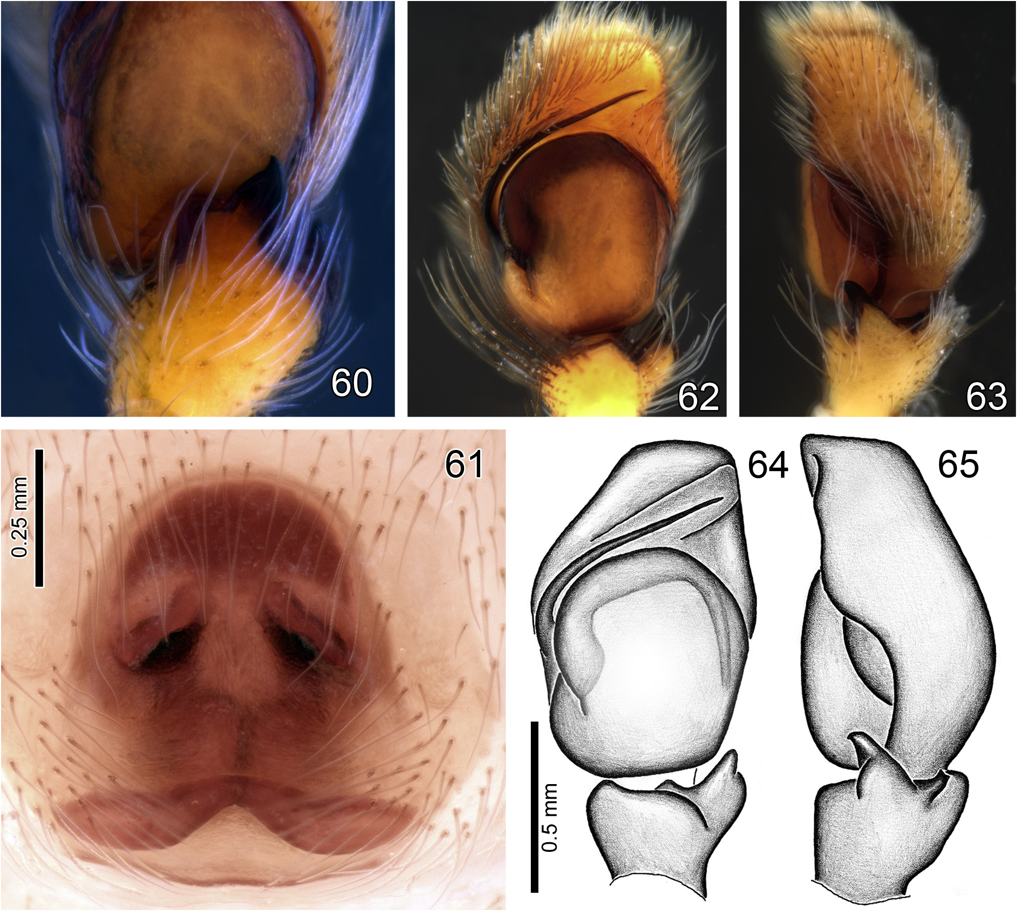
FIGURES 60–65. Matinta spp., left male palps and epigyne. 60 M. vicana Simon, male palp, retroventral view, detail of RvTA. 61 M. silvae Crane, epigyne, ventrral view. 62–65 M. tatianae sp. nov., left male palp (62 ventral view; 63 retrolateral view; 64 ventral view; 65 retrolateral view).

FIGURES 66–68. M. tatianae sp. nov. 66–67 male holotype (66 dorsal view; 67 ventral view). 68 male paratype from Paragominas, laterofrontal view.
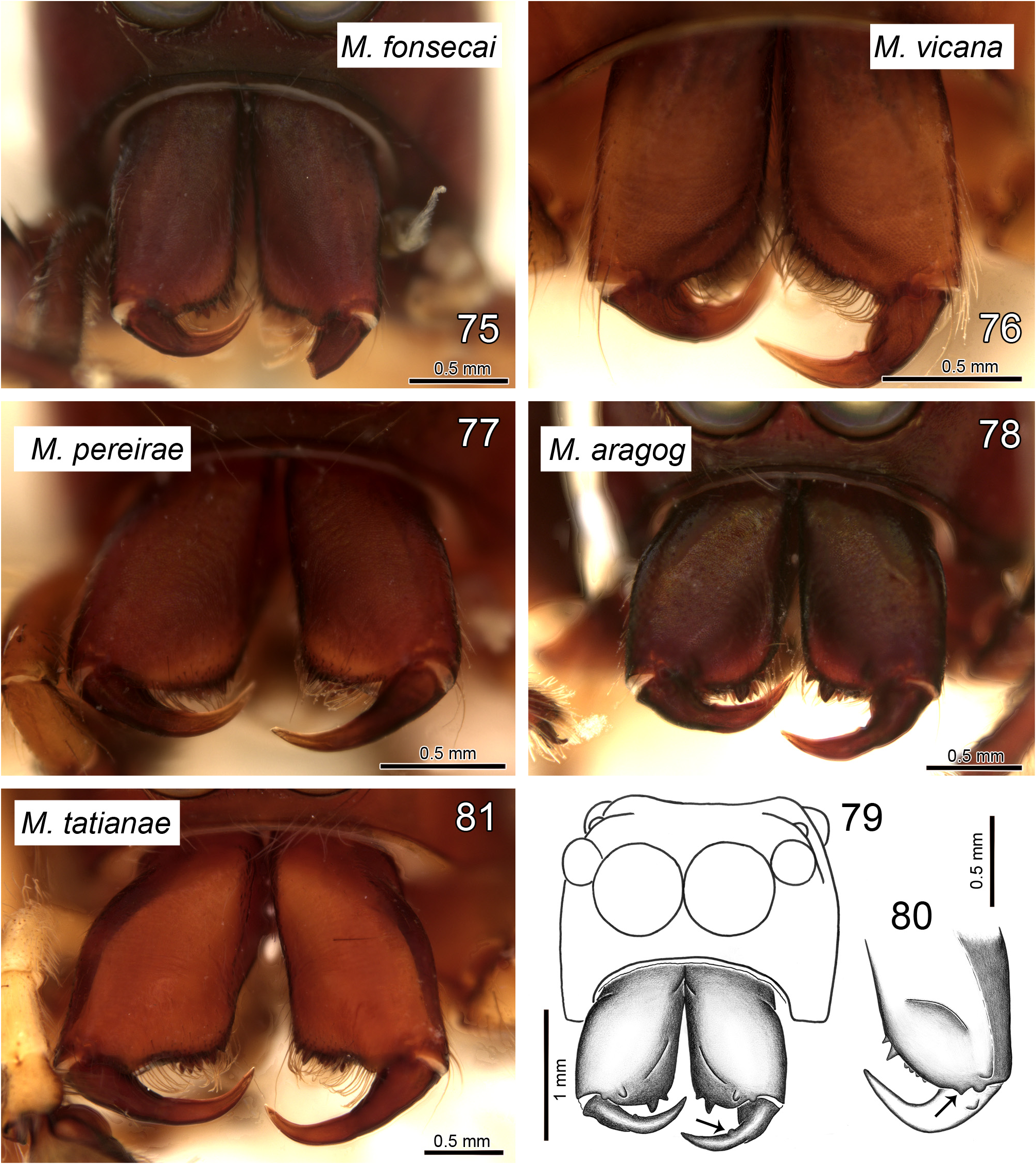
FIGURES 75–81. Matinta spp., male chelicerae, frontal view. 75 M. fonsecai Soares & Camargo. 76 M. vicana Simon. 77 M. pereirae sp. nov. 78–79 M. aragog sp. nov. 80 M. maddisoni sp. nov. 81 M. tatianae sp. nov. Arrow in 79 shows bump on fang and in 80 the paracondylic projection.
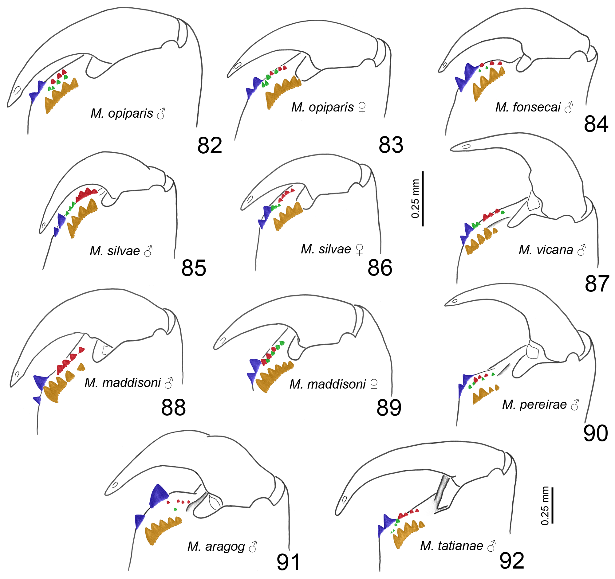
FIGURES 82–92. Matinta spp., left chelicera teeth, retrolateral view. 82–83 M. opiparis Simon (82 male; 83 female). 84 M. fonsecai Soares & Camargo, male. 85–86 M. silvae Crane (85 male; 86 female). 87 M. vicana Simon, male. 88–89 M. maddisoni sp. nov. (88 male; 89 female). 90 M. pereirae sp. nov., male. 91 M. aragog sp. nov., male. 92 M. tatianae sp. nov., male. All illustrations in same scale, except for M. tatianae (92). Colors indicate tooth rows: promarginal (blue), retromarginal (orange), prointermarginal (red) and retrointermarginal (green).
No known copyright restrictions apply. See Agosti, D., Egloff, W., 2009. Taxonomic information exchange and copyright: the Plazi approach. BMC Research Notes 2009, 2:53 for further explanation.
|
Kingdom |
|
|
Phylum |
|
|
Class |
|
|
Order |
|
|
Family |
|
|
Genus |