
Matinta aragog, Matos & Ruiz, 2023
|
publication ID |
https://doi.org/10.11646/zootaxa.5343.2.2 |
|
publication LSID |
lsid:zoobank.org:pub:63876BA9-D973-40E1-BB4B-03F60CD088A7 |
|
DOI |
https://doi.org/10.5281/zenodo.8329843 |
|
persistent identifier |
https://treatment.plazi.org/id/03D587F3-F150-FFDA-FF79-A6AAFC0BEB4A |
|
treatment provided by |
Plazi (2023-09-07 07:04:19, last updated 2024-11-27 01:47:46) |
|
scientific name |
Matinta aragog |
| status |
sp. nov. |
Matinta aragog sp. nov.
Figs 29–30 View FIGURES 27–30 , 42–43 View FIGURES 31–43 , 49 View FIGURES 44–49 , 55 View FIGURES 50–55 , 78–79 View FIGURES 75–81 , 91 View FIGURES 82–92
Type material. Holotype: ♁ from French Guiana, Réserve Naturelle National de la Trinité (4.583⁰N 53.3⁰W), 25.X.2008, C. Courtial leg. ( MNHN).
Etymology. The epithet is given in apposition honoring the giant spider present in Harry Potter’s series.
Diagnosis. The male of M. aragog is similar to those of the species within the vicana species-group, especially those of M. maddisoni for having mastidia, separate prolateral cheliceral teeth, a bump on the cheliceral fang and a more symmetrical palpal bulb. It can be distinguished from M. maddisoni for having the bifid embolus tip ( Figs 42 View FIGURES 31–43 , 49 View FIGURES 44–49 ) and a triangular RTA in retrolateral view ( Figs 43 View FIGURES 31–43 , 55 View FIGURES 50–55 ; squared in M. maddisoni ).
Description. Male ( Figs 29–30 View FIGURES 27–30 ). Total length: 6.02. Carapace dark reddish-brown, 3.11 long, 2.27 wide and 1.63 high. Ocular area 1.27 long, with white scales between posterior lateral and median eyes. Anterior eye row 1.92 and posterior 1.74 wide. Chelicera dark brown, with a mastidion and a bump on the fang ( Figs 78–79 View FIGURES 75–81 ); PMT: 2, RMT: 5, PIMT: 4, RIMT: 1 ( Fig. 91 View FIGURES 82–92 ). Palp ( Figs 42–43 View FIGURES 31–43 , 49 View FIGURES 44–49 , 55 View FIGURES 50–55 ) light brown. Sternum yellow. Legs 1324; I: dark brown; II–III: femur proximally yellow, distally dark brown, other articles dark brown; IV: brown. Leg length: I 6.72 (femur: 2.06; patella: 0.88; tibia: 1.90; metatarsus: 1.10; tarsus: 0.78); II 5.56 (1.72; 1.02; 1.38; 0.82; 0.62); III 6.33 (2.23; 1.00; 1.33; 1.17; 0.60); IV 5.47 (1.70; 0.70; 1.27; 1.19; 0.61). Leg spination: femur I d1-1-1, p0-1-2, r0-0-2; II d1-1-1, p0-0-2, r0-0-2; III d1-1-1, p0-1-2, r0-0-1; IV d1-1-1, p0-0-1, r0-0-1; patella I–II p0-1-0; III–IV p0-1-0, r0-1-0; tibia I v2-1r-2, p1-0-1, r0; II v1r-2-2, p1-0-1, r0; III v2-0-2, p1-0-0, r1-1-1; IV v1p-0-2; p1-0-0, r1-1-1; metatarsus I–II v2-2; III v2-0-2, p2-0-2, r2-0-2; IV v0-2, p2-0-2, r1-1-2. The dorsal region of the abdomen cream-colored with sparse dark brown markings ( Fig. 29 View FIGURES 27–30 ); ventrally with wide longitudinal dark brown stripe ( Fig. 30 View FIGURES 27–30 ). Spinnerets brown.
Female. Unknown. Note. Matinta fasciata (Mello-Leit„o, 1940) was described from Guyana, near the border with Venezuela. Given the distance between the two type localities, separated by 700–800 km, and the more conspicuous abdominal pattern in M. fasciata (see Mello-Leit„o 1940: fig. 25), we are describing the male of M. aragog as a new taxon. Distribution. Known only from type locality (French Guiana).
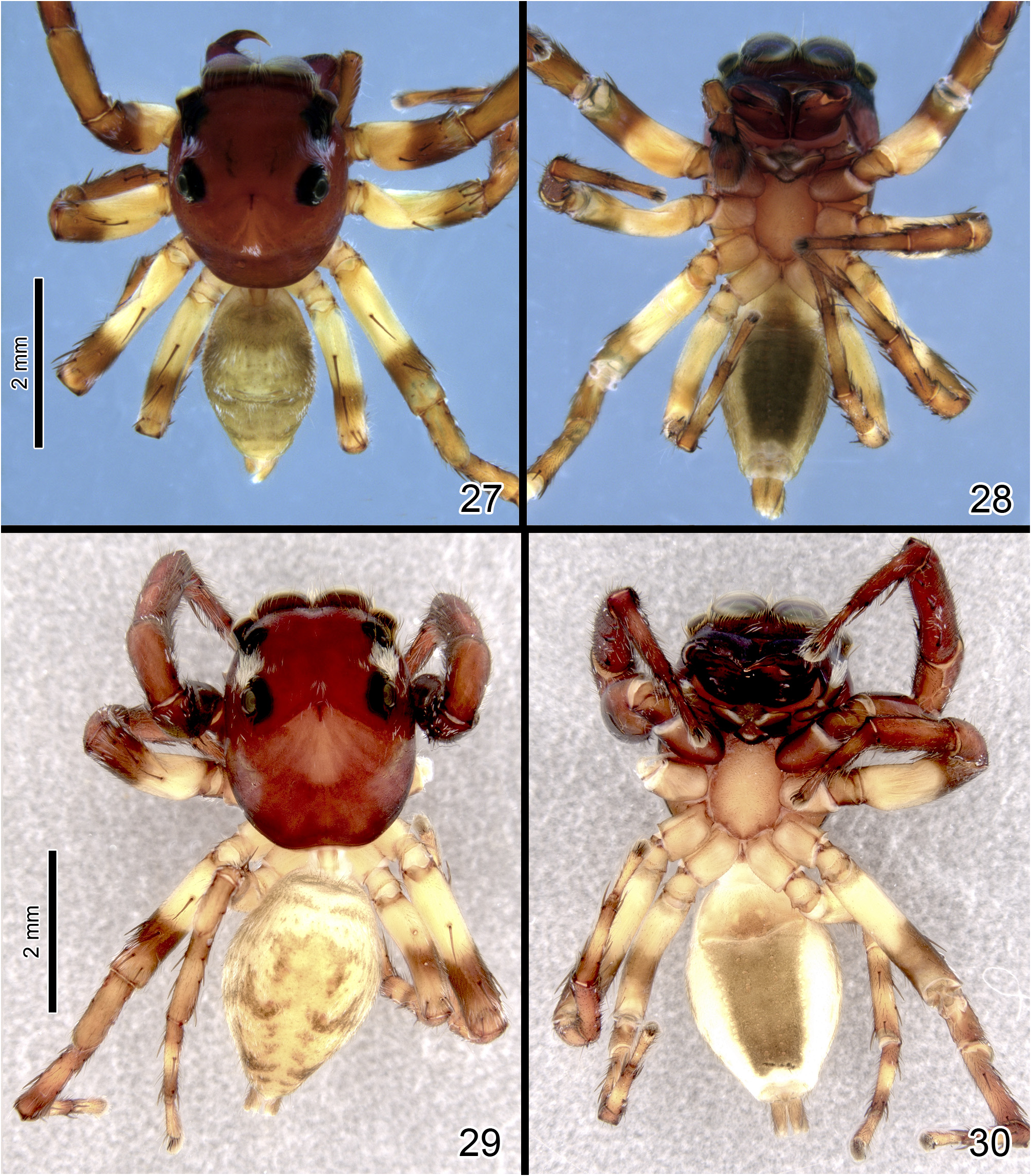
FIGURES 27–30. Matinta spp. 27–28 Matinta pereirae sp. nov., male (27 dorsal view; 28 ventral view). 29–30 Matinta aragog sp. nov., male (29 dorsal view; 30 ventral view).

FIGURES 31–43. Matinta spp., left male palps. 31–33 M. vicana Simon (31 dorsal view; 32 ventral view; 33 retrolateral view). 34–35 M. pereirae sp. nov. (34 ventral view; 35 retrolateral view). 36–37 M. maddisoni sp. nov. (36 ventral view; 37 retrolateral view). 38–39 M. silvae Crane (38 ventral view; 39 retrolateral view). 40–41 M. fonsecai Soares & Camargo (40 ventral view; 41 retrolateral view). 42–43 M. aragog sp. nov. (42 ventral view; 43 retrolateral view).

FIGURES 44–49. Matinta spp., left male palps, ventral view. 44 M. vicana Simon. 45 M. pereirae sp. nov. 46 M. maddisoni sp. nov. 47 M. silvae Crane. 48 M. fonsecai Soares & Camargo. 49 M. aragog sp. nov.

FIGURES 50–55. Matinta spp., left male palps, retrolateral view. 50 M. vicana Simon. 51 M. pereirae sp. nov. 52 M. maddisoni sp. nov. 53 M. silvae Crane. 54 M. fonsecai Soares & Camargo. 55 M. aragog sp. nov.
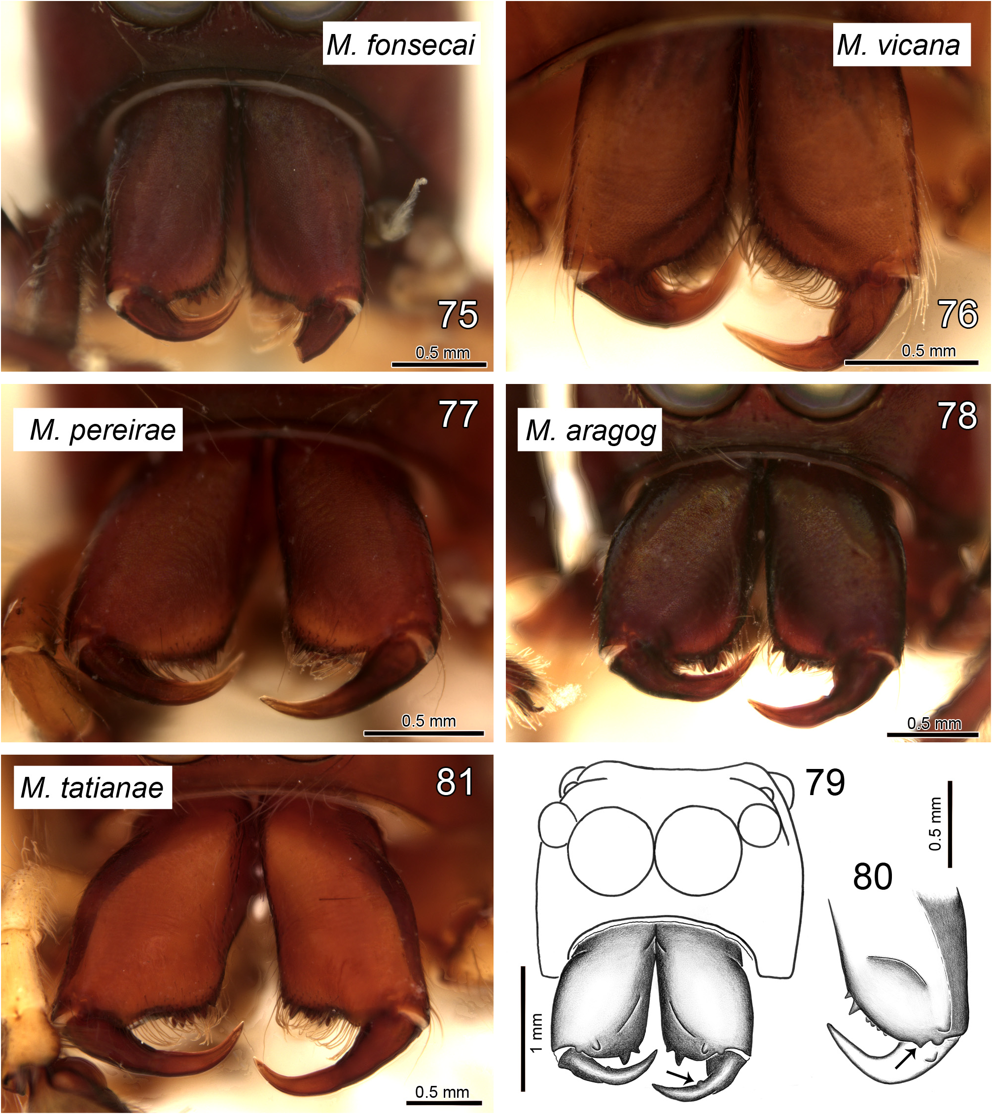
FIGURES 75–81. Matinta spp., male chelicerae, frontal view. 75 M. fonsecai Soares & Camargo. 76 M. vicana Simon. 77 M. pereirae sp. nov. 78–79 M. aragog sp. nov. 80 M. maddisoni sp. nov. 81 M. tatianae sp. nov. Arrow in 79 shows bump on fang and in 80 the paracondylic projection.
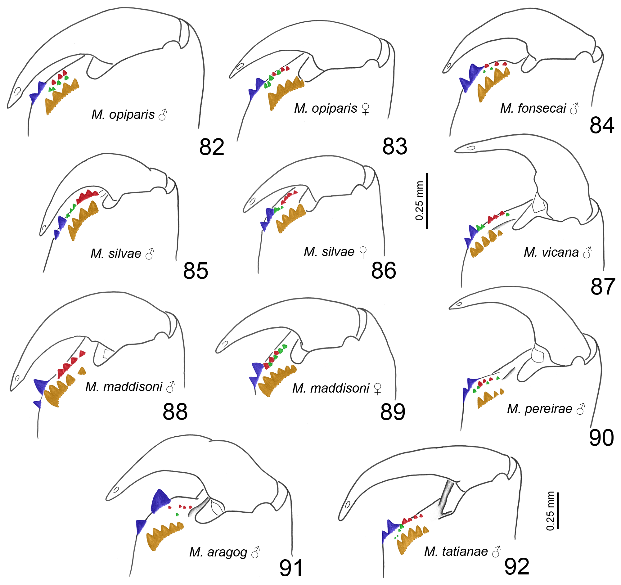
FIGURES 82–92. Matinta spp., left chelicera teeth, retrolateral view. 82–83 M. opiparis Simon (82 male; 83 female). 84 M. fonsecai Soares & Camargo, male. 85–86 M. silvae Crane (85 male; 86 female). 87 M. vicana Simon, male. 88–89 M. maddisoni sp. nov. (88 male; 89 female). 90 M. pereirae sp. nov., male. 91 M. aragog sp. nov., male. 92 M. tatianae sp. nov., male. All illustrations in same scale, except for M. tatianae (92). Colors indicate tooth rows: promarginal (blue), retromarginal (orange), prointermarginal (red) and retrointermarginal (green).
| MNHN |
Museum National d'Histoire Naturelle |
No known copyright restrictions apply. See Agosti, D., Egloff, W., 2009. Taxonomic information exchange and copyright: the Plazi approach. BMC Research Notes 2009, 2:53 for further explanation.
|
Kingdom |
|
|
Phylum |
|
|
Class |
|
|
Order |
|
|
Family |
|
|
Genus |