
Matinta vicana ( Simon, 1900 )
|
publication ID |
https://doi.org/10.11646/zootaxa.5343.2.2 |
|
publication LSID |
lsid:zoobank.org:pub:63876BA9-D973-40E1-BB4B-03F60CD088A7 |
|
DOI |
https://doi.org/10.5281/zenodo.8324683 |
|
persistent identifier |
https://treatment.plazi.org/id/03D587F3-F14E-FFD8-FF79-A47AFC11EABA |
|
treatment provided by |
Plazi (2023-09-07 07:04:19, last updated 2024-11-27 01:47:46) |
|
scientific name |
Matinta vicana ( Simon, 1900 ) |
| status |
|
Matinta vicana ( Simon, 1900) View in CoL View at ENA
Figs 25–26 View FIGURES 23–26 , 31–33 View FIGURES 31–43 , 44 View FIGURES 44–49 , 50 View FIGURES 50–55 , 60 View FIGURES 60–65 , 76 View FIGURES 75–81 , 87 View FIGURES 82–92
Mago vicanus Simon, 1900: 57 View in CoL (Male lectotype designated by M.E. Galiano from S„o Paulo de Olivença, Amazonas, Brazil, de Mathan leg., deposited in MNHN, not examined); Galiano, 1963: 394, pl. XXVII, figs 1–3.
Matinta vicana: Ruiz et al., 2019: 136 View in CoL ; World Spider Catalog, 2023.
Note. The species was identified using Galiano’s (1963) illustrations of the lectotype.
Material examined. BRAZIL: Amazonas: Juruá, Tamaniquá, Lago Tamaniquá (2.734⁰S 65.725⁰W): 1 ♁, 19.IX.2003, F.N.A.A. Rego et al. leg. ( IBSP 98224 View Materials ) .
Diagnosis. The male of M. vicana is similar to those of the species within the vicana species-group, especially those of M. silvae , M. fonsecai and M. pereirae for having no mastidia, a bump on the retrolateral, proximal portion of palpal tibia, and the prolateral portion of tegulum (embolus base) strongly asymmetrical proximally when compared to retrolateral portion. It can be distinguished from those of M. fonsecai and M. silvae for not having a bifid embolus tip, and from M. pereirae for having the embolus tip more slender ( Figs 32 View FIGURES 31–43 , 44 View FIGURES 44–49 ).
Description. Male ( Figs 25–26 View FIGURES 23–26 ). Total length: 4.38. Carapace light brown, with white scales behind fovea and between posterior lateral and median eyes, 2.42 long, 1.78 wide and 1.24 high. Ocular area 1.41 long. Anterior eye row 1.54 and posterior 1.52 wide. Chelicera dark brown, with no mastidion or frontal keel, with paracondylic projection and projection on fang ( Fig. 76 View FIGURES 75–81 ); PMT: 2, RMT: 5, PIMT: 3, RIMT: 3; intermarginal area with keel ( Fig. 87 View FIGURES 82–92 ). Palp ( Figs 31–33 View FIGURES 31–43 , 44 View FIGURES 44–49 , 50 View FIGURES 50–55 , 60 View FIGURES 60–65 ) light brown. Sternum yellow. Legs 3421; I: light brown; II–III: femur proximally yellow and distally dark brown, other articles yellow; IV: brown. Leg length I 7.20 (femur: 2.34; patella: 1.07; tibia: 1.87; metatarsus: 1.25; tarsus: 0.67); II 10.01 (3.80; 1.39; 2.20; 1.54; 1.08); III 12.22 (4.44; 1.82; 2.72; 2.08; 1.16); IV 10.47 (2.02; 1.35; 2.92; 2.70; 1.48). Leg spination: femur I d1-1-1, p0-0-2, r0; II–IV d1-1-1, p0-0-2, r0- 0-1; patella I–II p1; III–IV p1, r1; tibia I v2-2-2, p1-0-0, r0; II v2-2-1r, p1-1-0, r0; III v1p-0-2, p1-1-1, r1-1-1; IV v0-1p-2; p1-1-1, r1-1-1; metatarsus I–II v2-2; III v2-2, p1-0-2, r1-0-2; IV v2-2, p1-0-2, r1-1-2. Abdomen dorsally cream-colored, with dark brown irregular mark ( Fig. 25 View FIGURES 23–26 ); ventrally with three thin longitudinal dark brown stripes, which join at the distal part of the abdomen ( Fig. 26 View FIGURES 23–26 ). Spinnerets brown.
Female. Unknown.
Distribution. Known only from the state of Amazonas ( Brazil).
Galiano, M. E. (1963) Las especies americanas de aranas de la familia Salticidae descriptas por Eugene Simon: Redescripciones basadas en los ejemplares tipicos, Revista de la Sociedad Argentina de Ciencias Naturales, Series C, 23, 273 - 470.
Ruiz, G. R. S., Maddison, W. P. & Galiano, M. E. (2019) A revision of the concept of Mago O. Pickard-Cambridge, 1882, and proposal of a new genus (Araneae: Salticidae: Amycini). Zootaxa, 4658 (1), 124 - 140. https: // doi. org / 10.11646 / zootaxa. 4658.1.5
Simon, E. (1900) Etudes arachnologiques. 30 e Memoire. XLVII. Descriptions d'especes nouvelles de la famille des Attidae. Annales de la Societe Entomologique de France, 69, 27 - 61.
World Spider Catalog (2023) World Spider Catalog. Version 24.5. Natural History Museum Bern, Bern. Available from: http: // wsc. nmbe. ch (accessed 24 August 2023)
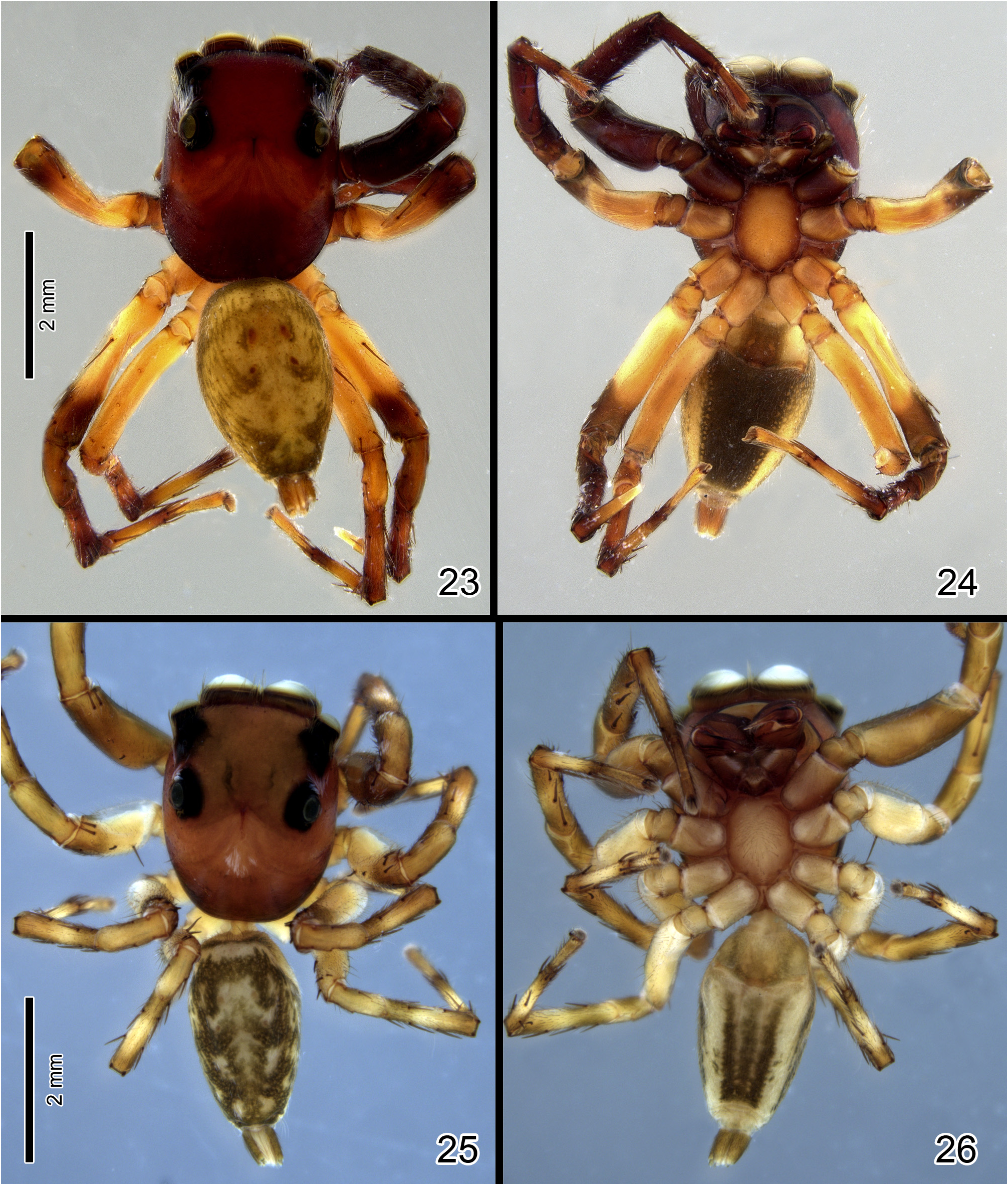
FIGURES 23–26. Matinta spp. 23–24 Matinta fonsecai Soares & Camargo, male (23 dorsal view; 24 ventral view). 25–26 Matinta vicana Simon, male (25 dorsal view; 26 ventral view).

FIGURES 31–43. Matinta spp., left male palps. 31–33 M. vicana Simon (31 dorsal view; 32 ventral view; 33 retrolateral view). 34–35 M. pereirae sp. nov. (34 ventral view; 35 retrolateral view). 36–37 M. maddisoni sp. nov. (36 ventral view; 37 retrolateral view). 38–39 M. silvae Crane (38 ventral view; 39 retrolateral view). 40–41 M. fonsecai Soares & Camargo (40 ventral view; 41 retrolateral view). 42–43 M. aragog sp. nov. (42 ventral view; 43 retrolateral view).

FIGURES 44–49. Matinta spp., left male palps, ventral view. 44 M. vicana Simon. 45 M. pereirae sp. nov. 46 M. maddisoni sp. nov. 47 M. silvae Crane. 48 M. fonsecai Soares & Camargo. 49 M. aragog sp. nov.

FIGURES 50–55. Matinta spp., left male palps, retrolateral view. 50 M. vicana Simon. 51 M. pereirae sp. nov. 52 M. maddisoni sp. nov. 53 M. silvae Crane. 54 M. fonsecai Soares & Camargo. 55 M. aragog sp. nov.
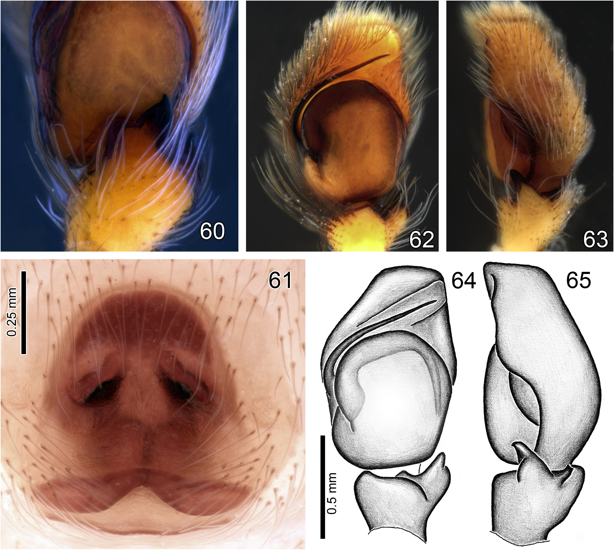
FIGURES 60–65. Matinta spp., left male palps and epigyne. 60 M. vicana Simon, male palp, retroventral view, detail of RvTA. 61 M. silvae Crane, epigyne, ventrral view. 62–65 M. tatianae sp. nov., left male palp (62 ventral view; 63 retrolateral view; 64 ventral view; 65 retrolateral view).
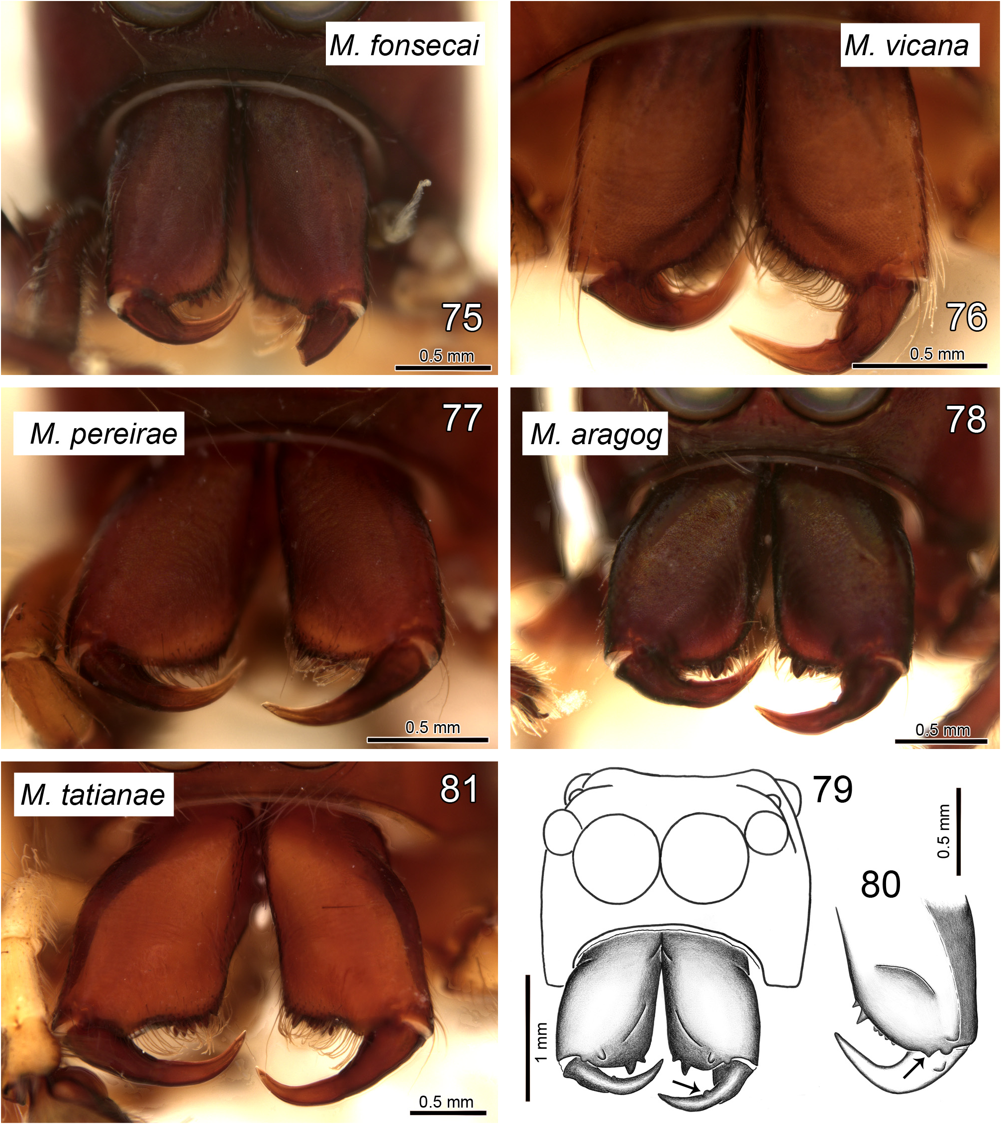
FIGURES 75–81. Matinta spp., male chelicerae, frontal view. 75 M. fonsecai Soares & Camargo. 76 M. vicana Simon. 77 M. pereirae sp. nov. 78–79 M. aragog sp. nov. 80 M. maddisoni sp. nov. 81 M. tatianae sp. nov. Arrow in 79 shows bump on fang and in 80 the paracondylic projection.
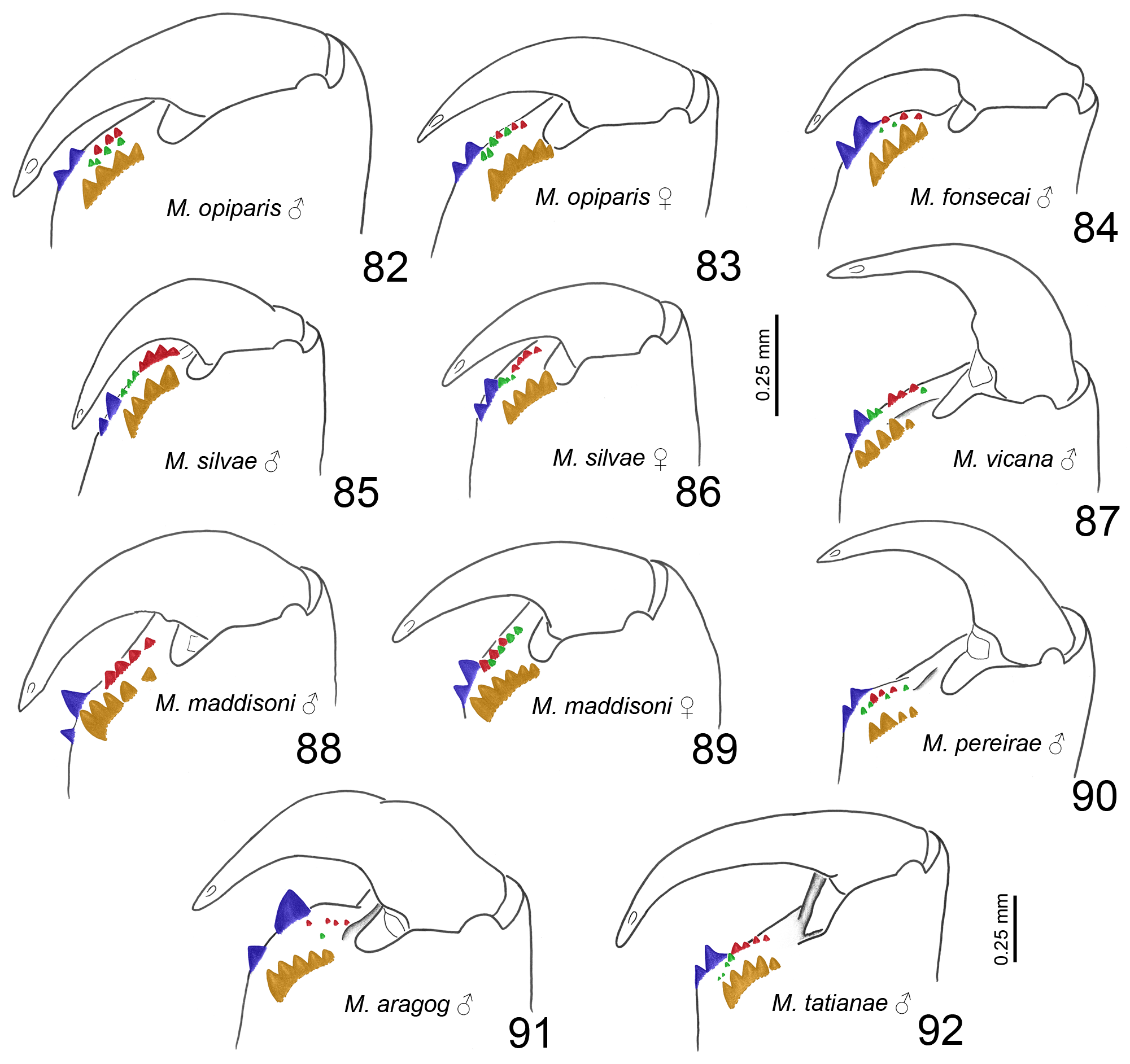
FIGURES 82–92. Matinta spp., left chelicera teeth, retrolateral view. 82–83 M. opiparis Simon (82 male; 83 female). 84 M. fonsecai Soares & Camargo, male. 85–86 M. silvae Crane (85 male; 86 female). 87 M. vicana Simon, male. 88–89 M. maddisoni sp. nov. (88 male; 89 female). 90 M. pereirae sp. nov., male. 91 M. aragog sp. nov., male. 92 M. tatianae sp. nov., male. All illustrations in same scale, except for M. tatianae (92). Colors indicate tooth rows: promarginal (blue), retromarginal (orange), prointermarginal (red) and retrointermarginal (green).
No known copyright restrictions apply. See Agosti, D., Egloff, W., 2009. Taxonomic information exchange and copyright: the Plazi approach. BMC Research Notes 2009, 2:53 for further explanation.
|
Kingdom |
|
|
Phylum |
|
|
Class |
|
|
Order |
|
|
Family |
|
|
Genus |
Matinta vicana ( Simon, 1900 )
| Matos, Tainá D. S. & Ruiz, Gustavo R. S. 2023 |
Matinta vicana:
| Ruiz, G. R. S. & Maddison, W. P. & Galiano, M. E. 2019: 136 |
Mago vicanus
| Galiano, M. E. 1963: 394 |
| Simon, E. 1900: 57 |