
Matinta fonsecai ( Soares & Camargo, 1948 )
|
publication ID |
https://doi.org/10.11646/zootaxa.5343.2.2 |
|
publication LSID |
lsid:zoobank.org:pub:63876BA9-D973-40E1-BB4B-03F60CD088A7 |
|
DOI |
https://doi.org/10.5281/zenodo.8329839 |
|
persistent identifier |
https://treatment.plazi.org/id/03D587F3-F148-FFC6-FF79-A452FC70EB6E |
|
treatment provided by |
Plazi (2023-09-07 07:04:19, last updated 2024-11-27 01:47:46) |
|
scientific name |
Matinta fonsecai ( Soares & Camargo, 1948 ) |
| status |
|
Matinta fonsecai ( Soares & Camargo, 1948) View in CoL View at ENA
Figs 23–24 View FIGURES 23–26 , 40–41 View FIGURES 31–43 , 48 View FIGURES 44–49 , 54 View FIGURES 50–55 , 75 View FIGURES 75–81 , 84 View FIGURES 82–92
Mago fonsecai Soares & Camargo, 1948: 440 View in CoL , figs 35–37 (Male holotype from Fazenda da Floresta, Matipoó river, Minas Gerais, Brazil, VIII.1919, J.P da Fonseca leg., deposited in MZUSP, not examined); Galiano, 1968: 339, figs 62–63. Matinta fonsecai: Ruiz et al., 2019: 136 View in CoL ; World Spider Catalog, 2023.
Note. The species was identified using the illustrations and descriptions by Galiano (1968: figs 62–63).
Material examined. BRAZIL: Acre: Senador Guimard, Reserva Extrativista de Catuaba [10.067⁰S 67.633⁰W]: 1 ♁, 2003, E. Morato leg. ( IBSP 161435 View Materials ) .
Diagnosis. The male of M. fonsecai is similar to those of the species within the vicana species-group, especially those of M. vicana , M. silvae and M. pereirae for not having mastidia, and by having a bump on the retrolateral, proximal portion of palpal tibia ( Figs 34, 38, 40 View FIGURES 31–43 ), and the prolateral portion of tegulum (embolus base) strongly asymmetrical proximally when compared to the retrolateral portion. It can be distinguished from those of M. vicana and M. pereirae for having a bifid embolus tip ( Figs 40 View FIGURES 31–43 , 48 View FIGURES 44–49 ), and from M. silvae for having a bifid, short RvTA ( Figs 41 View FIGURES 31–43 , 54 View FIGURES 50–55 ). The male also resembles those of M. aragog for having the bifid embolus tip (developed membranous portion), but can be distinguished for having joint prolateral cheliceral teeth, no bump on fang, no mastidia, besides the shape of tibia and palpal bulb.
Description. Male ( Figs 23–24 View FIGURES 23–26 ). Total length: 6.28. Carapace dark reddish-brown, 3.26 long, 2.30 wide and 1.83 high. Ocular area 1.51 long, laterally with white scales.Anterior eye row 2.12 and posterior 1.74 wide. Chelicera dark brown, with no mastidion or keel, with paracondylic projection ( Fig. 75 View FIGURES 75–81 ); PMT: 2, RMT: 4, PIMT: 3, RIMT: 2 ( Fig. 84 View FIGURES 82–92 ). Palp ( Figs 40–41 View FIGURES 31–43 , 48 View FIGURES 44–49 , 54 View FIGURES 50–55 ) dark brown. Sternum yellow. Legs 3412; I: dark brown; II–IV: dark brown, with the exception of proximal femur and tarsus, yellow. Leg length I 6.04 (femur: 1.86; patella: 0.95; tibia: 1.67; metatarsus: 0.99; tarsus: 0.57); II 5.72 (1.79; 0.98; 1.50; 0.92; 0.53); III 6.81 (2.40; 0.86; 1.46; 1.34; 0.75); IV 6.28 (2.15; 0.70; 1.23; 1.44; 0.76). Leg spination: femur I–II d1-1-1, p0-0-2, r0-0-1; III–IV d1-1-1, p0-1-2, r0-0-1; patella I–II 0; III–IV p0-1-0, r0-1-0; tibia I v2-2-2, p0-0-1, r0; II v1p-2-2, p0-0-1, r0; III v1p-0-2, p1-0-1, r1-1-1; IV v1p-0-2; p1-0-1, r1-1-1; metatarsus I–II v2-2; III v2-0-2, p1-0-2, r1-0-2; IV v2-0-2, p1-1-2, r1-1-2. Abdomen dorsally cream-colored with irregular light brown marks ( Fig. 23 View FIGURES 23–26 ); ventrally black ( Fig. 24 View FIGURES 23–26 ). Spinnerets light brown.
Female. Unknown.
Distribution. Known from the states of Acre and Minas Gerais ( Brazil).
Galiano, M. E. (1968) Revision de los generos Acragas, Amycus, Encolpius, Hypaeus, Mago y Noegus (Salticidae, Araneae). Revista del Museo Argentino de Ciencias Naturales Bernardino Rivadavia, Entomologia, 2, 267 - 360.
Ruiz, G. R. S., Maddison, W. P. & Galiano, M. E. (2019) A revision of the concept of Mago O. Pickard-Cambridge, 1882, and proposal of a new genus (Araneae: Salticidae: Amycini). Zootaxa, 4658 (1), 124 - 140. https: // doi. org / 10.11646 / zootaxa. 4658.1.5
Soares, B. A. M. & Camargo, H. F. de A. (1948) Alguns novos salticidas do Brasil (Araneae, Salticidae). Revista Brasileira de Biologia, 8, 421 - 443.
World Spider Catalog (2023) World Spider Catalog. Version 24.5. Natural History Museum Bern, Bern. Available from: http: // wsc. nmbe. ch (accessed 24 August 2023)
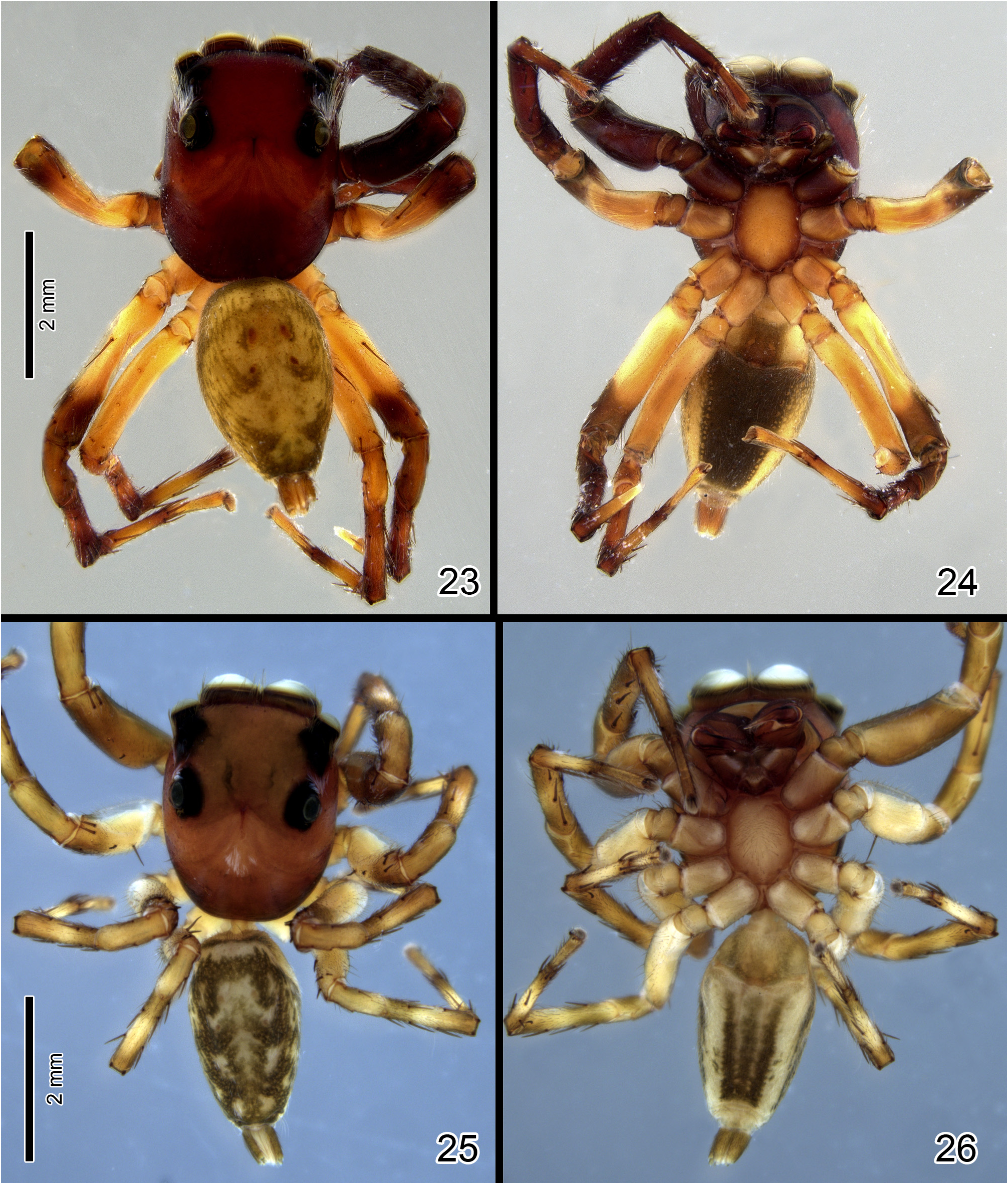
FIGURES 23–26. Matinta spp. 23–24 Matinta fonsecai Soares & Camargo, male (23 dorsal view; 24 ventral view). 25–26 Matinta vicana Simon, male (25 dorsal view; 26 ventral view).

FIGURES 31–43. Matinta spp., left male palps. 31–33 M. vicana Simon (31 dorsal view; 32 ventral view; 33 retrolateral view). 34–35 M. pereirae sp. nov. (34 ventral view; 35 retrolateral view). 36–37 M. maddisoni sp. nov. (36 ventral view; 37 retrolateral view). 38–39 M. silvae Crane (38 ventral view; 39 retrolateral view). 40–41 M. fonsecai Soares & Camargo (40 ventral view; 41 retrolateral view). 42–43 M. aragog sp. nov. (42 ventral view; 43 retrolateral view).

FIGURES 44–49. Matinta spp., left male palps, ventral view. 44 M. vicana Simon. 45 M. pereirae sp. nov. 46 M. maddisoni sp. nov. 47 M. silvae Crane. 48 M. fonsecai Soares & Camargo. 49 M. aragog sp. nov.

FIGURES 50–55. Matinta spp., left male palps, retrolateral view. 50 M. vicana Simon. 51 M. pereirae sp. nov. 52 M. maddisoni sp. nov. 53 M. silvae Crane. 54 M. fonsecai Soares & Camargo. 55 M. aragog sp. nov.
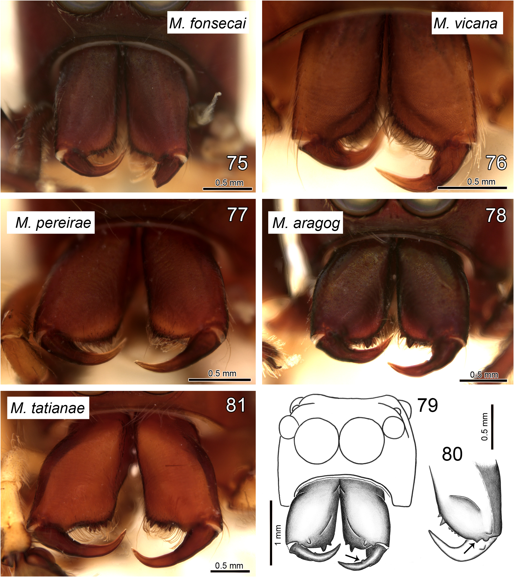
FIGURES 75–81. Matinta spp., male chelicerae, frontal view. 75 M. fonsecai Soares & Camargo. 76 M. vicana Simon. 77 M. pereirae sp. nov. 78–79 M. aragog sp. nov. 80 M. maddisoni sp. nov. 81 M. tatianae sp. nov. Arrow in 79 shows bump on fang and in 80 the paracondylic projection.
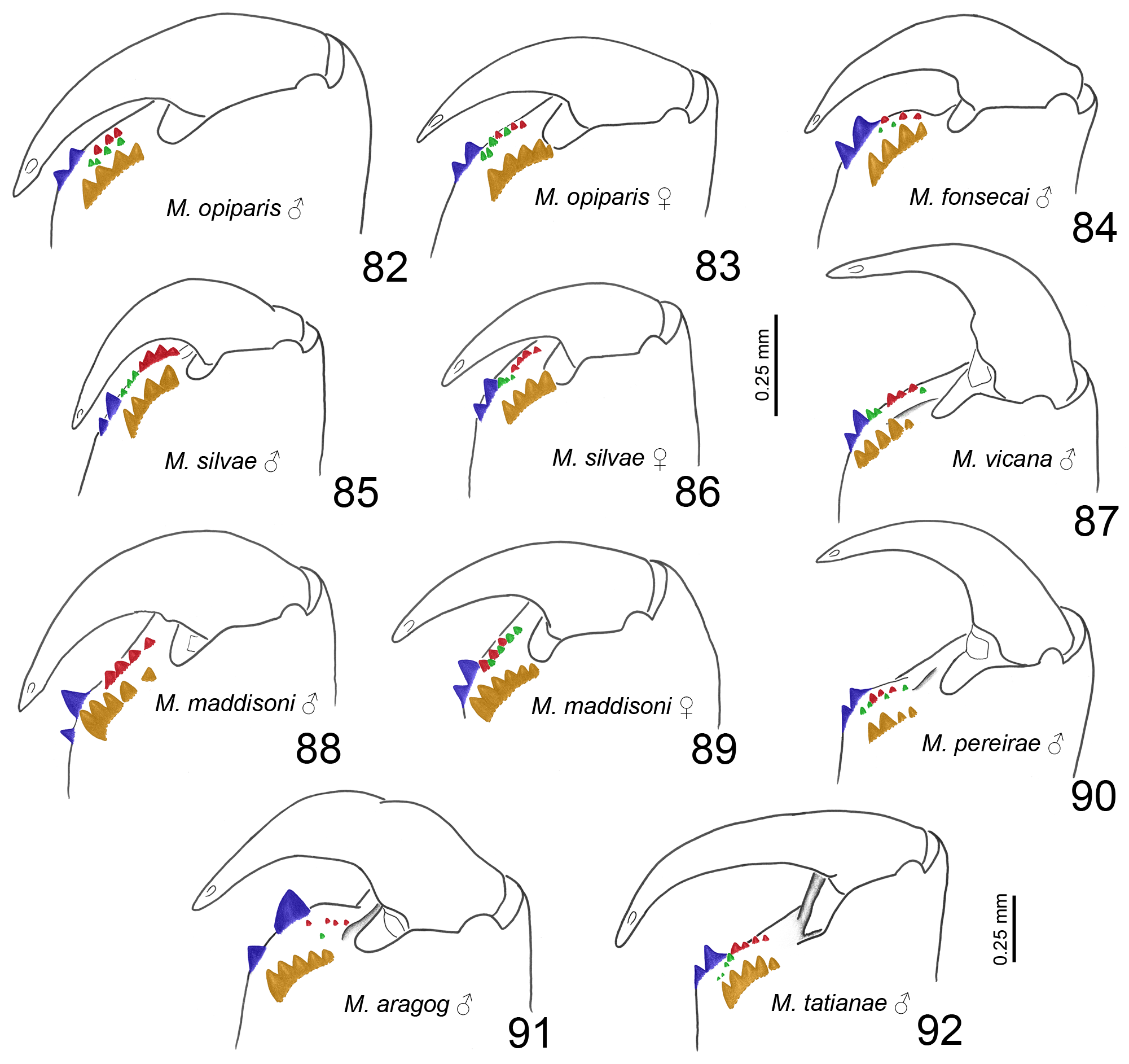
FIGURES 82–92. Matinta spp., left chelicera teeth, retrolateral view. 82–83 M. opiparis Simon (82 male; 83 female). 84 M. fonsecai Soares & Camargo, male. 85–86 M. silvae Crane (85 male; 86 female). 87 M. vicana Simon, male. 88–89 M. maddisoni sp. nov. (88 male; 89 female). 90 M. pereirae sp. nov., male. 91 M. aragog sp. nov., male. 92 M. tatianae sp. nov., male. All illustrations in same scale, except for M. tatianae (92). Colors indicate tooth rows: promarginal (blue), retromarginal (orange), prointermarginal (red) and retrointermarginal (green).
No known copyright restrictions apply. See Agosti, D., Egloff, W., 2009. Taxonomic information exchange and copyright: the Plazi approach. BMC Research Notes 2009, 2:53 for further explanation.
|
Kingdom |
|
|
Phylum |
|
|
Class |
|
|
Order |
|
|
Family |
|
|
Genus |
Matinta fonsecai ( Soares & Camargo, 1948 )
| Matos, Tainá D. S. & Ruiz, Gustavo R. S. 2023 |
Mago fonsecai
| Soares, B. A. M. & Camargo, H. F. de 1948: 440 |