
Epipleoneura janirae Machado, 2005
|
publication ID |
https://doi.org/10.11646/zootaxa.3872.3.1 |
|
publication LSID |
lsid:zoobank.org:pub:72ACE4FF-9A41-4D26-A201-01E020439899 |
|
DOI |
https://doi.org/10.5281/zenodo.5296820 |
|
persistent identifier |
https://treatment.plazi.org/id/03D3FD33-FFB2-FFFB-4E8F-7326F37AF96E |
|
treatment provided by |
Plazi (2016-04-17 21:01:09, last updated 2023-10-27 12:13:27) |
|
scientific name |
Epipleoneura janirae Machado, 2005 |
| status |
|
Epipleoneura janirae Machado, 2005 View in CoL
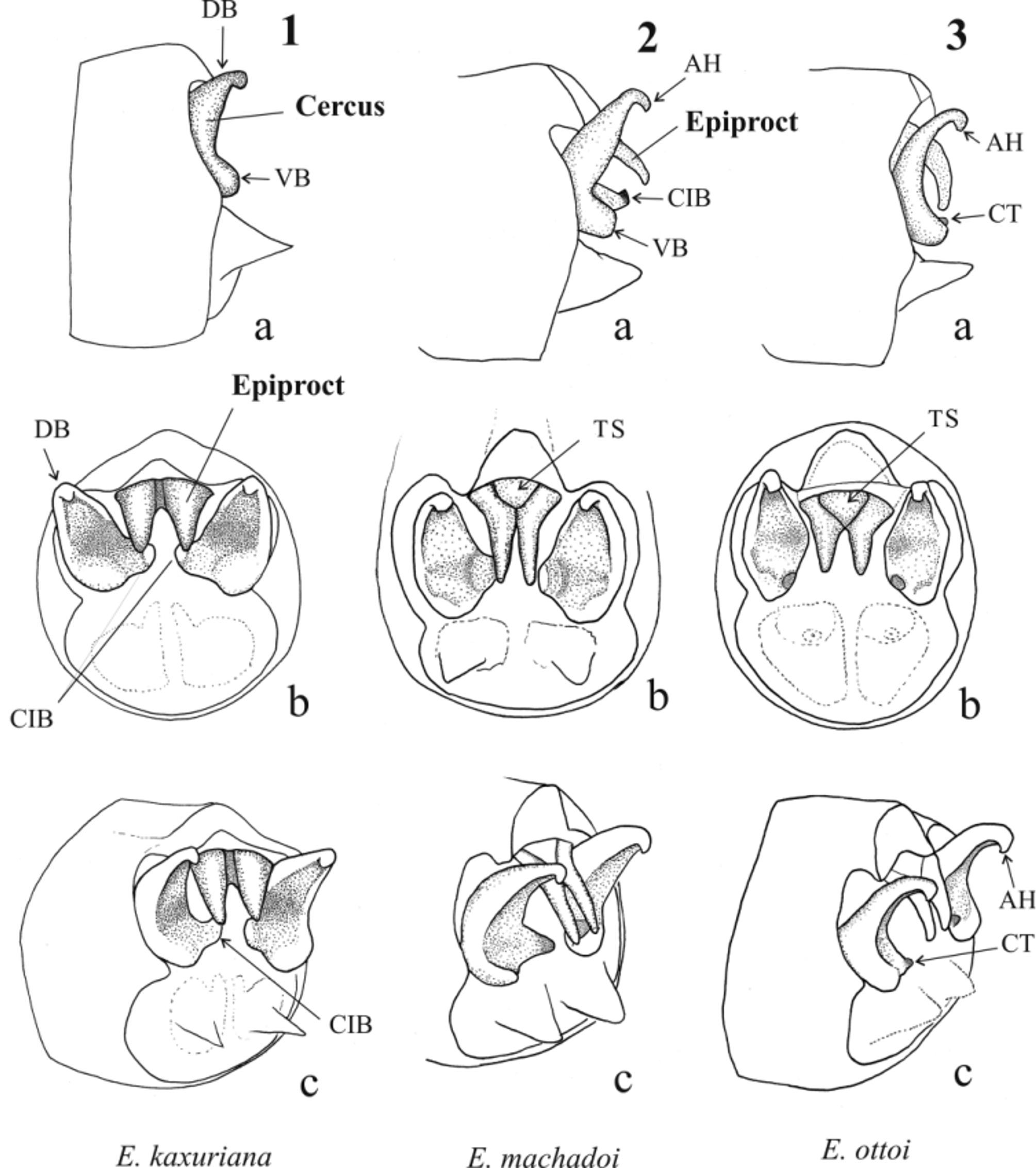
( Figs. 14 View FIGURES 13 – 15 , 37 View FIGURES 28 – 47 , 65 View FIGURES 53 – 66 )
Epipleoneura janirae Machado 2005: 47 View in CoL –48, Figs. 1–2 View FIGURES 1 – 3 (new species, male and female description, illustrations of male S 10 in lateral and posterior view and female prothorax in dorsal view, type material at ABMM). Garrison et al. 2010: 352, 354 (included in list of species, illustrations of epiproct in posterior view). Pessacq et al. 2012: 4 (included in list of Brazilian Protoneuridae View in CoL ).
Specimens examined. 2 ♂, 1 ♀. Holotype male, Brazil, Pará State, Santarém, Belterra (Porto Novo) (Santarém: 03º08’41”S, 55º02’42”W), leg. A.B.M. Machado & P. Pereira, ii 1957. Allotype ♀ and 1 paratype ♂, same data as holotype. ABMM.
Diagnosis. Male cercus resembles that of E. capilliformis . The cercus has a well-developed inner-basal tooth. The epiproct consists of two long narrow branches, tapering toward apex and fused along almost their entire length with only their tips free, thus resembling that of E. uncinata De Marmels, 1989 , but in E. janirae there is a triangular swollen structure between branches base and the apex is comparatively thicker. Epipleoneura janirae further differs from that species by the different genital ligula morphology ( Fig. 37 View FIGURES 28 – 47 ); E. janirae is similar to that described for E. humeralis . Female posterior margin of pronotum ( Fig. 65 View FIGURES 53 – 66 ) is composed of a wide erected lobe with a medial very shallow V depression.
Distribution. Pará State, northern Brazil (type locality Santarém, Belterra [02 º 38’14” S, 54 º 09’96” W]).
De Marmels, J. (1989) Odonata or dragonflies from Cerro de la Neblina. Academia de las Ciencias Fisicas, Matematicas y Naturales, Caracas, Venezuela, 25, 1 - 78.
Garrison, R. W., von Ellenrieder, N. & Louton, J. A. (2010) Damselfly genera of the New World. An Illustrated and Annotated Key to the Zygoptera. The Johns Hopkins University Press, Baltimore, 490 pp.
Machado, A. B. M. (2005) Studies on Neotropical Protoneuridae. 18. Epipleoneura janirae sp. n. from the Amazonian region of Brazil (Odonata: Protoneuridae). Lundiana, 6 (1), 47 - 48.
Pessacq, P., Santos, T. C. & Costa, JM. (2012) Checklist and updated distribution of Protoneuridae from Brazil. International Journal of Odonatology, 15 (1), 1 - 15. http: // dx. doi. org / 10.1080 / 13887890.2012.672158

FIGURES 13 – 15. Male caudal appendages, a: lateral view; b: posterior view, c: latero-posterior view. CIB: cercus inner-basal branch. CT: cercus inner-basal tooth.
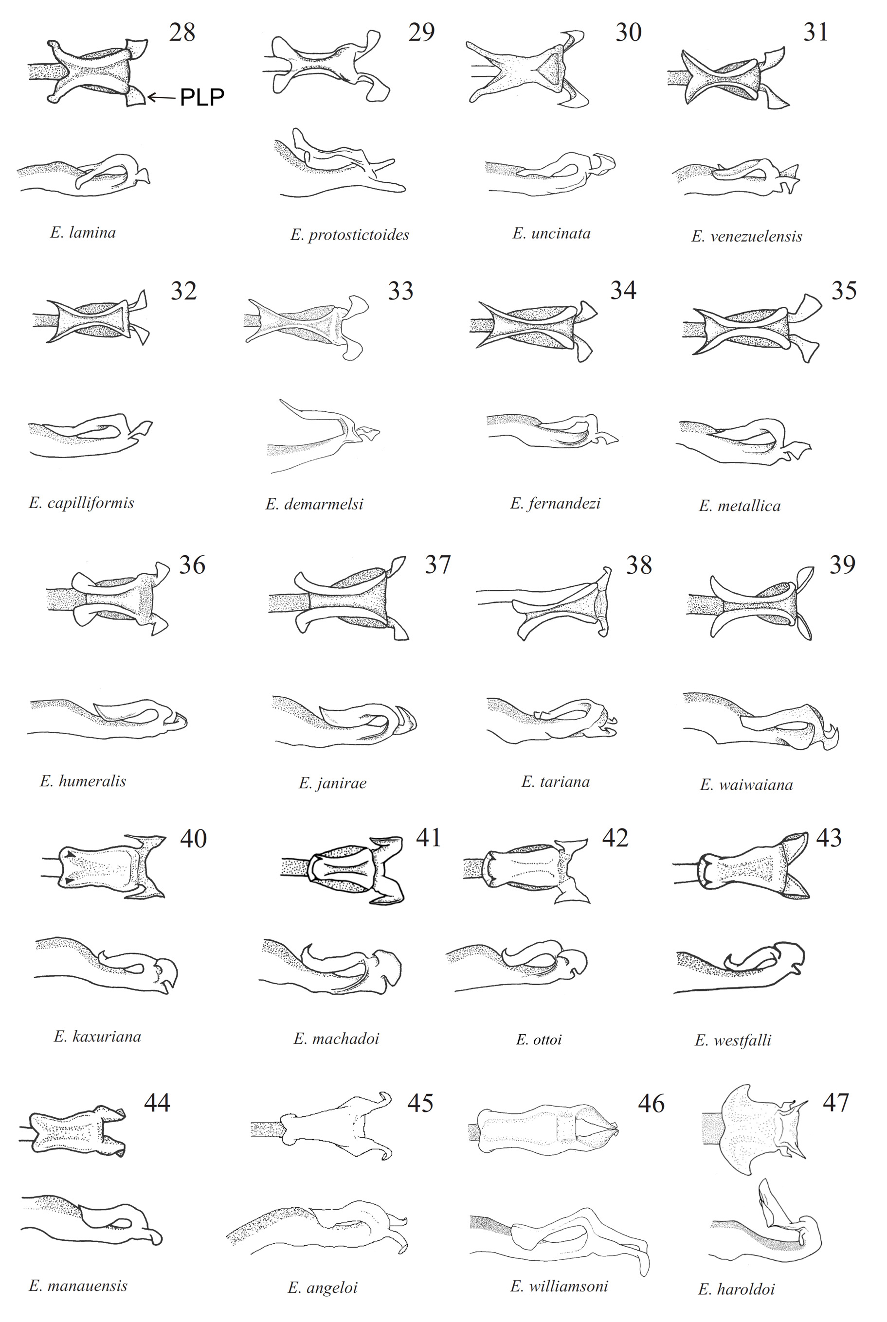
FIGURES 28 – 47. Genital ligula, lateral and ectal view. Figures 29, 33, 47 modified from Garrison et al. (2010). PLP: posterolateral projection.

FIGURES 53 – 66. Female pronotum in lateral, dorsal, antero-lateral, anterior postero-lateral and posterior view. Figures 57, 64 and 65 modified from Garrison et al. (2010), Rácenis (1960) and Machado (2005) respectively.
No known copyright restrictions apply. See Agosti, D., Egloff, W., 2009. Taxonomic information exchange and copyright: the Plazi approach. BMC Research Notes 2009, 2:53 for further explanation.
|
Kingdom |
|
|
Phylum |
|
|
Class |
|
|
Order |
|
|
Family |
|
|
Genus |
Epipleoneura janirae Machado, 2005
| Pessacq, Pablo 2014 |
Epipleoneura janirae
| Pessacq 2012: 4 |
| Garrison 2010: 352 |
| Machado 2005: 47 |
1 (by plazi, 2016-04-17 21:01:09)
2 (by ImsDioSync, 2017-01-05 11:53:45)
3 (by ImsDioSync, 2017-01-05 11:54:52)
4 (by ImsDioSync, 2017-02-09 17:50:15)
5 (by ImsDioSync, 2017-06-19 22:51:31)
6 (by ExternalLinkService, 2019-09-26 13:52:28)
7 (by ExternalLinkService, 2021-08-27 09:25:53)
8 (by ExternalLinkService, 2021-08-27 22:53:31)
9 (by ExternalLinkService, 2022-01-30 06:43:05)