
Caayguara, Rheims, Cristina A., 2010
|
publication ID |
https://doi.org/ 10.5281/zenodo.198317 |
|
DOI |
https://doi.org/10.5281/zenodo.6209402 |
|
persistent identifier |
https://treatment.plazi.org/id/03CF8789-FFA1-0708-FF22-FA3C1547E04F |
|
treatment provided by |
Plazi |
|
scientific name |
Caayguara |
| status |
gen. nov. |
Caayguara View in CoL View at ENA gen. nov.
Etymology. The generic name is a noun taken from the Tupi Indian language that means “forest dweller” (caá = forest; yguara = dweller). The gender is neutral.
Type species: Olios albus Mello-Leitão, 1918
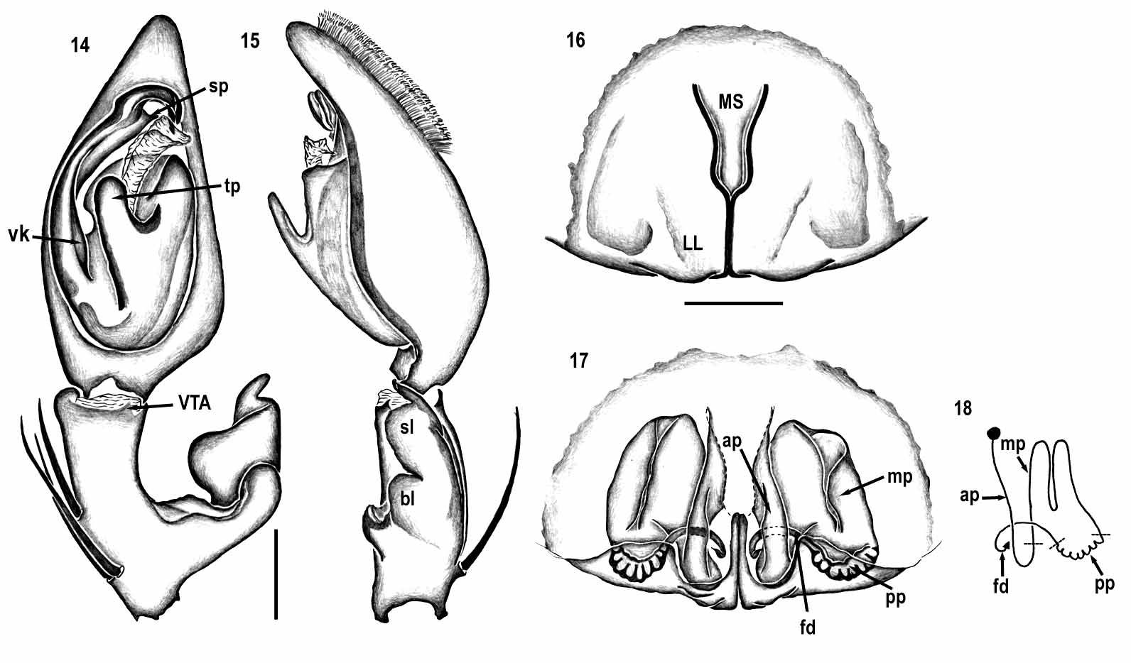
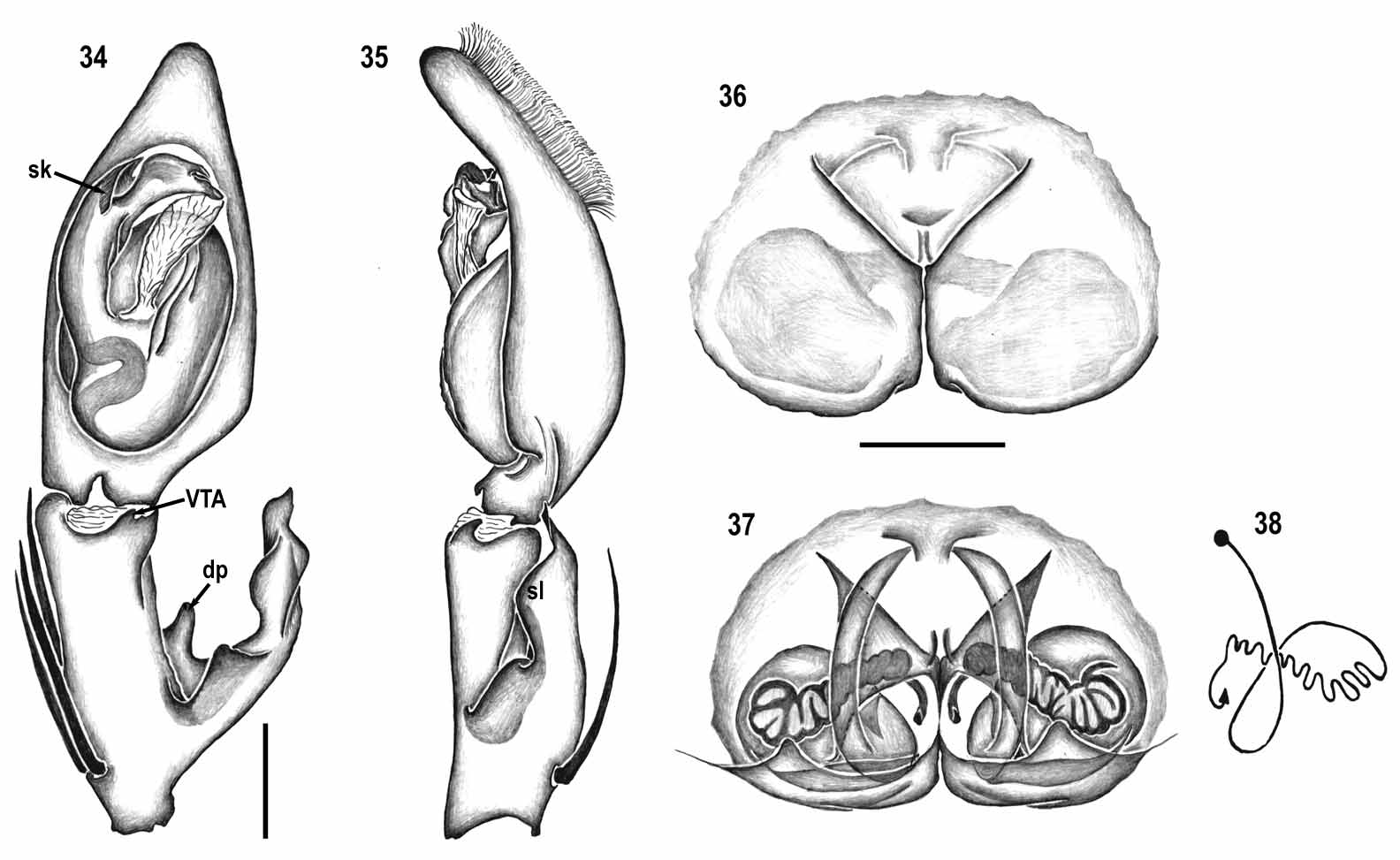
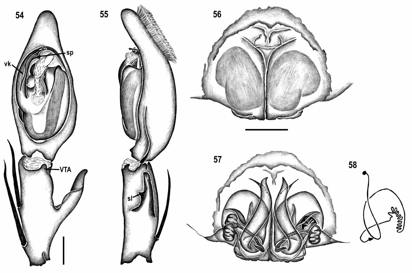
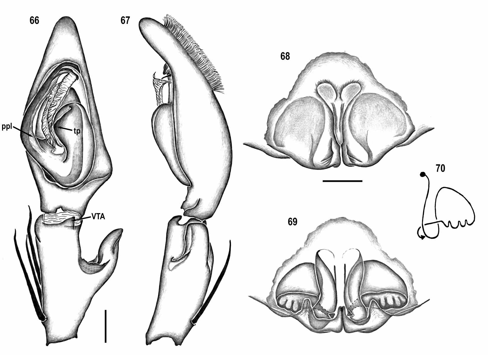
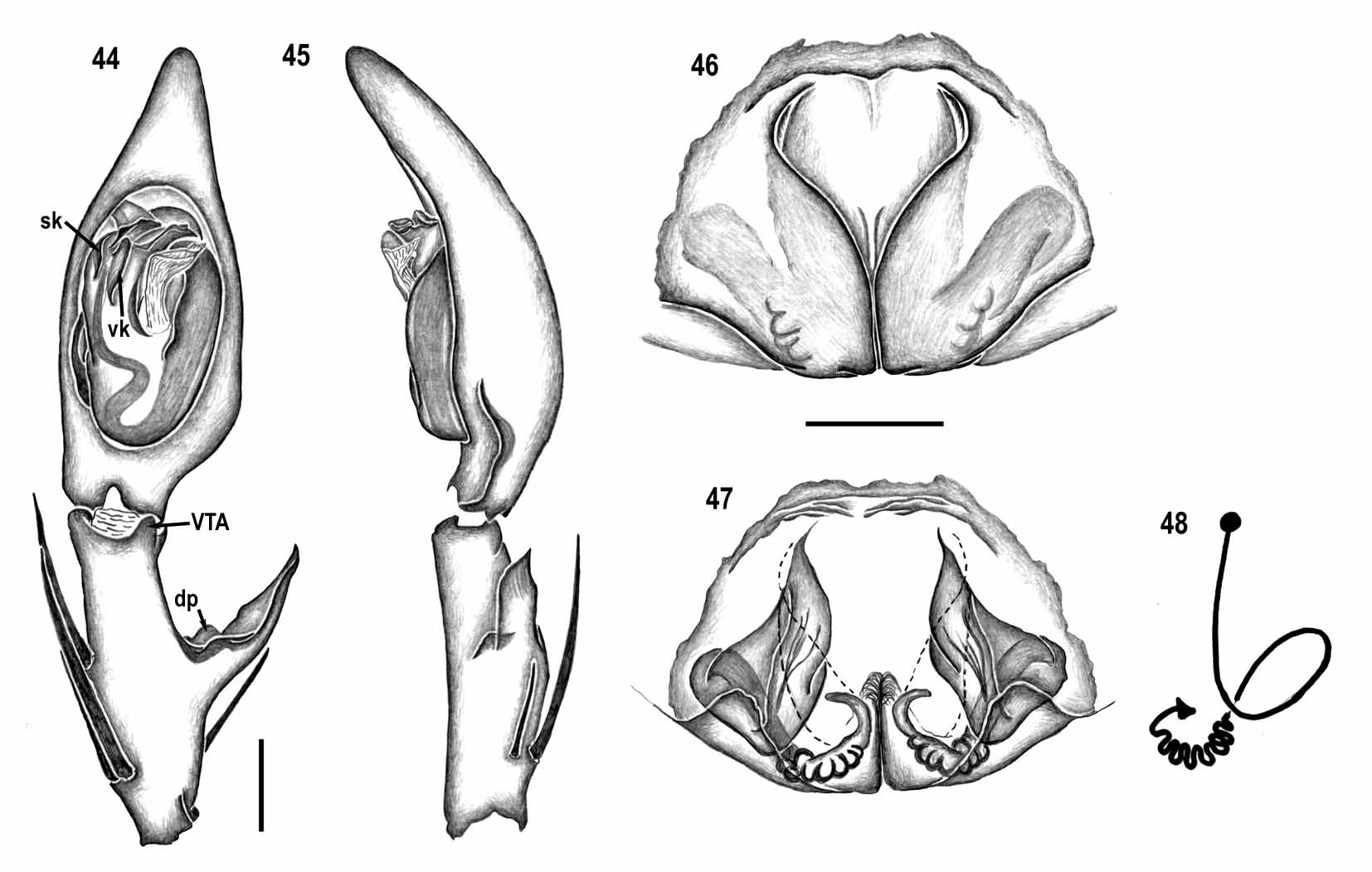
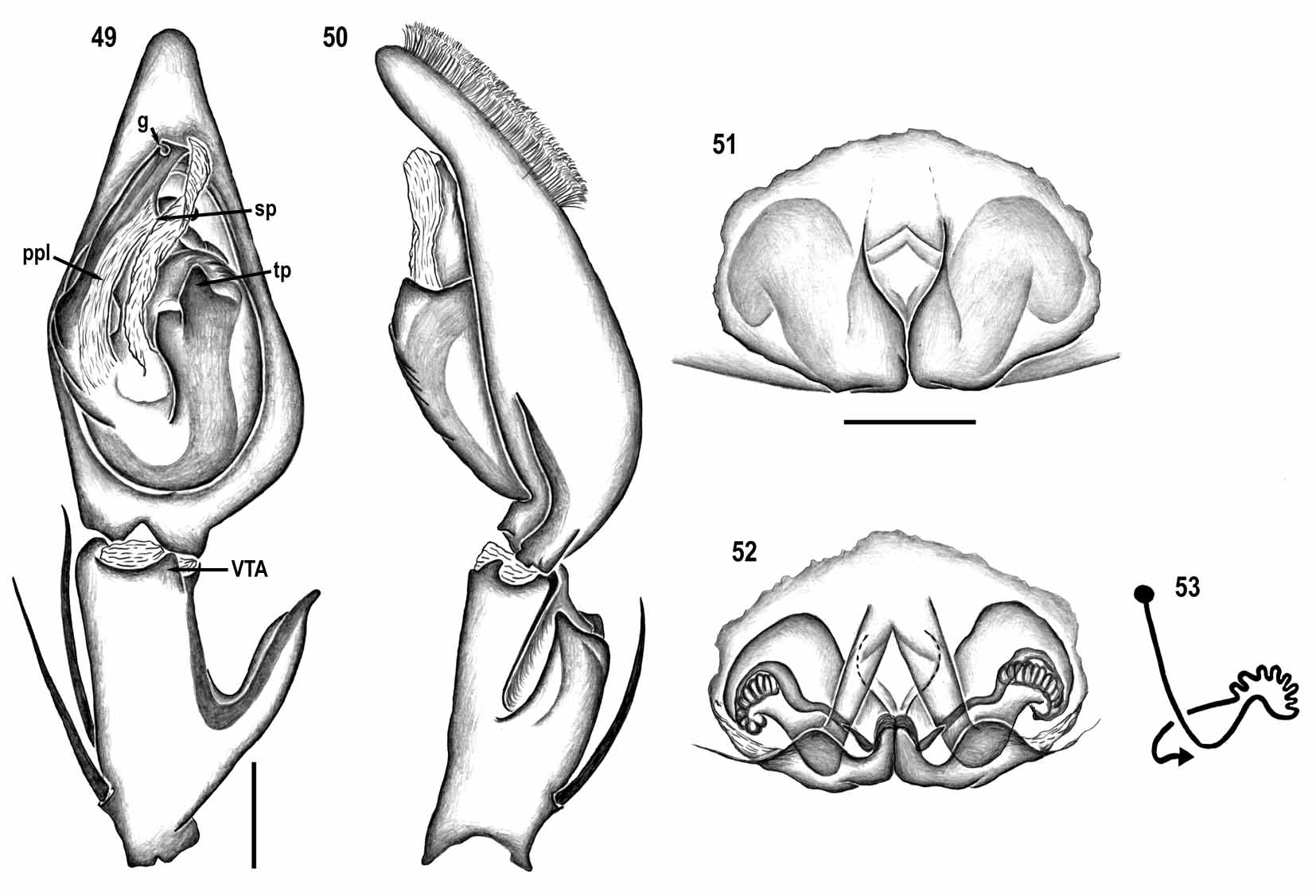
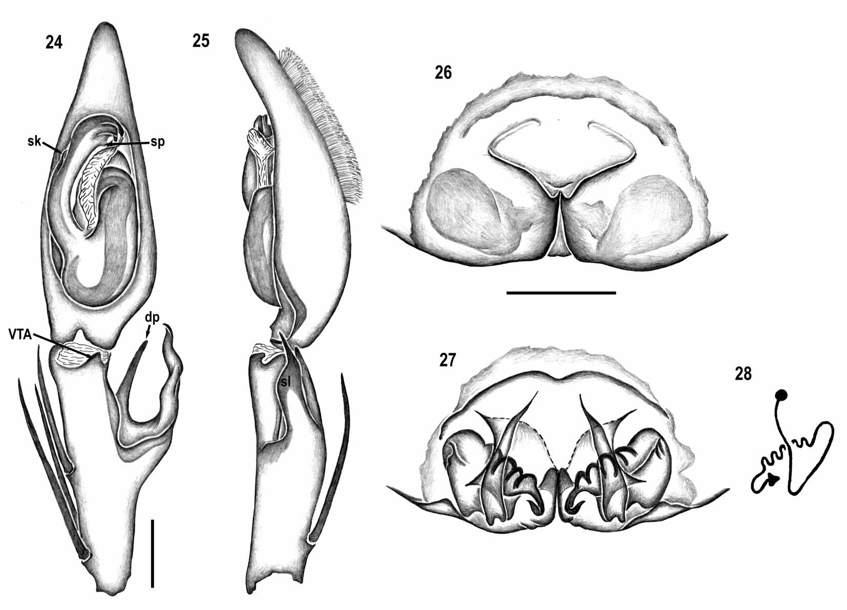
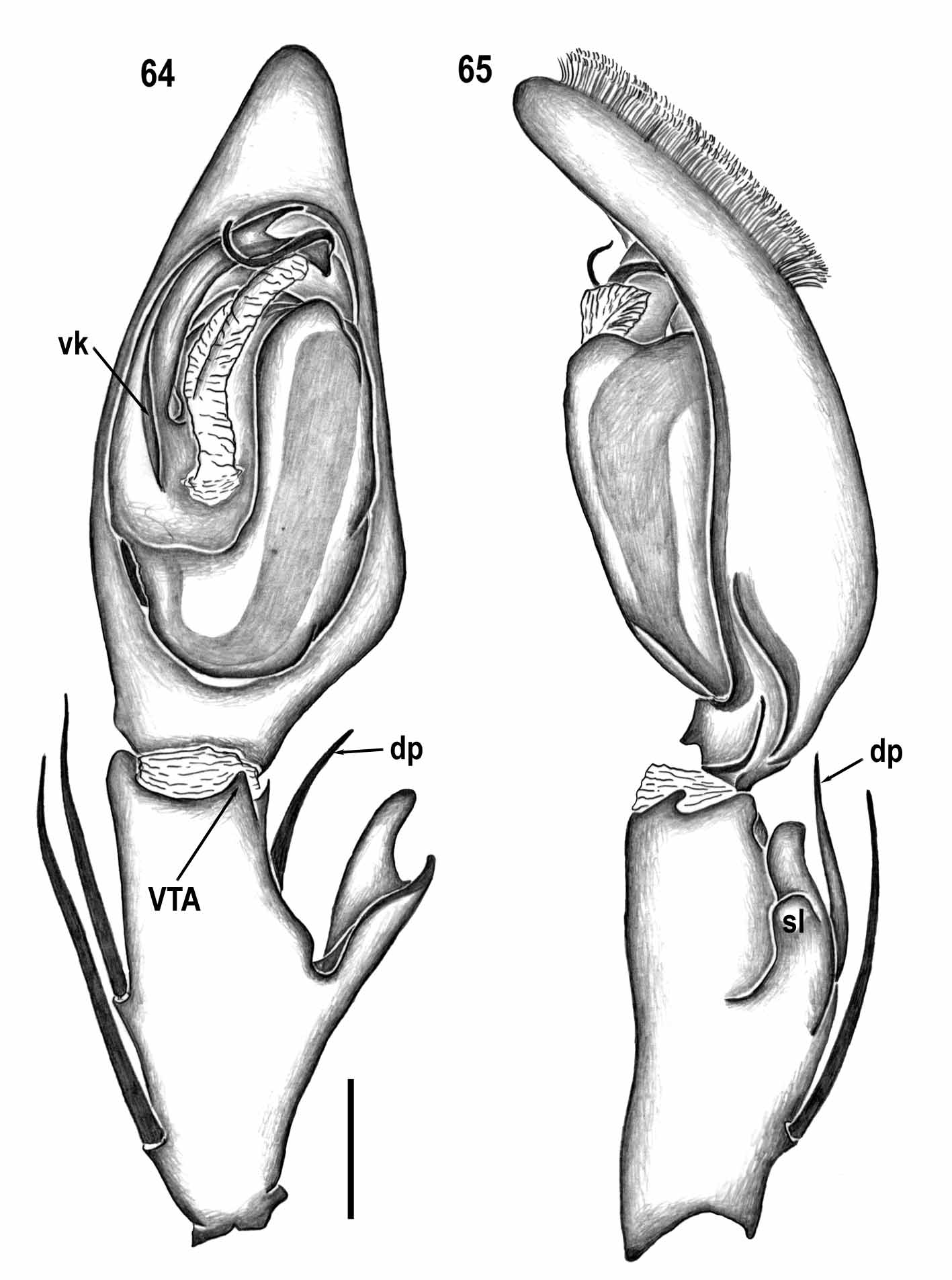
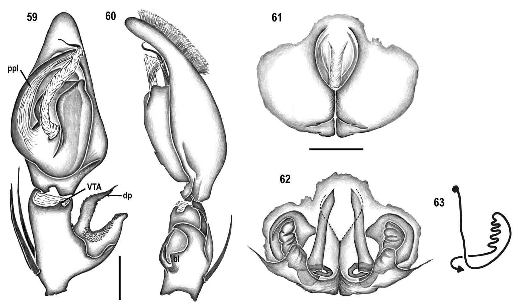
Diagnosis. Species of the genus Caayguara gen. nov. are distinguished from those of other Sparassidae genera by the combination of the following characters: intermarginal denticles on the chelicerae ( Fig. 2 View FIGURES 2 – 7 ), only two pairs of spines on ventral side of tibiae and one lateral spine on metatarsi I–II; male palp with RTA arising from medial to basal retrolateral tibia, and embolus with keels and projections (e.g., Figs 14 View FIGURES 14 – 18 , 29 View FIGURES 29 – 33 , 34 View FIGURES 34 – 38 , 54 View FIGURES 54 – 58 , 66 View FIGURES 66 – 70 ); and female vulva with median part of copulatory ducts expanded, forming a sac-like structure, and posterior part with several linearly arranged lobes (e.g., Figs 17–18 View FIGURES 14 – 18 , 32–33 View FIGURES 29 – 33 ; 52–53, 57–58, 62–63).
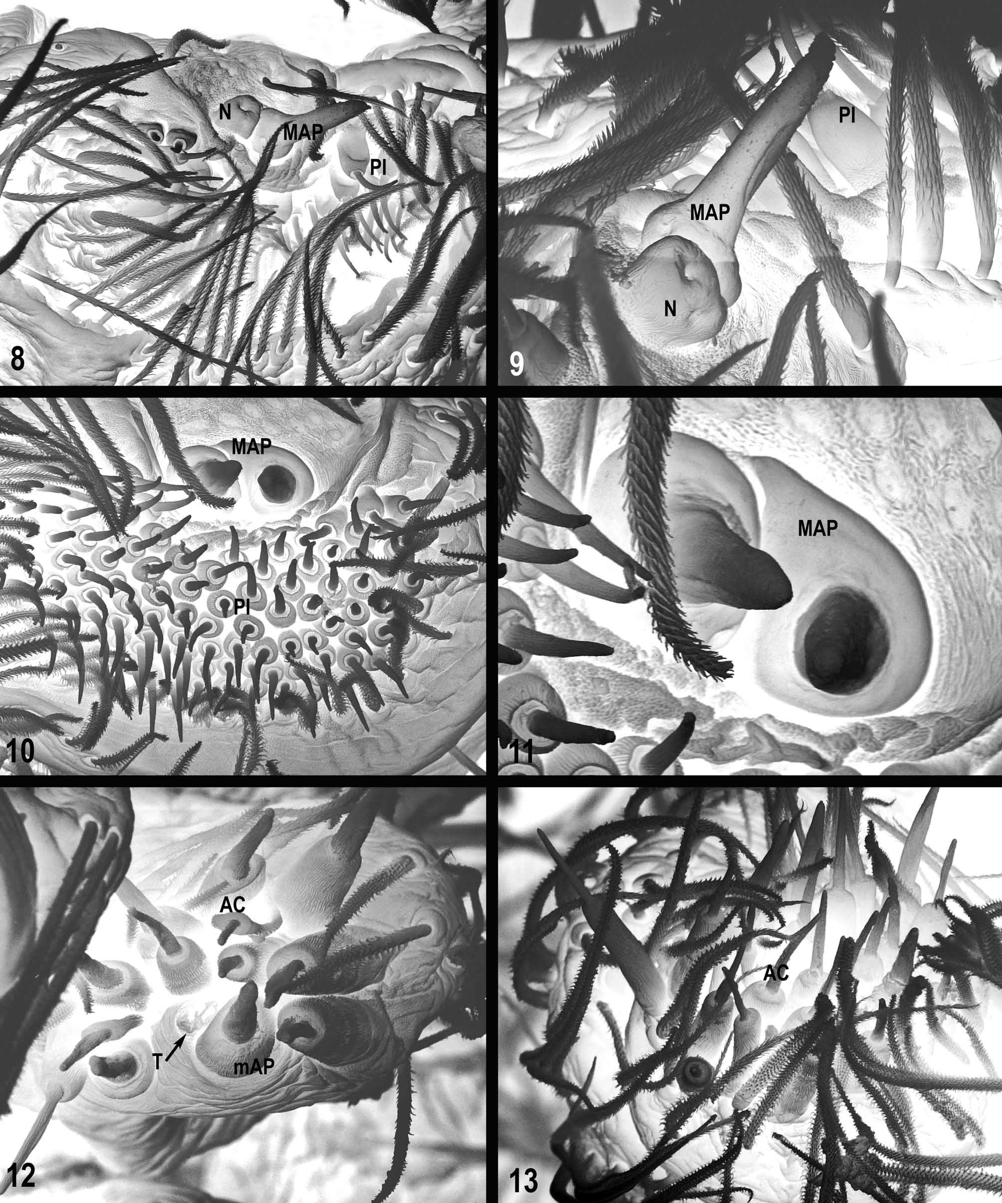
Description. Total length of males 5.9–12.0, of females 6.3–12.8. Prosoma slightly longer than wide; cephalic region slightly higher than thoracic region, gradually flattening posteriorly; fovea conspicuous on posterior third of prosoma. Eight eyes arranged in two rows, the anterior slightly recurved or straight, the posterior straight; AME very slightly larger than ALE and more distant from each other than from ALE; PME smaller than PLE and equidistant. Clypeus low, less than AME diameter. Chelicerae longer than wide with 3 promarginal teeth, the median one largest, and four retromarginal teeth, the most basal one smallest. Intermarginal denticles present mostly at the base of the furrow ( Fig. 2 View FIGURES 2 – 7 ). One single strong seta at the base of fang ( Fig. 2 View FIGURES 2 – 7 ). Labium slightly longer than wide. Endites slightly convergent, with dense scopulae on internal margin. Serrula with a single row of denticles. Sternum as long as wide, very slightly projected between coxae IV. Legs laterigrade (2143). Spination in males: femora I–III: p1-1-1; d0-1-1; r1-1-1; femur IV: p1-1-1; d0-1- 1; r0-0-1; tibiae I–II: p1-0-1; d1-0-1; r1-0-1; v2-2 -0; tibiae III–IV: p1-0-1; d0-0-1; r1-0-1; v2-2 -0; metatarsi I– II: p1-0-0; r1-0-0; v2-2 -0; metatarsus III: p1-1-0; r1-1-0; v2-2 -0; metatarsus IV: p1-1-2; r1-1-1; v2-2 -0. Spination in females as in males except tibiae I–II: d0-0-1; tibia IV: d0. Metatarsi I–IV distally with dorsal trilobate membrane with median hook slightly larger than lateral projections ( Fig. 3 View FIGURES 2 – 7 ). Tarsi and anterior half of metatarsi scopulate. Trichobothria present on dorsal side of tibiae, metatarsi and tarsi, arranged in two or more rows on tarsi and one on metatarsi. Bothrium with dorsal plate with one distal grove, projected over smooth basal plate ( Fig. 4 View FIGURES 2 – 7 ). Tarsal organ capsulate with slightly oval opening ( Fig. 5 View FIGURES 2 – 7 ), located dorsally on distal third of metatarsi. Leg tarsi with pair of pectinate claws with 12–15 very slightly curved teeth and claw tufts ( Fig. 6 View FIGURES 2 – 7 ). Female pedipalp claw with 4–5 short, slightly curved teeth ( Fig. 7 View FIGURES 2 – 7 ). Opisthosoma oval, longer than wide. Six spinnerets: ALS contiguous, conical and bi-segmented, distal segment with several piriform gland spigots, one major ampullate gland spigot and one nubbin in male ( Figs 8–9 View FIGURES 8 – 13 ) and two major ampullate gland spigots in female ( Figs 10–11 View FIGURES 8 – 13 ); AMS short and truncated with one minor ampullate gland spigot, one tartipore, 6–8 cylindrical gland spigots and 5–6 aciniform gland spigots ( Fig. 12 View FIGURES 8 – 13 ); PLS conical and bi-segmented, distal segment with many aciniform gland spigots ( Fig. 13 View FIGURES 8 – 13 ). Male palp: tibia slightly elongate, as long as cymbium, with three prolateral spines and one dorsal spine; VTA small and slightly dislocated retrolaterally (e.g., Figs 14 View FIGURES 14 – 18 , 19, 54, 59, 66); RTA, arising from medial to basal retrolateral tibia, being simple (e.g., Figs 40 View FIGURES 39 – 43 , 45 View FIGURES 44 – 48 , 50 View FIGURES 49 – 53 , 67 View FIGURES 66 – 70 ) or having ventral lobes, one subdistal and one at base ( Figs 15 View FIGURES 14 – 18 , 20), only one subdistal ( Figs 25 View FIGURES 24 – 28 , 30 View FIGURES 29 – 33 , 35 View FIGURES 34 – 38 , 55 View FIGURES 54 – 58 , 65 View FIGURES 64 – 65 ), or only one at base ( Fig. 60 View FIGURES 59 – 63 ); dorsal projection, when present, spine-like, almost as long or longer than RTA ( Figs 24 View FIGURES 24 – 28 , 59 View FIGURES 59 – 63 , 64 View FIGURES 64 – 65 ) or shorter ( Figs 30 View FIGURES 29 – 33 , 34 View FIGURES 34 – 38 , 39 View FIGURES 39 – 43 , 44 View FIGURES 44 – 48 ); cymbium with large rounded alveolus and dorsal scopula; subtegulum smooth, slightly prolateral; tegulum rounded, smooth ( Figs 24 View FIGURES 24 – 28 , 29 View FIGURES 29 – 33 , 34 View FIGURES 34 – 38 , 44 View FIGURES 44 – 48 , 64 View FIGURES 64 – 65 ) or with projections ( Figs 14 View FIGURES 14 – 18 , 19, 39, 49, 59, 66); conductor hyaline arising medially from tegulum; embolus long and slightly curved retrolaterally, arising between 7–8 o’clock (e.g., Figs 29 View FIGURES 29 – 33 , 34 View FIGURES 34 – 38 , 54 View FIGURES 54 – 58 , 59 View FIGURES 59 – 63 , 66 View FIGURES 66 – 70 ), with pars pendula inconspicuous ( Figs 14 View FIGURES 14 – 18 , 24 View FIGURES 24 – 28 , 29 View FIGURES 29 – 33 , 34 View FIGURES 34 – 38 , 39 View FIGURES 39 – 43 , 44 View FIGURES 44 – 48 , 54 View FIGURES 54 – 58 , 64 View FIGURES 64 – 65 ) or well developed (Figs 19, 49, 59, 66). Female epigynum: epigynal field slightly rounded, wider than long or as wide as long, divided into lateral lobes and median septum; lateral lobes (LL) simple, without projections, touching each other posteriorly; median septum (MS) with pair of anterior copulatory openings (e.g. Figs 16 View FIGURES 14 – 18 , 21, 26). Vulva: duct system with anterior part (ap), slender and elongate, running from anterior to posterior; median part (mp) dilated, running in a lateral loop posteriad to ap; posterior part (pp) slender and multilobed, running transversely (e.g. Figs 17–18 View FIGURES 14 – 18 , 42–43 View FIGURES 39 – 43 ), or longitudinally (e.g. Figs 57–58 View FIGURES 54 – 58 , 62–63 View FIGURES 59 – 63 ) towards hook-shaped fertilization ducts.
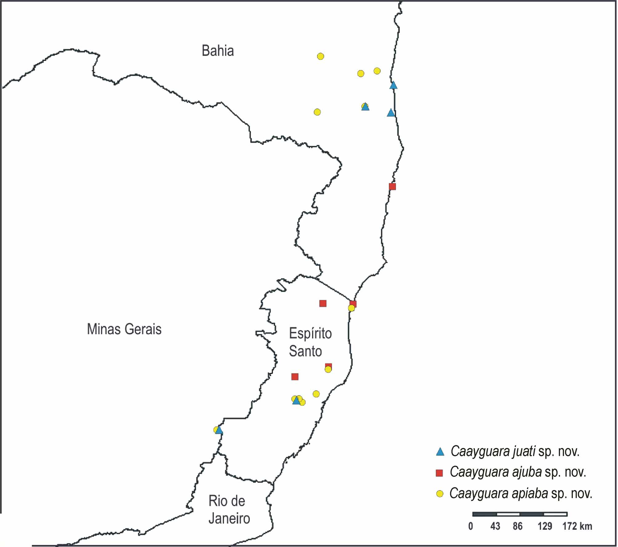
Distribution. Brazilian Atlantic forest, from southern Bahia to Rio Grande do Sul ( Figs 83−87 View FIGURE 83 View FIGURE 84 View FIGURE 85 View FIGURE 86 View FIGURE 87 ).
Composition. Twelve species: Caayguara albus (Mello-Leitão) comb. nov.; C. pinda sp. nov.; C. ajuba sp. nov.; C. juati sp. nov.; C. apiaba sp. nov.; C. cupepemassu sp. nov.; C. cupepemayri sp. nov.; C. itajucamussi sp. nov.; C. ybyratyriguara sp. nov.; C. atyaia sp. nov.; C. poi sp. nov.; C. catuoca sp. nov..
Remarks. Although no cladistic analysis has been tackled for the family as a whole and not much can be said about its subfamilial limits and relationships, the genus seems to be closely related to both Sparassinae Bertkau and Heteropodinae Thorell. It shares with Sparassinae the presence of only two pairs of spines on ventral tibia (Rheims et al. 2008) and an expanded, membranous region of the copulatory ducts (subepigyneal sac sensu Järvi, 1912). It shares with Heteropodinae the presence of three promarginal teeth and intermarginal denticles at the chelicerae ( Jäger 1998) but lacks the long toothed female pedipalp claws, characteristic for this subfamily.


No known copyright restrictions apply. See Agosti, D., Egloff, W., 2009. Taxonomic information exchange and copyright: the Plazi approach. BMC Research Notes 2009, 2:53 for further explanation.