
Anthonomus, Germar, 1817
|
publication ID |
https://doi.org/10.11646/zootaxa.5032.4.1 |
|
publication LSID |
lsid:zoobank.org:pub:0C2AFB68-79AE-4A9E-B09F-341828F93792 |
|
persistent identifier |
https://treatment.plazi.org/id/03C6E508-FF92-DC4A-A3E7-C9482DB6FE82 |
|
treatment provided by |
Plazi (2021-09-09 15:12:58, last updated 2023-11-08 01:17:13) |
|
scientific name |
Anthonomus |
| status |
|
Key to known immature stages of the Palaearctic species of the genus Anthonomus View in CoL
The larva and pupa of A. rectirostris and the pupae of A. pedicularius and A. pyri have not been studied by me and are included according to the works of Burke (1968) and Ahmad & Burke (1972). It should be borne in mind that according to the works of Marvaldi (1997; 1999), des 3 in the sense of Ahmad and Burke should be called des 4, while des 4, respectively, des 3, pds 5 on meso- and metathorax are alar setae (as). I did not include the larva of A. bisignifer described by Lee & Morimoto (1996) in the key (see discussion).
Larvae ( Figs. 1–11 View FIGURES 1–5 View FIGURES 6–11 , 18–28 View FIGURES 18–22 View FIGURES 23–28 , 37–47 View FIGURES 37–41 View FIGURES 42–47 , 51–61 View FIGURES 51–55 View FIGURES 56–61 , 67–77 View FIGURES 67–71 View FIGURES 72–77 , 84–94 View FIGURES 84–88 View FIGURES 89–94 )
1. Epicranium with four dorsal setae (des 4 absent) ( Figs. 56 View FIGURES 56–61 , 72 View FIGURES 72–77 ).................................................. 2
- Epicranium with five dorsal setae (des 4 present) ( Figs. 6 View FIGURES 6–11 , 23 View FIGURES 23–28 , 42 View FIGURES 42–47 , 89 View FIGURES 89–94 )............................................ 5
2. Endocarinal line is more than 2/3 as long as frons. Epipharynx with two als. Meso- and metathorax with short pds 1 and long pds 2. Pda 5 very short............................................................... A. (Furcipus) rectirostris View in CoL
- Endocarinal line is about 2/3 as long as frons ( Figs. 56 View FIGURES 56–61 , 72 View FIGURES 72–77 ). Epipharynx with three als ( Figs. 59 View FIGURES 56–61 , 75 View FIGURES 72–77 ). Meso- and metathorax ( Figs. 53 View FIGURES 51–55 , 69 View FIGURES 67–71 ) with long pds 1 and short pds 2. Pda 5 minute..................................................... 3
3. On meso- (Th2) and metathorax (Th3) ( Fig. 69 View FIGURES 67–71 ) pds 1 and pds 3 —long, pds 2 and pds 4 —short, as —long...... A. (s. str.) latior View in CoL
- On meso- (Th2) and metathorax (Th3) ( Fig. 53 View FIGURES 51–55 ) pds 1, pds 3, pds 4 —long, pds 2 —short, as —short....................... 4
4. Body length: 2.62–3.16 (mean 2.89). Pedal lobe with one long, two medium and two minute pda ( Fig. 53 View FIGURES 51–55 ). Mala of maxilla on ventral side with four vms ( Fig. 61 View FIGURES 56–61 )...................................................... A. (s. str.) conspersus View in CoL
- Body length: 3.04–3.78 (mean 3.34). Pedal lobe with two long, two short and one minute pda. Mala of maxilla on ventral side with five vms ...................................................................... A. (s. str.) pedicularius View in CoL
5. Pms 1 — long, about 2.5x as long as pms 3 ( Fig. 47 View FIGURES 42–47 ). Abdominal segments ( Fig. 40 View FIGURES 37–41 ) with long e ps 1, long ps, and medium lsts ( Fig. 18 View FIGURES 18–22 )................................................................................................ 6
- Pms 1 — short or medium, as long as pms 3, or slightly longer than pms 3 ( Figs. 11 View FIGURES 6–11 , 28 View FIGURES 23–28 , 94 View FIGURES 89–94 ). Abdominal segments with short e ps 1, short ps, and very short lsts ( Figs. 4 View FIGURES 1–5 , 21 View FIGURES 18–22 , 87 View FIGURES 84–88 )................................................................ 7
6. Body length: 3.78–4.78 (mean 4.25). Host plants: Malus View in CoL , Pyrus View in CoL ................................. A. (s. str.) pomorum View in CoL
- Body length: 2.87–4.11 (mean 3.49). Host plants: Padus , Padellus , Cerasus ....................... A. (s. str.) incurvus View in CoL
7. Head dark brown ( Fig. 1 View FIGURES 1–5 ). Four epipharyngeal sensilla combined in a single median cluster (esc) ( Fig. 8 View FIGURES 6–11 ). Prothorax (Th1) ( Fig. 3 View FIGURES 1–5 ) with ten prns; prns 1 medium. Pedal lobe ( Fig. 3 View FIGURES 1–5 ) contains only two long pda (the rest of the pda short or minute).................................................................................... A. (Anthomorphus) pinivorax View in CoL
- Head pale yellow or brownish yellow ( Figs. 18 View FIGURES 18–22 , 85 View FIGURES 84–88 ). Four epipharyngeal sensilla combined in two clusters with two pores in each ( Figs. 26 View FIGURES 23–28 , 93 View FIGURES 89–94 ). Prothorax (Th1) ( Figs. 20 View FIGURES 18–22 , 87 View FIGURES 84–88 ) with eleven prns; prns 1 short. Pedal lobe ( Figs. 20 View FIGURES 18–22 , 87 View FIGURES 84–88 ) contains either three or four long pda (the rest of the pda short or minute)......................................................... 8
8. Pedal lobe ( Fig. 20 View FIGURES 18–22 ) with four very long pda. Eus 1 on thoracic segments (Th1–3) long ( Fig. 20 View FIGURES 18–22 ). Mala of the maxilla on the dorsal side with six dms, on the ventral side with four vms ( Fig. 28 View FIGURES 23–28 ).......... A. (Anthonomidius) rubripes Gyllenhal, 1835 View in CoL
- Pedal lobe ( Fig. 86 View FIGURES 84–88 ) with three very long and one very short pda. Eus 1 on thoracic segments (Th1–3) short ( Fig. 86 View FIGURES 84–88 ). Mala of the maxilla on the dorsal side with five or six dms, on the ventral side with five vms ( Fig. 94 View FIGURES 89–94 )..................................................................................................... A. (s. str.) rubi (Herbst, 1795) View in CoL
Ahmad, M. & Burke, H. R. (1972) Larvae of the weevil tribe Anthonomini (Coleoptera: Curculionidae). Miscellaneous Publications of the Entomological Society of America, 8 (2), 33 - 81.
Burke, H. R. (1968) Pupae of the weevil tribe Anthonomini (Coleoptera, Curculionidae). Technical Monographs Texas Agricultural Experiment Station, 5, 1 - 92.
Lee, Ch. - Y. & Morimoto, K. (1996) Larvae of the Weevil Family Curculionidae of Japan Part 3. Ramphinae to Curculioninae (Insecta: Coleoptera). Journal of the Faculty of Agriculture, Kyushu University, 40 (3 - 4), 287 - 306. https: // doi. org / 10.5109 / 24115
Marvaldi, A. E. (1997) Higher level phylogeny of Curculionidae (Coleoptera: Curculionoidea) based mainly on larval characters, with special reference to broad-nosed weevils. Cladistics, 13, 285 - 312. https: // doi. org / 10.1111 / j. 1096 - 0031.1997. tb 00321. x
Marvaldi, A. E. (1999) Morfologia larval en Curculionidae. Acta Zoologica Lilloana, 45, 7 - 24.

FIGURES 1–5. Anthonomus pinivorax, mature larva. 1—habitus of the body and frontal view of head, 2—thoracic spiracle, 3—lateral view of thoracic segments, 4—lateral view of abdominal segment I, 5—lateral view of abdominal segments VIII–X (setae: as—alar, ds—dorsal, eps—epipleural, eus—eusternal, lsts—laterosternal, pda—pedal, pds—postdorsal, prns—pronotal, prs—prodorsal, ps—pleural, ss—spiracular, sts—sternal).
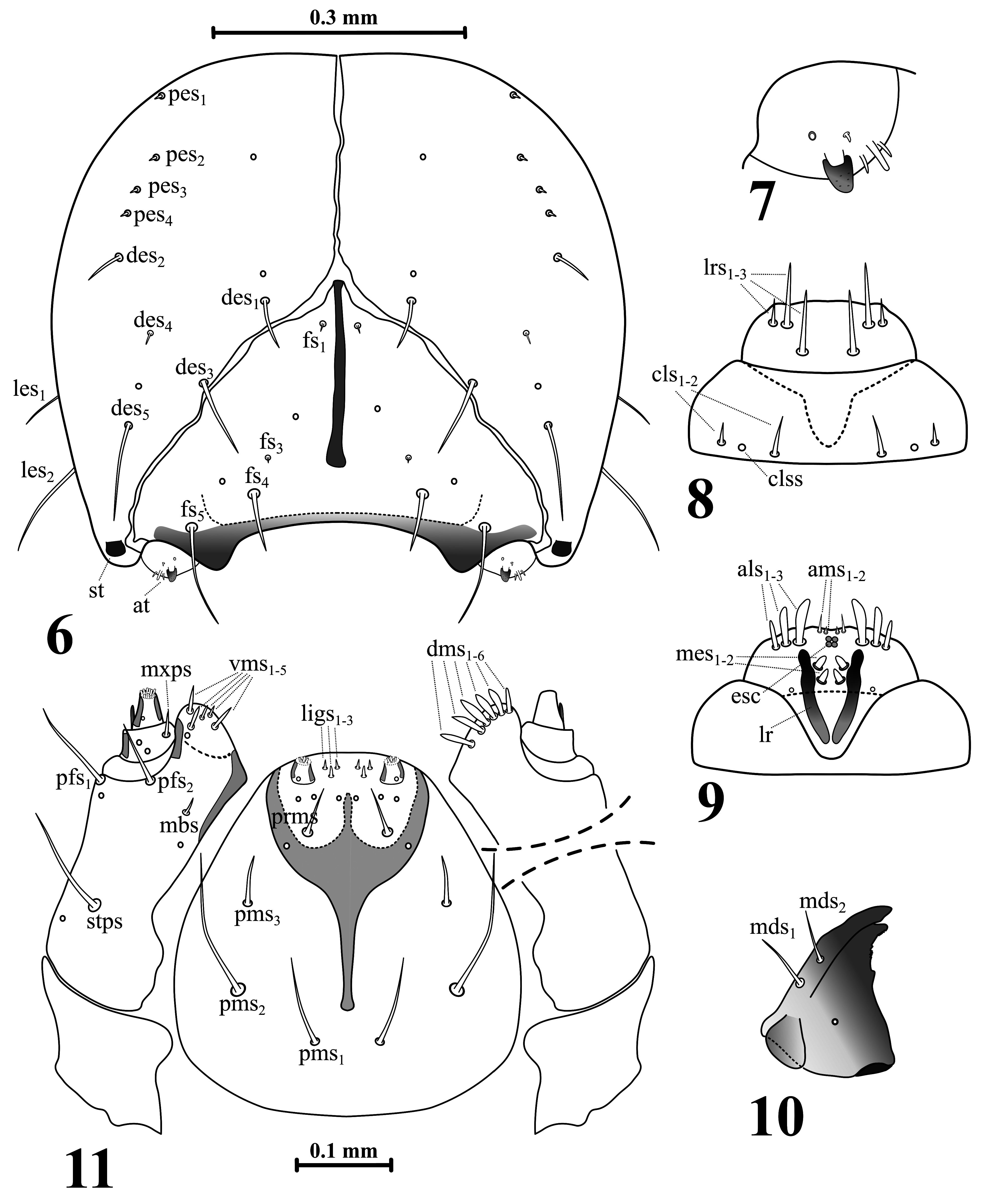
FIGURES 6–11. Anthonomus pinivorax, mature larva head and mouthparts. 6—head, frontal view, 7—antenna, 8—clypeus and labrum, 9—epipharynx, 10—left mandible, 11—maxillolabial complex, ventral aspect (apical part of the right maxilla is shown in dorsal aspect) (at—antenna, clss—clypeal sensilla, esc—epipharyngeal sensilla cluster, lr—labral rods, st—stemma; setae: als—anterolateral, ams—anteromedial, cls—clypeal, des—dorsal epicranial, dms—dorsal malar, fs—frontal, ligs—ligular, lrs—labral, ls—lateral epicranial, mbs—basiventral malar, mds—mandibular, mes—median, mxps—maxillary palp, pes— postepicranial, pfs—palpiferal, pms—postlabial, prms—prelabial, stps—stipal, vms—ventral malar).
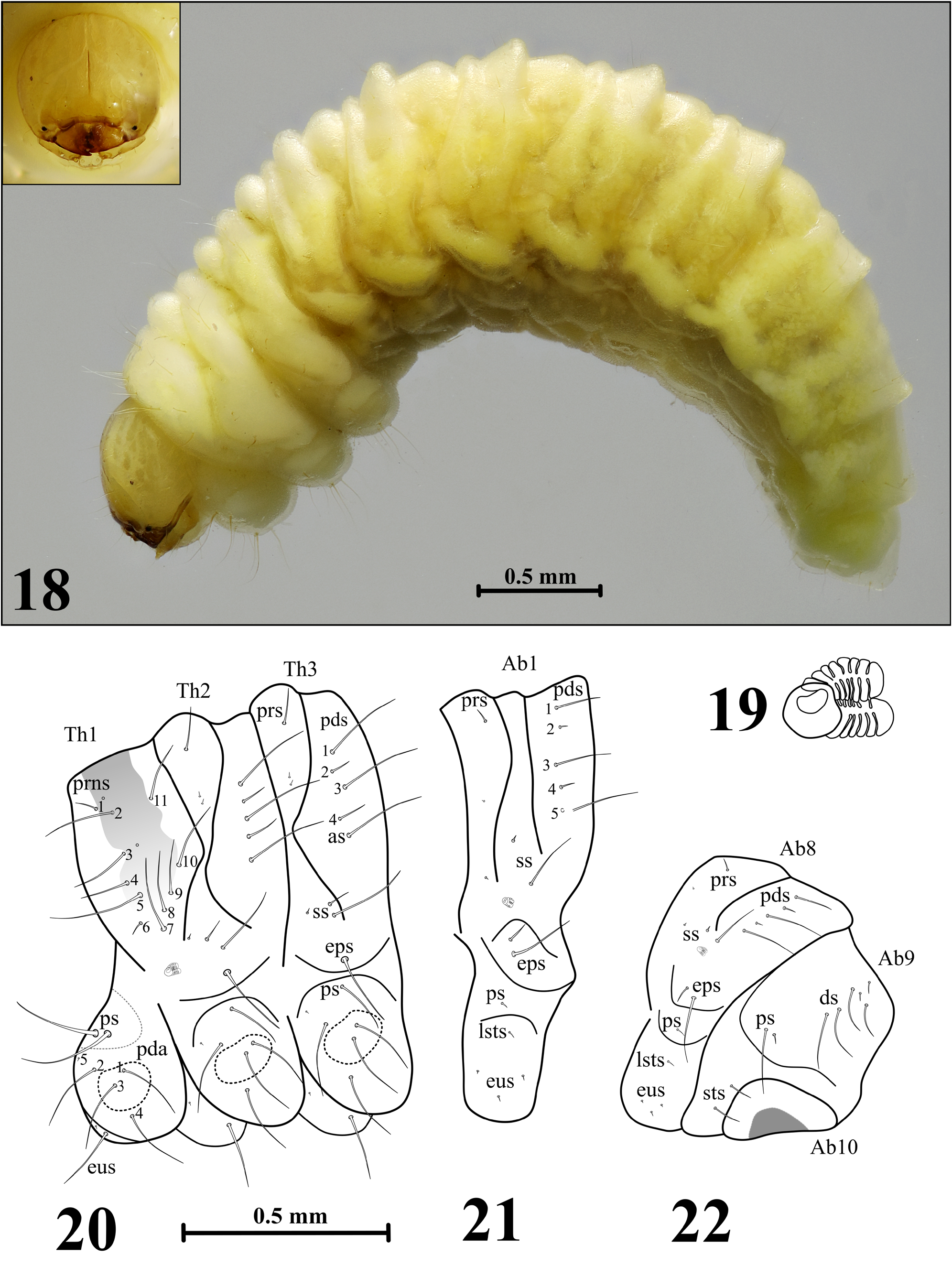
FIGURES 18–22. Anthonomus rubripes, mature larva. 18—habitus of the body and frontal view of head, 19—thoracic spiracle, 20—lateral view of thoracic segments, 21—lateral view of abdominal segment I, 22—lateral view of abdominal segments VIII–X (setae: as—alar, ds—dorsal, eps—epipleural, eus—eusternal, lsts—laterosternal, pda—pedal, pds—postdorsal, prns— pronotal, prs—prodorsal, ps—pleural, ss—spiracular, sts—sternal).

FIGURES 23–28. Anthonomus rubripes, mature larva head and mouthparts. 23—head, frontal view, 24—antenna, 25—clypeus and labrum, 26—epipharynx, 27—left mandible, 28—maxillolabial complex, ventral aspect (apical part of the right maxilla is shown in dorsal aspect) (at—antenna, clss—clypeal sensilla, lr—labral rods, esc—epipharyngeal sensilla cluster, st—stemma; setae: als—anterolateral, ams—anteromedial, cls—clypeal, des—dorsal epicranial, dms—dorsal malar, fs—frontal, ligs—ligular, lrs—labral, ls—lateral epicranial, mbs—basiventral malar, mds—mandibular, mes—median, mxps—maxillary palp, pes— postepicranial, pfs—palpiferal, pms—postlabial, prms—prelabial, stps—stipal, vms—ventral malar).
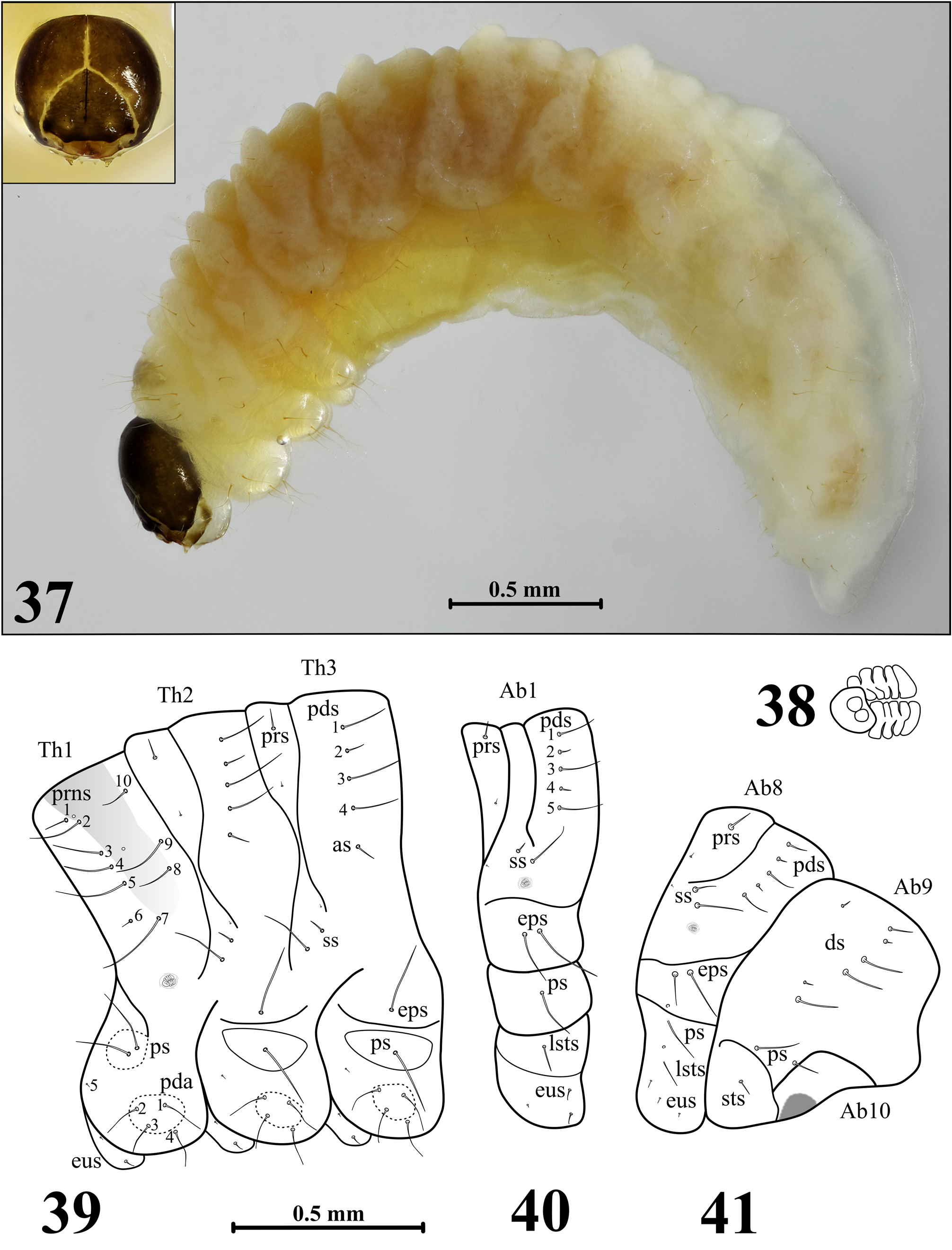
FIGURES 37–41. Anthonomus incurvus, mature larva. 37—habitus of the body and frontal view of head, 38—thoracic spiracle, 39—lateral view of thoracic segments, 40—lateral view of abdominal segment I, 41—lateral view of abdominal segments VIII–X (setae: as—alar, ds—dorsal, eps—epipleural, eus—eusternal, lsts—laterosternal, pda—pedal, pds—postdorsal, prns— pronotal, prs—prodorsal, ps—pleural, ss—spiracular, sts—sternal).
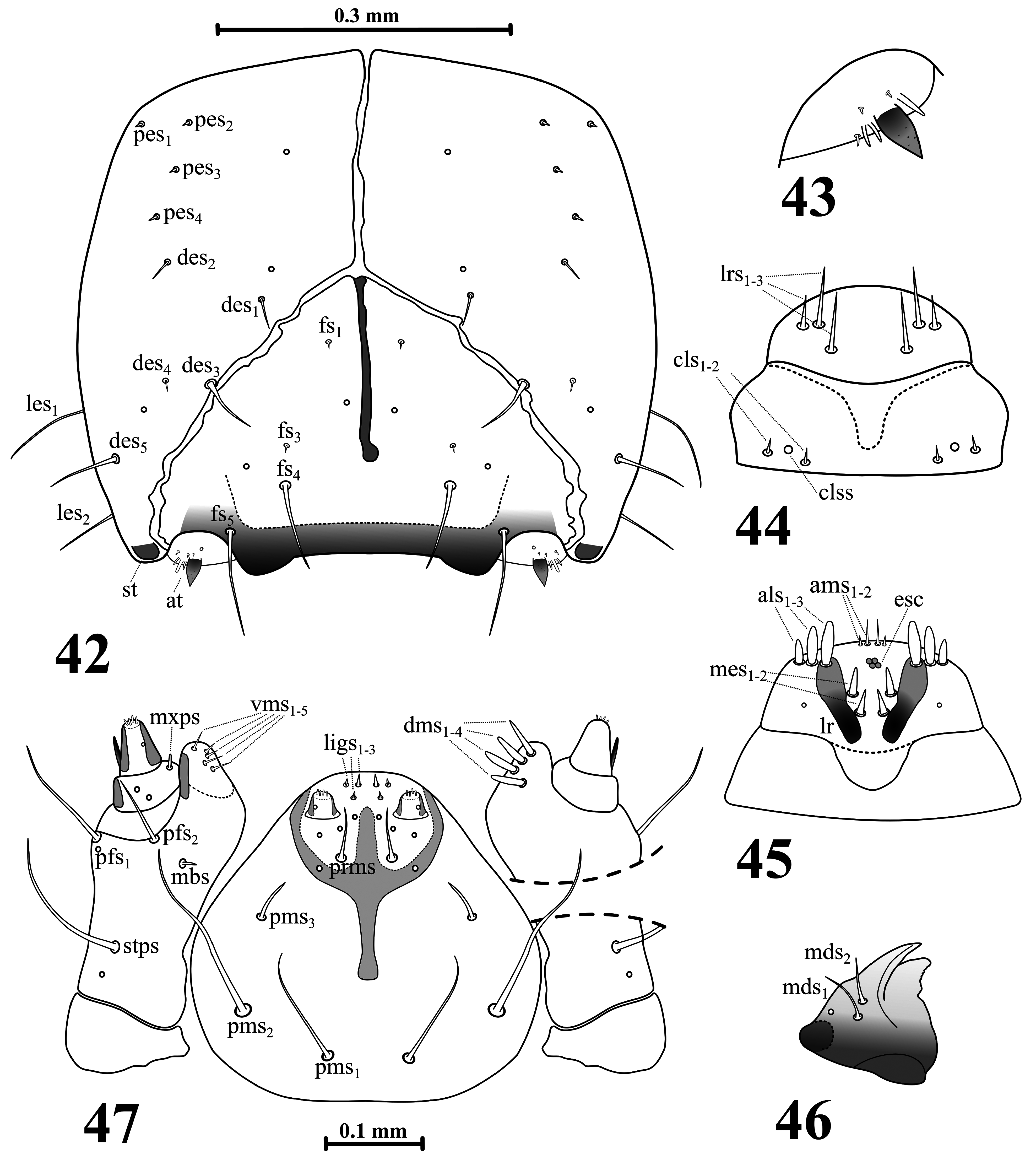
FIGURES 42–47. Anthonomus incurvus, mature larva head and mouthparts. 42—head, frontal view, 43—antenna, 44—clypeus and labrum, 45—epipharynx, 46—left mandible, 47—maxillolabial complex, ventral aspect (apical part of the right maxilla is shown in dorsal aspect) (at—antenna, clss—clypeal sensilla, esc—epipharyngeal sensilla cluster, lr—labral rods, st—stemma; setae: als—anterolateral, ams—anteromedial, cls—clypeal, des—dorsal epicranial, dms—dorsal malar, fs—frontal, ligs—ligular, lrs—labral, ls—lateral epicranial, mbs—basiventral malar, mds—mandibular, mes—median, mxps—maxillary palp, pes— postepicranial, pfs—palpiferal, pms—postlabial, prms—prelabial, stps—stipal, vms—ventral malar).
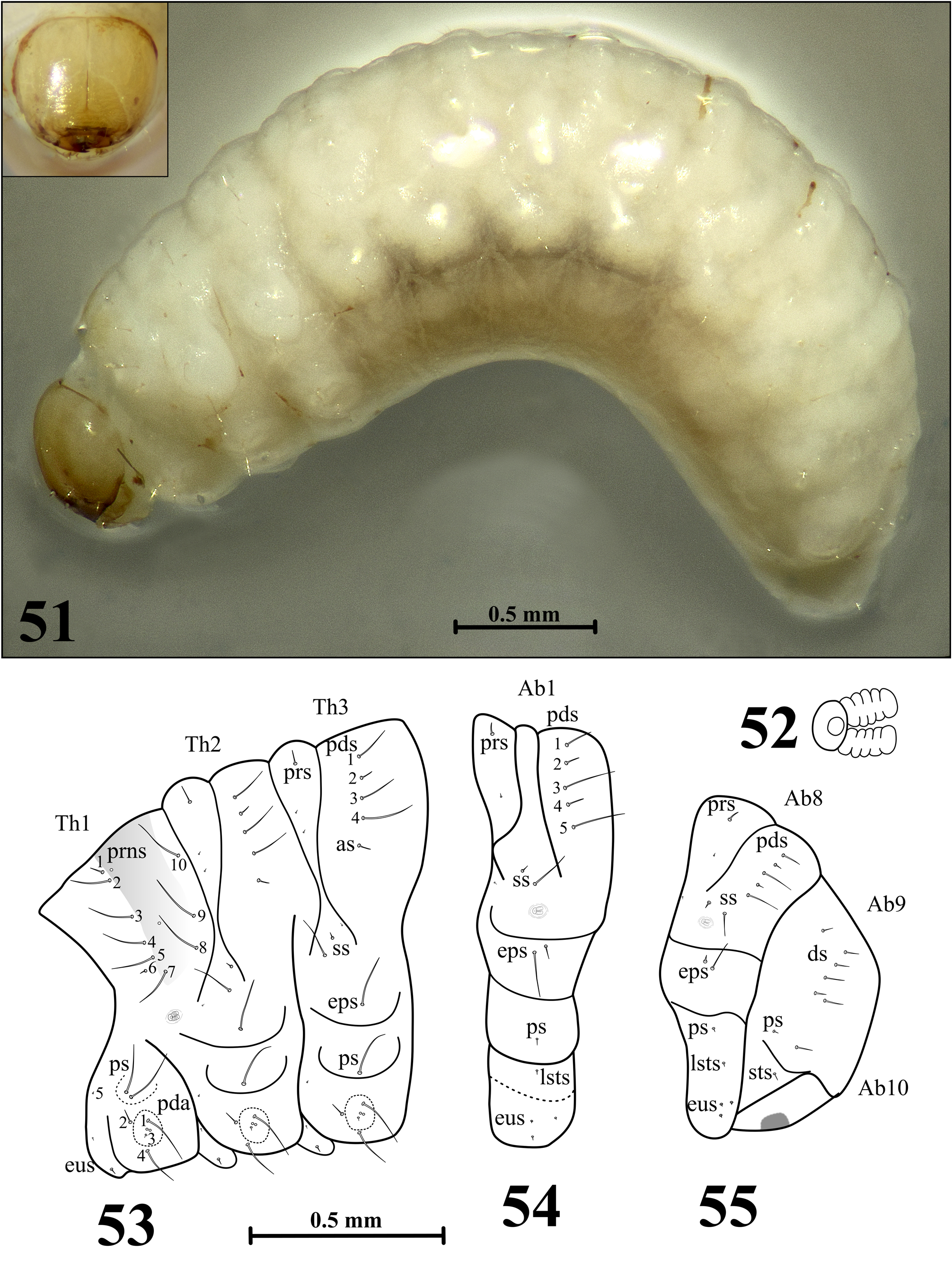
FIGURES 51–55. Anthonomus conspersus, mature larva. 51—habitus of the body and frontal view of the head, 52—thoracic spiracle, 53—lateral view of thoracic segments, 54—lateral view of abdominal segment I, 55—lateral view of abdominal segments VIII–X (setae: as—alar, ds—dorsal, eps—epipleural, eus—eusternal, lsts—laterosternal, pda—pedal, pds—postdorsal, prns—pronotal, prs—prodorsal, ps—pleural, ss—spiracular, sts—sternal).
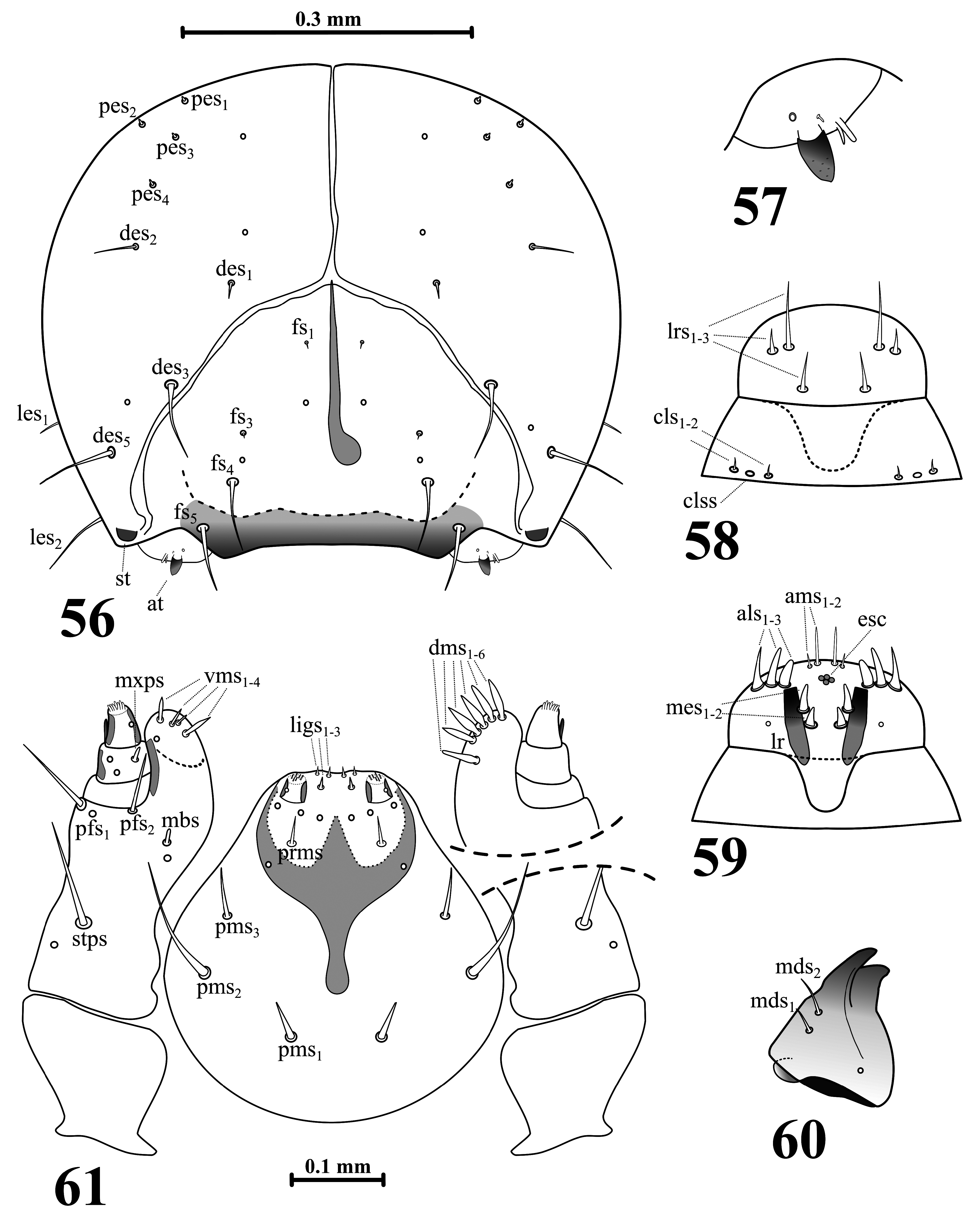
FIGURES 56–61. Anthonomus conspersus, mature larva head and mouthparts. 56—head, frontal view, 57—antenna, 58— clypeus and labrum, 59—epipharynx, 60—left mandible, 61—maxillolabial complex, ventral aspect (apical part of the right maxilla is shown in dorsal aspect) (at—antenna, clss—clypeal sensilla, lr—labral rods, st—stemma; setae: als—anterolateral, ams—anteromedial, cls—clypeal, des—dorsal epicranial, dms—dorsal malar, fs—frontal, ligs—ligular, lrs—labral, ls—lateral epicranial, mbs—basiventral malar, mds—mandibular, mes—median, mxps—maxillary palp, pes—postepicranial, pfs—palpiferal, pms—postlabial, prms—prelabial, stps—stipal, vms—ventral malar).
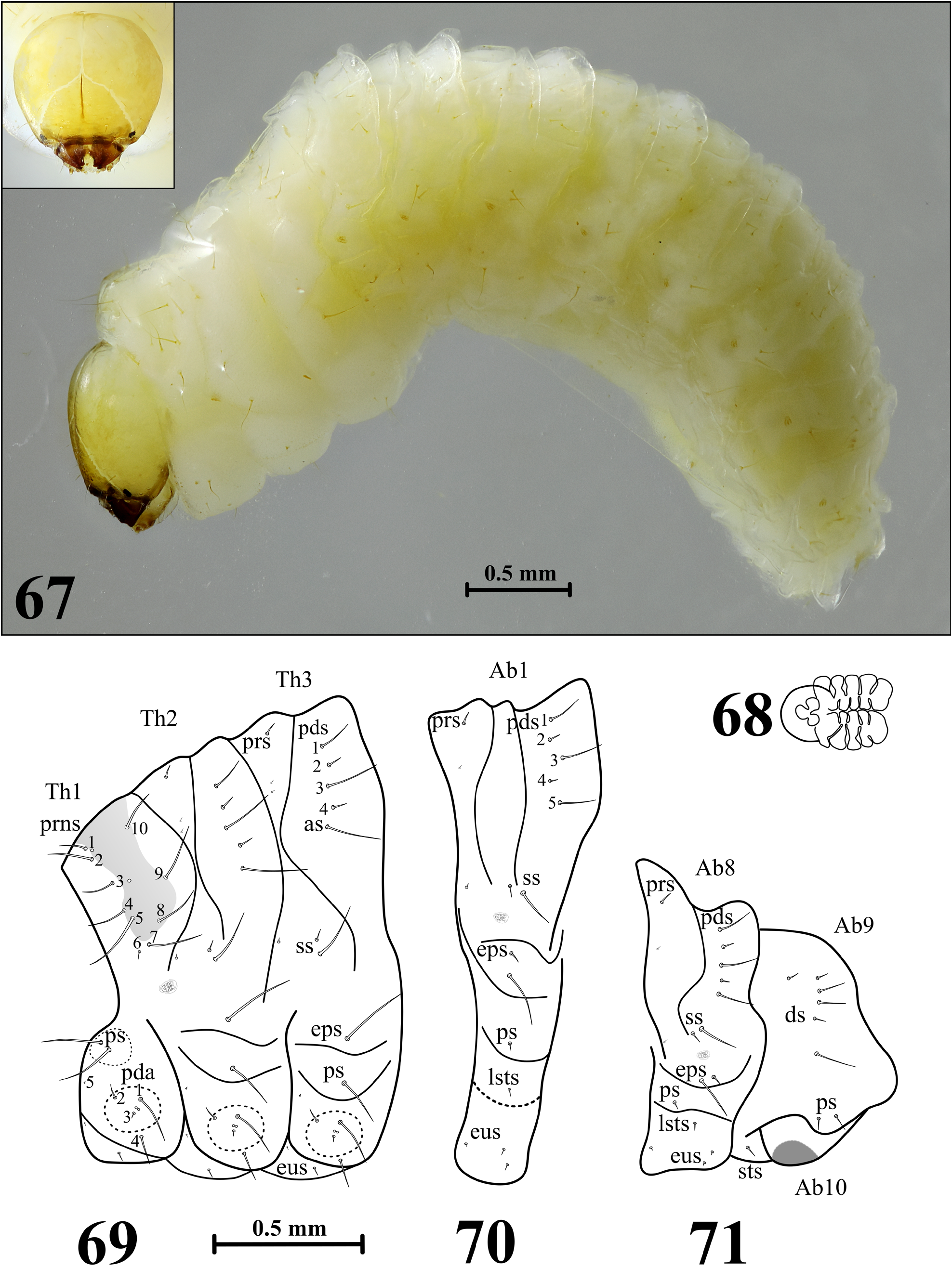
FIGURES 67–71. Anthonomus latior, mature larva. 67—habitus of the body and frontal view of head, 68—thoracic spiracle, 69—lateral view of thoracic segments, 70—lateral view of abdominal segment I, 71—lateral view of abdominal segments VIII–X (setae: as—alar, ds—dorsal, eps—epipleural, eus—eusternal, lsts—laterosternal, pda—pedal, pds—postdorsal, prns— pronotal, prs—prodorsal, ps—pleural, ss—spiracular, sts—sternal).
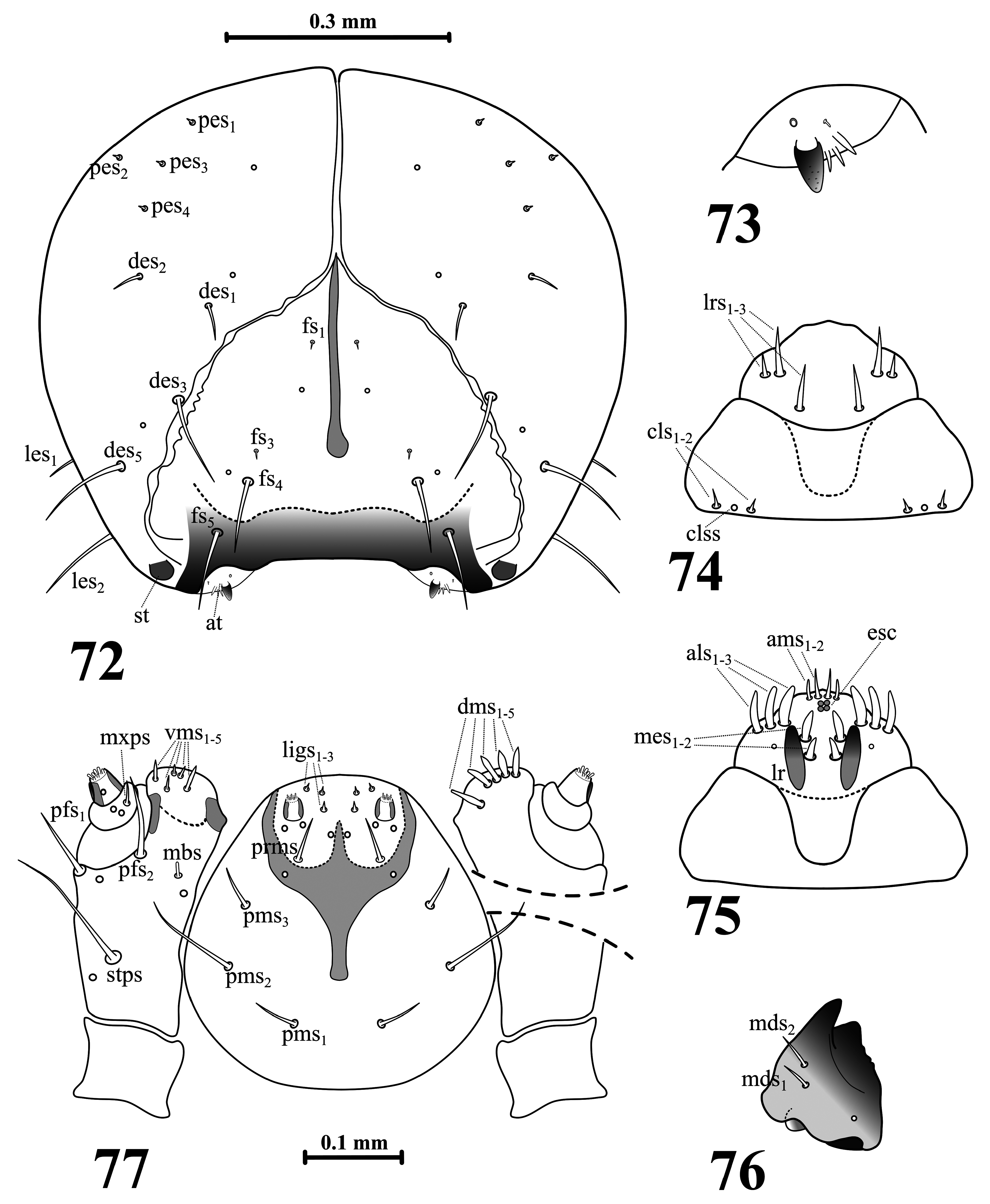
FIGURES 72–77. Anthonomus latior, mature larva head and mouthparts. 72—head, frontal view, 73—antenna, 74—clypeus and labrum, 75—epipharynx, 76—left mandible, 77—maxillolabial complex, ventral aspect (apical part of the right maxilla is shown in dorsal aspect) (at—antenna, clss—clypeal sensilla, lr—labral rods, st—stemma; setae: als—anterolateral, ams—anteromedial, cls—clypeal, des—dorsal epicranial, dms—dorsal malar, fs—frontal, ligs—ligular, lrs—labral, ls—lateral epicranial, mbs—basiventral malar, mds—mandibular, mes—median, mxps—maxillary palp, pes—postepicranial, pfs—palpiferal, pms—postlabial, prms—prelabial, stps—stipal, vms—ventral malar).
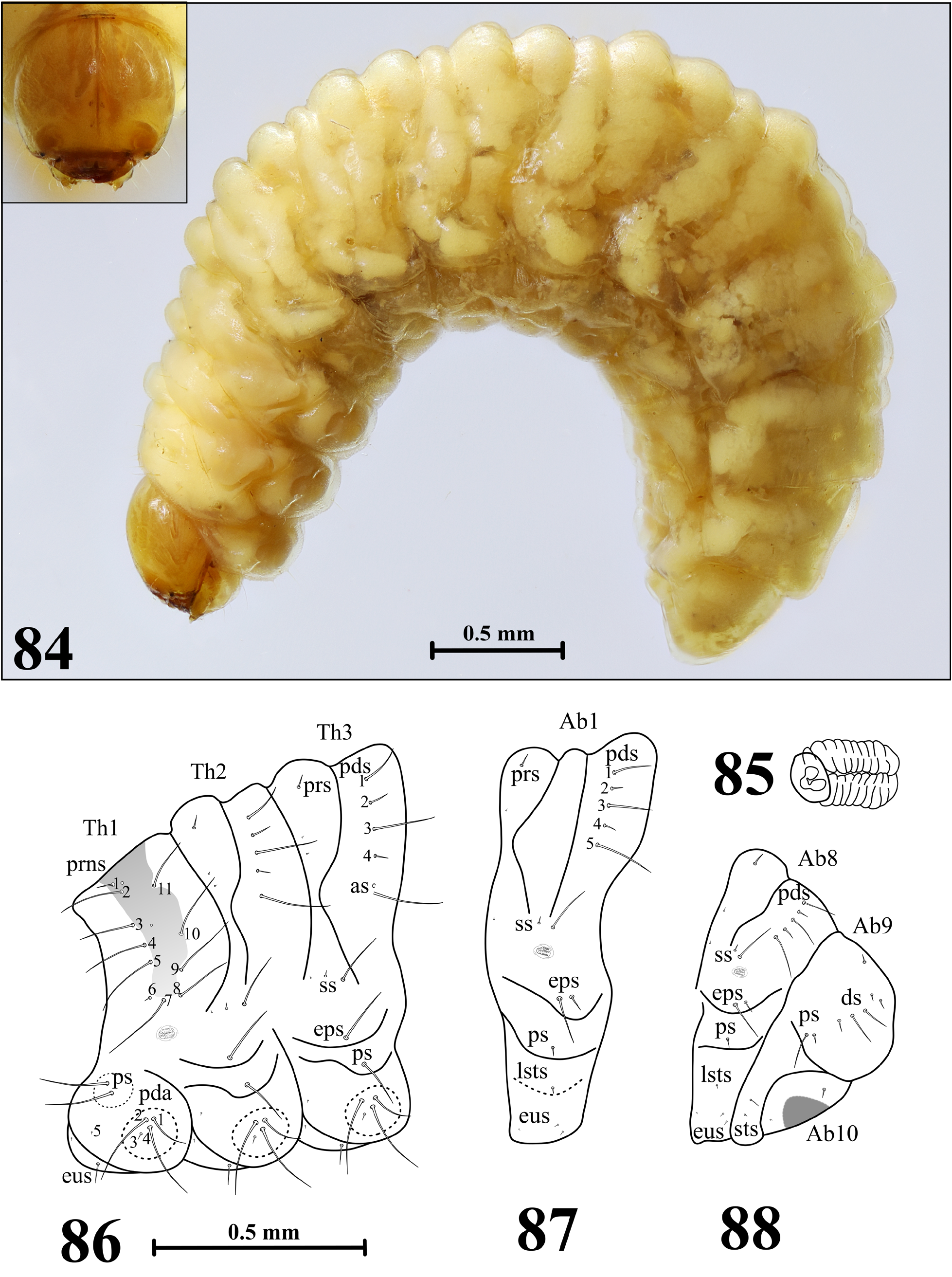
FIGURES 84–88. Anthonomus rubi, mature larva. 84—habitus of the body and frontal view of head, 85—thoracic spiracle, 86—lateral view of thoracic segments, 87—lateral view of abdominal segment I, 88—lateral view of abdominal segments VIII–X (setae: as—alar, ds—dorsal, eps—epipleural, eus—eusternal, lsts—laterosternal, pda—pedal, pds—postdorsal, prns— pronotal, prs—prodorsal, ps—pleural, ss—spiracular, sts—sternal).
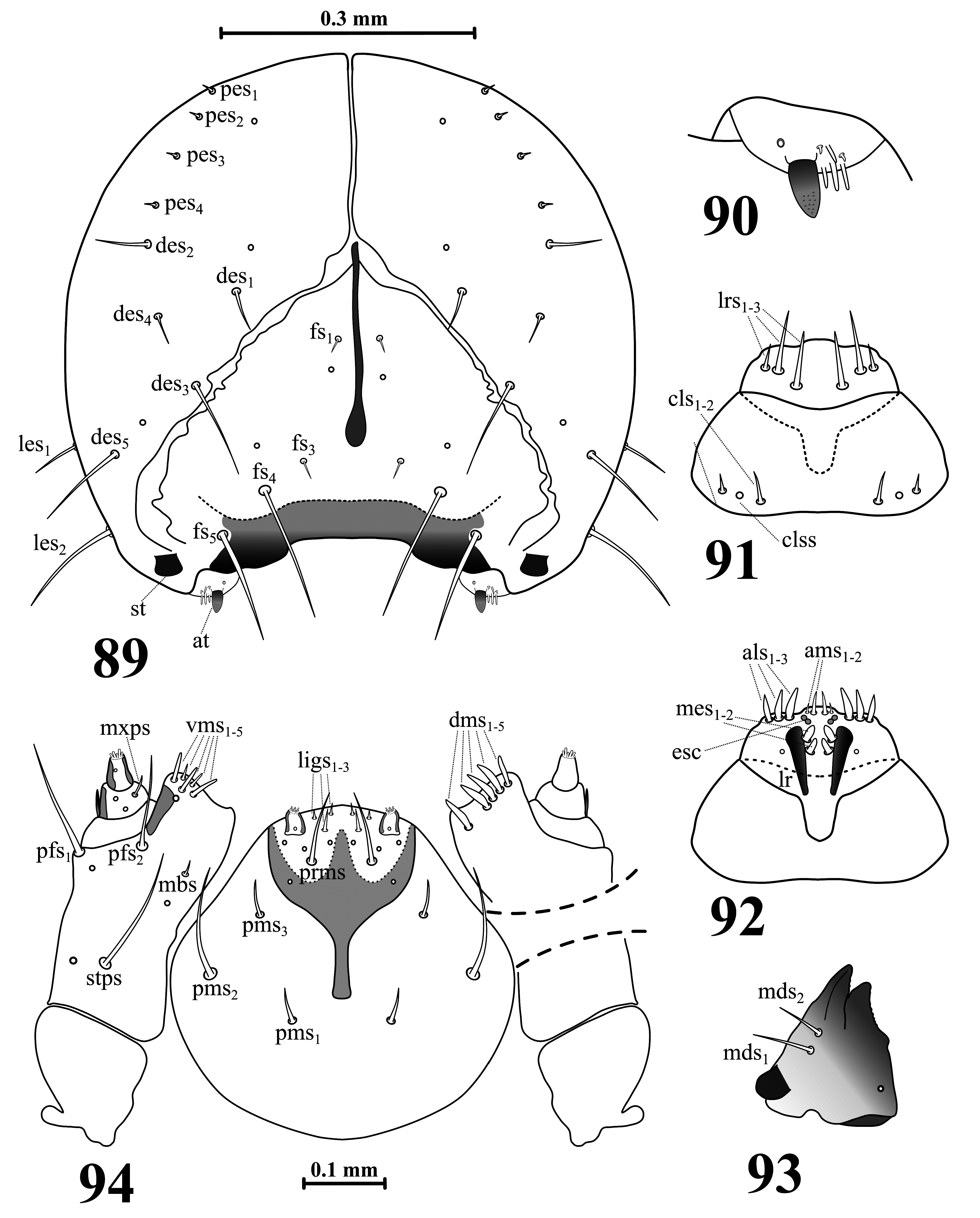
FIGURES 89–94. Anthonomus rubi, mature larva head and mouthparts. 89—head, frontal view, 90—antenna, 91—clypeus and labrum, 92—epipharynx, 93—left mandible, 94—maxillolabial complex, ventral aspect (apical part of the right maxilla is shown in dorsal aspect) (at—antenna, clss—clypeal sensilla, lr—labral rods, st—stemma; setae: als—anterolateral, ams—anteromedial, cls—clypeal, des—dorsal epicranial, dms—dorsal malar, fs—frontal, ligs—ligular, lrs—labral, ls—lateral epicranial, mbs—basiventral malar, mds—mandibular, mes—median, mxps—maxillary palp, pes—postepicranial, pfs—palpiferal, pms—postlabial, prms—prelabial, stps—stipal, vms—ventral malar).
No known copyright restrictions apply. See Agosti, D., Egloff, W., 2009. Taxonomic information exchange and copyright: the Plazi approach. BMC Research Notes 2009, 2:53 for further explanation.
|
Kingdom |
|
|
Phylum |
|
|
Class |
|
|
Order |
|
|
Family |
|
|
Genus |