
Surirella minuta Brébisson
|
publication ID |
https://doi.org/10.11646/phytotaxa.592.2.11 |
|
DOI |
https://doi.org/10.5281/zenodo.7840354 |
|
persistent identifier |
https://treatment.plazi.org/id/03C387BA-FFB5-FFF6-EFC6-FEA4FCCEFC7D |
|
treatment provided by |
Plazi (2023-04-18 10:55:46, last updated 2024-11-27 18:39:01) |
|
scientific name |
Surirella minuta Brébisson |
| status |
|
Surirella minuta Brébisson in Kützing 1849 (LM: Figs 27–42 View FIGURES 27–42 , SEM: Figs 43–54 View FIGURES 43–48 View FIGURES 49–54 , 104 View FIGURES 102–107 )
Observations:— LM: Valve outline long-ovate to ovate with broadly rounded headpole and cuneate footpole ( Figs 27–42 View FIGURES 27–42 ). Valve dimensions (n = 38): length 20–46 μm, width 8.4–11.0 μm (measured at widest part of the valves), length/width ratio 2.1–4.4. Costa-stria bundles (CSBs) evident, alternated with over-fibula costae (OFCs) from pole to pole. CSBs reaching valve margins. CSBs parallel at valve middle, radiate approaching apices. Fibulae evident, nearly extending to midline, 7–9 in 10 μm.
SEM: Each CSB mostly composed of three costae and four striae, or two costae and three striae ( Figs 43–46 View FIGURES 43–48 ). Striae multiseriate, composed of 3–5 rows of areolae, 25–27 in 10 μm (measured under SEM images, n = 3). Outside openings of areolae slit-like ( Figs 45–48 View FIGURES 43–48 ). Raphe canal located directly on mantle. Distal raphe endings simple and interrupting at both apices ( Figs 47, 48 View FIGURES 43–48 , two arrows respectively). Internally, wall of raphe canal only slightly growing into cell cavity such as the portulae are visible ( Figs 49, 50, 54 View FIGURES 49–54 , 104 View FIGURES 102–107 ). Fibulae medium-sized, occasionally bifurcate, extending to valve midline ( Figs 49–51 View FIGURES 49–54 ). Each fibula corresponds to a sinking in the mantle ( Fig. 51 View FIGURES 49–54 ). Raphe continuous at headpole ( Fig. 52 View FIGURES 49–54 , wavy arrow), but interrupted at footpole ( Fig. 53 View FIGURES 49–54 , two arrows). One portula produced between two adjacent fibulae ( Fig. 54 View FIGURES 49–54 , two arrows, Fig. 104 View FIGURES 102–107 , three arrows).
Distribution:— Surirella minuta was commonly found and was dominant in the assemblages collected on the stone surfaces in the river course. It was associated with S. liubingii , S. lacrimula and S. brebissonii var. kuetzingi . From the literature, this species has a global distribution ( Krammer & Lange-Bertalot 1987, English & Potapova 2012, Bey & Ector 2013, Dedić et al. 2015, Caglar et al. 2022).
Bey, M. - Y. & Ector, L. (2013) Atlas des diatomees des cours d'eau de la region Rhone-Alpes, Tome 6. Bacillariacees, Rhopalodiacees, Surirellacees. DREAL Rhone-Alpes, Lyon, France, pp. 973 - 1182.
Caglar, M., Bulut, H. & Selamoglu, Z. (2022) Epipsammic diatom fauna of Karabey Creek in Turkiye. Survey in Fisheries Sciences 9 (1): 65 - 77. https: // doi. org / 10.18331 / SFS 2022.9.1.6
Dedic, A., Plenkovic-Moraj, A., Kralj Borojevic, K. & Hafner, D. (2015) The first report on periphytic diatoms on artificial and natural substrate in the karstic spring Bunica, Bosnia and Herzegovina. Acta Botanica Croatica 74 (2): 393 - 406. https: // doi. org / 10.1515 / botcro- 2015 - 0029
English, J. D. & Potapova, M. G. (2012) Ontogenetic and interspecific valve shape variation in the Pinnatae group of the genus Surirella and the description of S. lacrimula sp. nov. Diatom Research 27 (1): 9 - 27. https: // doi. org / 10.1080 / 0269249 X. 2011.642950
Krammer, K. & Lange-Bertalot, H. (1987) Morphology and taxonomy of Surirella ovalis and related taxa. Diatom Research 2 (1): 77 - 95. https: // doi. org / 10.1080 / 0269249 X. 1987.9704986
Kutzing, F. T. (1849) Species algarum. Lipsiae [Leipzig]: F. A. Brockhaus. pp. [i] - vi, [1] - 922.

FIGURES 27–42. Surirella minuta, LM (1000×). Sixteen valves illustrating the size diminution series. Note the costa-stria bundles that nearly reach the valve margins. Scale bar = 10 μm (in Fig. 27).
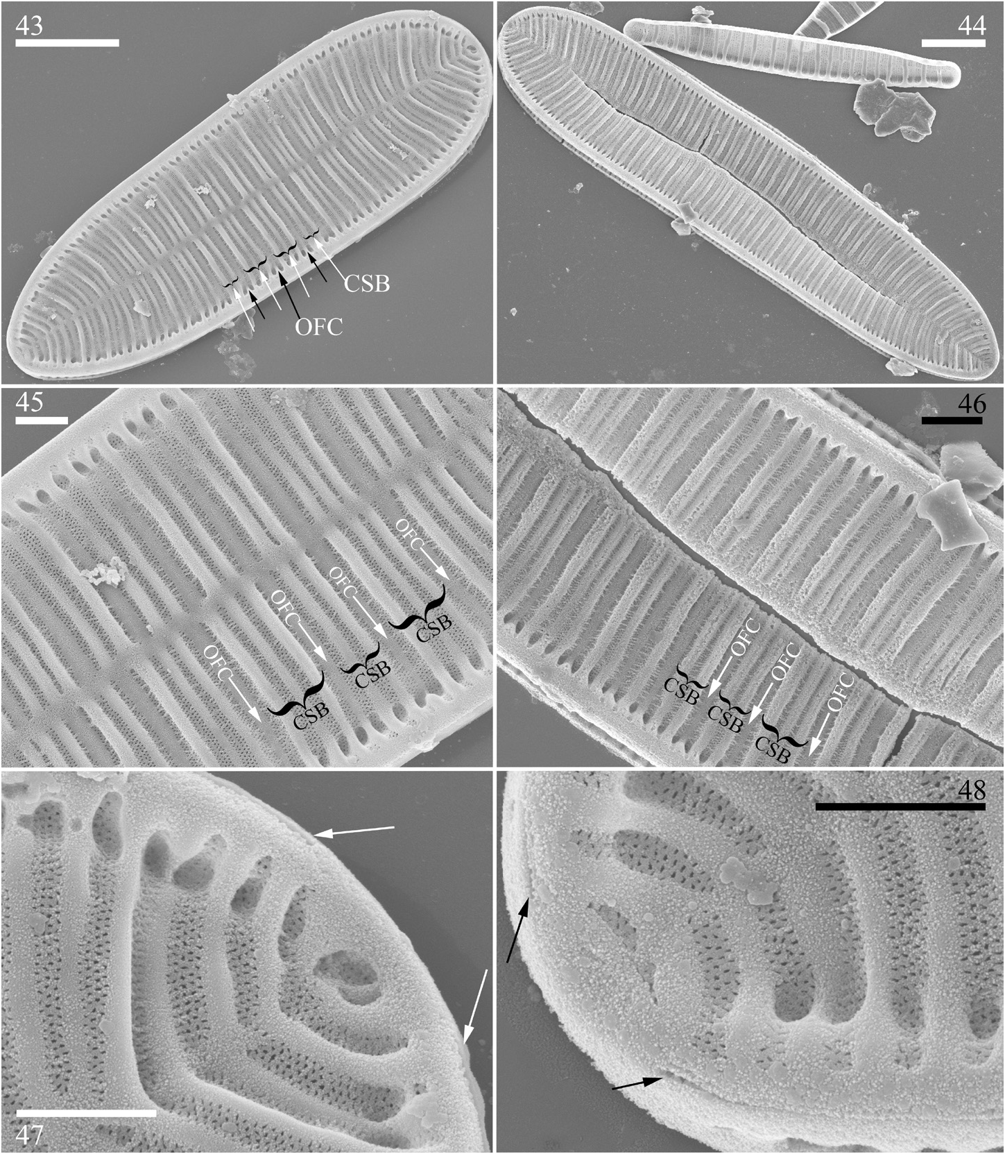
FIGURES 43–48. Surirella minuta, SEM, external view. 43, 44. Two valves showing the valve shape and the pattern of costa-stria bundles (CSB, labeled with white arrows in Fig. 43) alternated with over-fibula costae (OFC, labelled with black arrows in Fig. 43) from pole to pole. 45, 46. Details of the middle section of the valve (Fig. 45 from Fig. 43, Fig. 46 from Fig. 44), note the pattern of CSB alternated with OFC and multiseriate striae. 47, 48. Details of the apices from Fig. 43. Note the raphe endings interrupted at both the headpole (Fig. 47) and footpole (Fig. 48). Scale bars = 5 μm (Figs 43, 44), 1 μm (Figs 45–48).
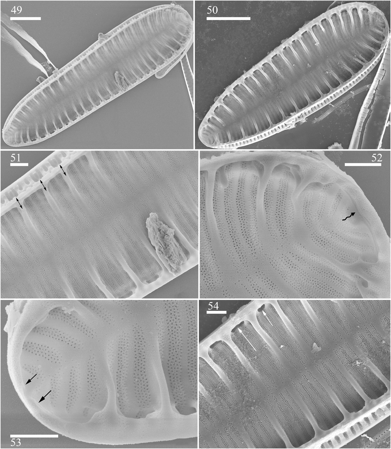
FIGURES 49–54. Surirella minuta, SEM, internal view. 49, 50.Two complete valves, note the portulae are clearly visible. 51. Middle part of the valve, details from Fig. 49. Note that each fibula corresponds to a sinking in the mantle (three double-headed arrows). 52. Headpole from Fig. 49, note the continuing raphe (wavy arrow). 53. Footpole from Fig. 49, note the interrupted raphe endings (two arrows). 54. Middle part from Fig. 50, note only one portula between two adjacent fibulae (two arrows). Scale bars = 5 μm (Figs 49–50), 1 μm (Figs 51–54).
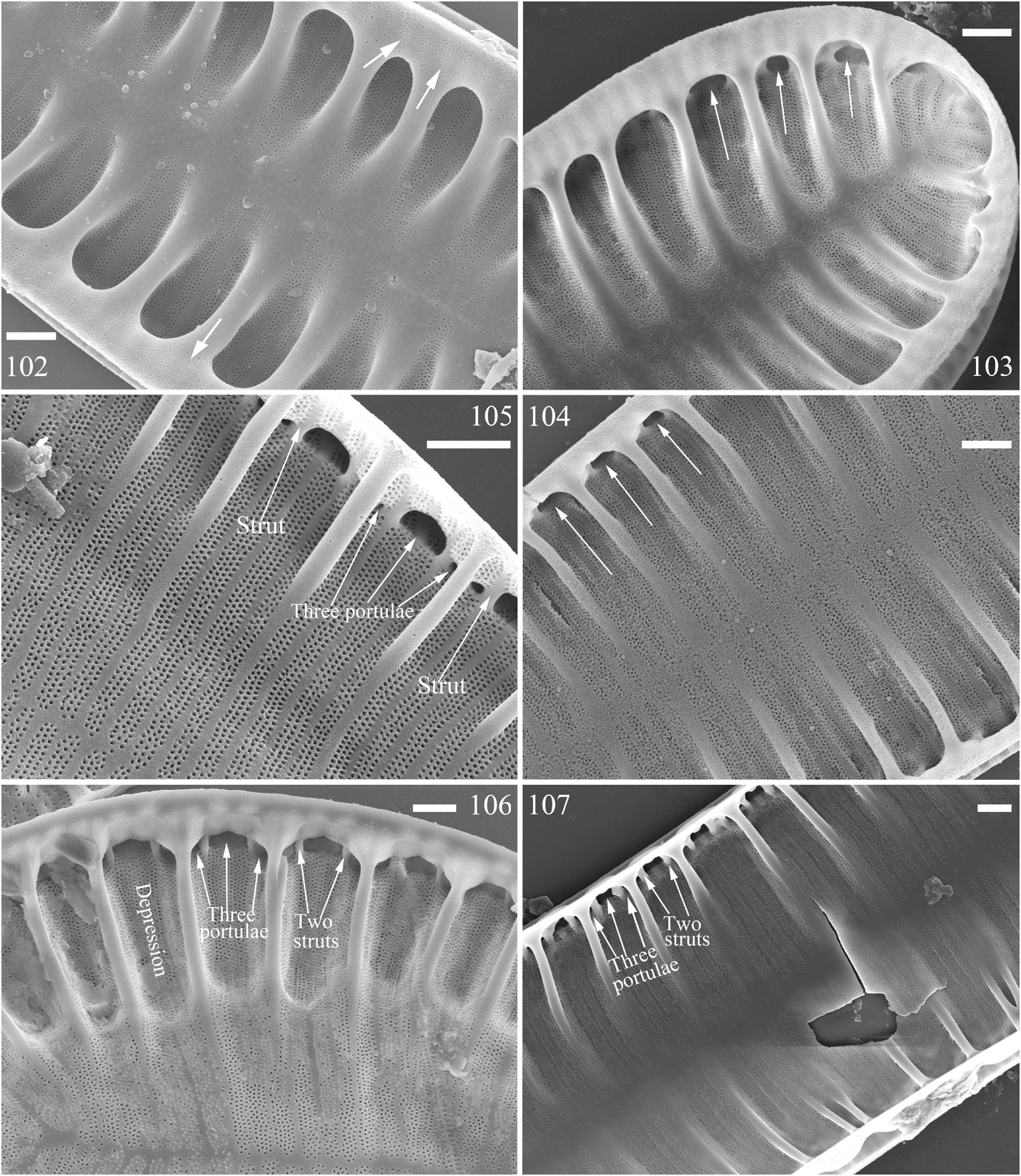
FIGURES 102–107. Characteristics of the portulae among five Surirella taxa, SEM, internal view. 102, 103. Portula structure of S. liubingii, note the striated internal raphe canal wall (Fig. 102, three arrows) and the presence of only one portula between two adjacent fibulae (Fig.103, three arrows). 104.Portula structure of S. minuta, note the occurrence of only one portula (three arrows). 105–107.Portula structure of S. lacrimula, S. brebissonii var. kuetzingii, and S. tientsinensis, respectively. Note that these three taxa have three portulae between two adjacent fibulae (three arrows respectively) but that a marginal trough-like depression is only produced in S. brebissonii var. kuetzingii (Fig. 106). Scale bars =1 μm (Figs 102–107).
No known copyright restrictions apply. See Agosti, D., Egloff, W., 2009. Taxonomic information exchange and copyright: the Plazi approach. BMC Research Notes 2009, 2:53 for further explanation.
|
Kingdom |
|
|
Phylum |
|
|
Class |
|
|
Order |
|
|
Family |
|
|
Genus |