
Phaelota Jacoby
|
publication ID |
https://doi.org/ 10.5281/zenodo.157817 |
|
publication LSID |
lsid:zoobank.org:pub:28D27114-DC98-4388-807D-E2FFF9FC7F3E |
|
DOI |
https://doi.org/10.5281/zenodo.6273444 |
|
persistent identifier |
https://treatment.plazi.org/id/03C21B4E-FFCC-FFD1-FEB6-FEA9FC3FFA04 |
|
treatment provided by |
Plazi |
|
scientific name |
Phaelota Jacoby |
| status |
|
Genus Phaelota Jacoby
Phaelota Jacoby, 1887 , 94 (Type species: Phaelota semifasciata Jacoby, 1887 , Sri Lanka, by monotypy); Maulik, 1926, 176, 280; Chen, 1936, 659; Heikertinger & Csiki, 1940, 511; Scherer, 1969, 8, 18, 218; Seeno & Wilcox, 1982, 144.
Distribution. South India, Sri Lanka.
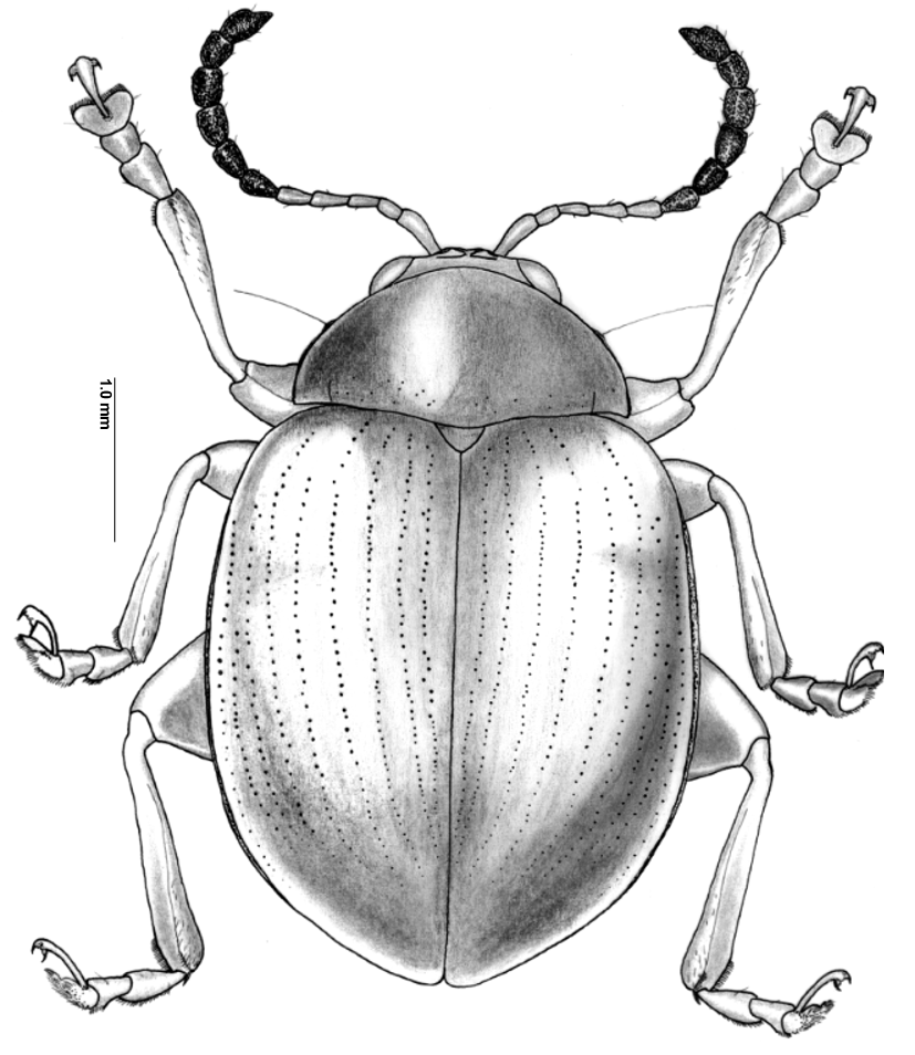
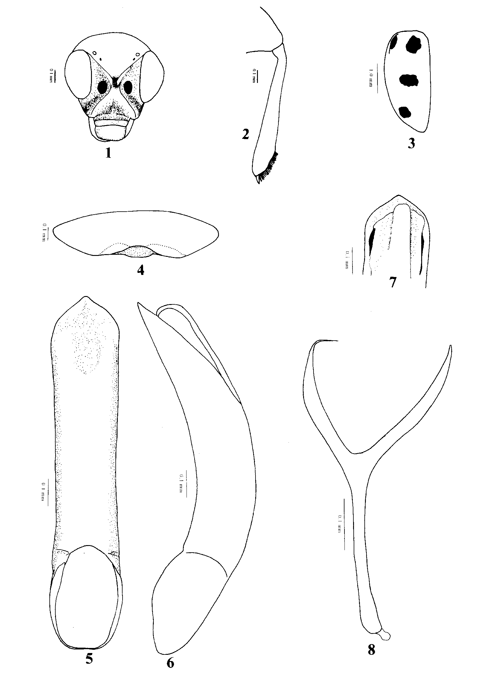
Description. Body length 2.8 to 5.2 mm, width 1.9 to 3.6 mm, ovoid, convex, narrowed posteriorly ( Fig. 15 View FIGURE 15 ). Colour non metallic, reddish or yellowish, sometimes with dark stripes or spots. Head ( Fig. 1 View FIGURES 1 – 8 ) hypognathous, partially withdrawn into prothorax, moderately flat in lateral view. Vertex moderately flat. Frons and vertex form nearly straight or slightly convex line in lateral view. Supraorbital pore well developed, circular, not surrounded by shallow groove. Antennal calli well developed, shining, impunctate, raised, oblique to transverse, curved, both ends narrowed, anterior ends triangular and entering into interantennal space. Antennal calli sharply delineated from vertex; separated from each other by a longitudinal midfrontal depression (midfrontal longitudinal depression very narrow in P. vaishakha ). Midfrontal sulcus vary (absent in P. sindhoori and P. semifasciata though antennal calli are separated by midfrontal longitudinal depression; traces of midfrontal sulcus present in midfrontal depression in P. jacobyi ; extremely short in P. vaishakha ). Orbital sulcus distinct. Supracallinal sulcus present, not deep. Supraorbital sulcus absent. Suprafrontal sulci vary (absent in P. sindhoori , ill defined in P. jacobyi , P. semifasciata and P. vaishakha ). Supraantennal sulcus distinct. Subgenal suture distinct above base of mandible. Distance between eye and adjacent antennal socket subequal to transverse diameter of a socket or slightly less; distance between antennal sockets 1.5 to 4.7 times diameter of a socket, less than transverse diameter of one eye. Eyes moderately large, lateral, separated by a distance of 1.6–2.3 times transverse diameter of one eye. Inner margin of eyes subparallel. Frontal ridge wide, flat, widening anteriorly. Frontal ridge elevated laterally forming a shallow antennal groove below eye. Length of frontal ridge from lower edge of antennal socket to clypeus less than width of frontal ridge anteriorly. Anterofrontal ridge lower than frontal ridge, flat. Frontoclypeal suture with four long setae. Labrum wider than long, slightly emarginate anteriorly, with a transverse row of four setiferous punctures. Distance between middle pair of setiferous punctures on labrum slightly more than distance between outermost puncture and nearest one. Mandible palmate bearing four teeth. Maxillary palpus with preapical palpomere shorter than apical. Apical palpomere pointed, slightly shorter than second. Palpomeres not incrassate.
Antennae extend slightly beyond humerus, not reaching middle of elytron. First antennomere club shaped, as long as next two combined; second slightly thicker than third; last six antennomeres thickened, thickly adorned with short hairs.
Pronotum transverse, 1.6–1.8 times wider than long, with poorly developed antebasal transverse impression delimited on either end by curved longitudinal impression (antebasal impression very poorly developed and indicated only by very faint longitudinal impressions on either side in P. sindhoori ). Pronotum proximally narrower than distally. Anterolateral callosity well developed, moderately long, convex, proximally higher than distally; pore well developed, situated at upper posterior face of callosity, seta long. Posterolateral callosity well developed, short, not longer than width of lateral margin, protruding, seta long but shorter than that on anterolateral callosity. Lateral margin proximally wider than distally. Posterior margin of pronotum weakly bisinuate. Scutellum well developed, triangular with rounded apex.
Procoxal cavity closed behind (narrowly open in P. sindhoori ). Distance between proximal part of prosternum to end of intercoxal prosternal process vary (1.0–1.3 times width of prosternal intercoxal process in the Indian species while the same is 1.6 times in P. semifasciata ). Distance between proximal part of prosternum to coxal cavity 4–10 times shorter than to end of intercoxal prosternal process. Intercoxal prosternal process slightly widened and truncate posteriorly. Mesosternal intercoxal process shorter and as wide as or wider than prosternal intercoxal process, its length subequal to or less than half of its width, width subequal to transverse diameter of mesocoxal cavity, emarginate posteriorly. Metasternum nearly two times as long as prosternum in the Indian species while it is slightly shorter than prosternum in P. semifasciata .
Elytra as wide as prothorax at base, widening at humerus, lateral sides moderately convex, distinctly narrowed posteriorly. Apical margin convex, apex obtusely angulate. Elytral punctures small, arranged in striae. Epipleuron horizontal to oblique, moderately wide, width of widest portion being slightly less than or subequal to width of midfemur. Epipleuron reaching beyond 4/5th of elytron.
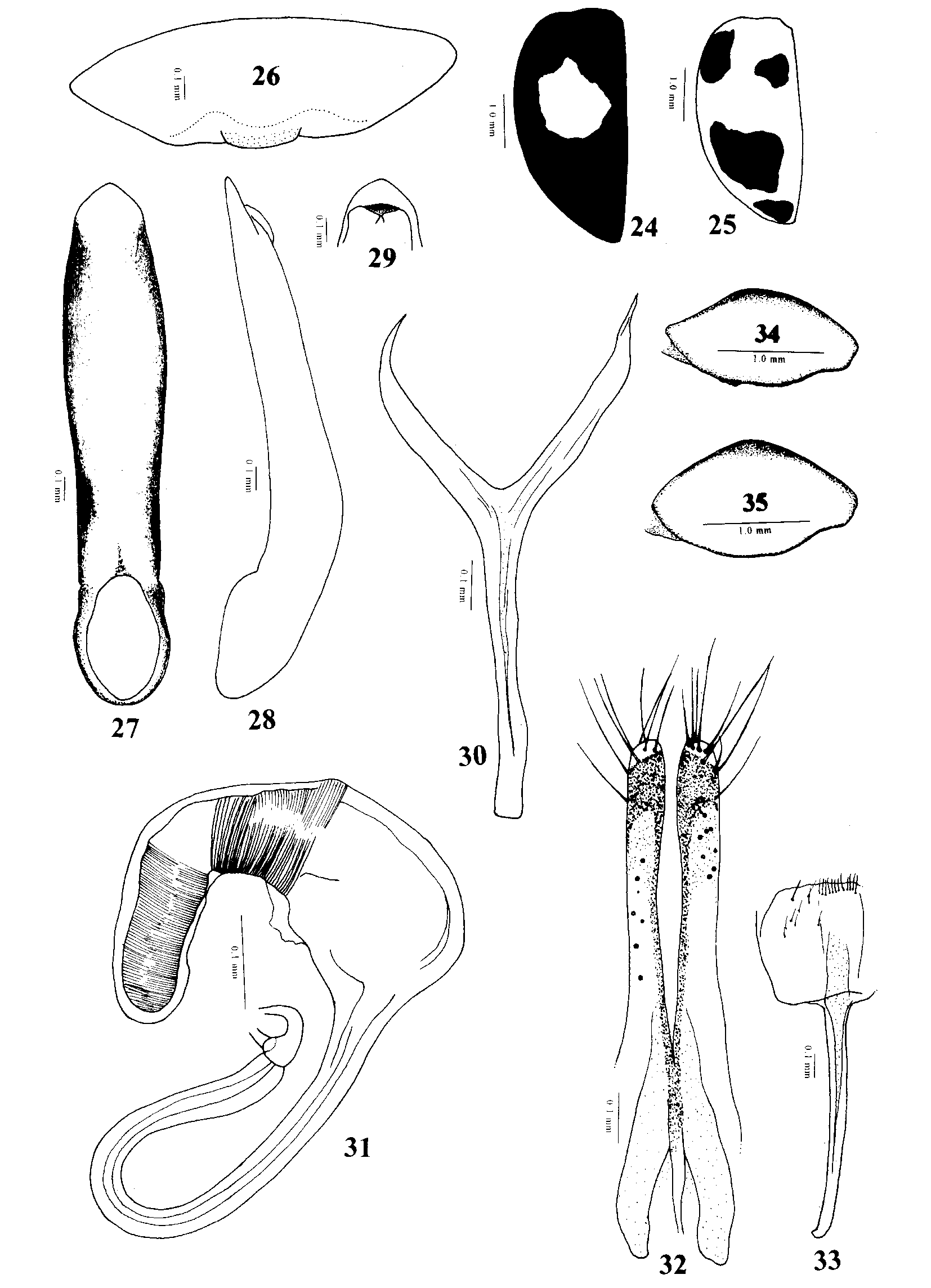
All coxae with a triangular, flat, acutely pointed, posteriorly directed denticle in front of and reaching trochanter. Pro and mesotibia widened from proximal end to distal end, apex being nearly two times as wide as base. Forelegs with basitarsomere longer than second tarsomere; second tarsomere narrower than third and first; claw tarsomere two times longer than third tarsomere. Dorsal surface of pro and mesotibia flat except near proximal end; apex with a row of strong bristles on either side dorsally. Metafemur incrassate. Metatibia subequal to metafemur in length, apically widened. Dorsal surface of metatibia flat or slightly concave. Mesal edge of dorsal surface of metatibia higher than lateral edge. In lateral view metatibia gradually widening from proximal end to about 5/6th distally and again narrowing to distal end; a row of strong bristles present on either margin of dorsal side from widest point to apex ( Fig.2 View FIGURES 1 – 8 ). Metatibial spur situated in middle at apex, sharp, dorsolaterally directed, shorter than or equal to tarsal claw. Metabasitarsus slightly longer than next two tarsomeres combined, narrower basally than apically; apically not wider than third metatarsomere; third bilobed; fourth subequal to twice the length of third. Claws appendiculate with a deep incision between the base of claw and appendix. In repose, metatibia received over a longitudinal ridge along mesal side of metafemur. Edge of this ridge distinctly serrulate in proximal half in male ( Fig. 34 View FIGURES 24 – 35 ). Serrulation absent in female ( Fig. 35 View FIGURES 24 – 35 ).
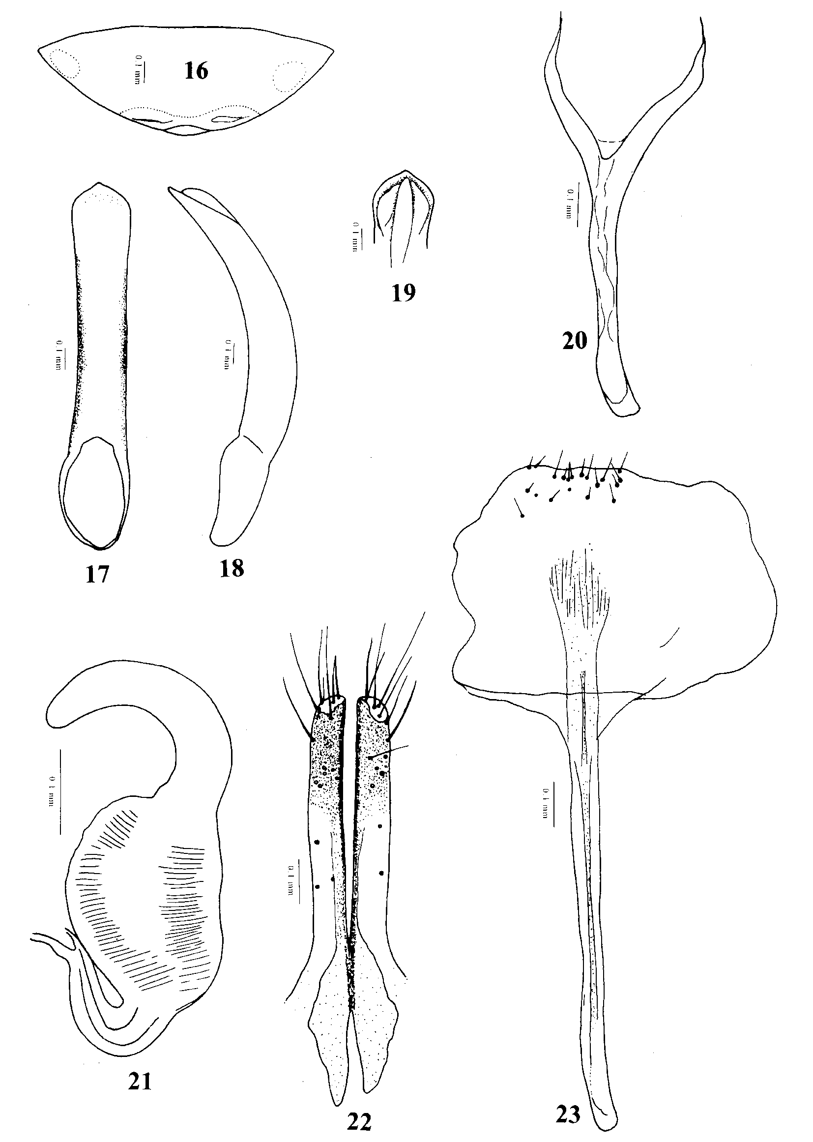
Last abdominal sternite with an internal folding at posterior margin. Median lobe of aedeagus simple tube, convex in lateral view, without transparent window. Tegmen Yshaped. Spermatheca with well developed pump, receptacle and duct. Duct without coils, making loop away from receptacle. Vaginal palpi elongate, fused before middle, apex transparent with long setae. Tignum elongate, with internal channel, posterior membranous region well developed with a few short setae.
Sexual dimorphism. All basitarsomeres slightly widened in male than in female. Longitudinal ridge along mesal side of metafemur is serrulate in male while smooth in female. Posterior margin of last abdominal sternite of male is bisinuate or nearly so with a distinct lobe in middle which is evident in macerated specimens ( Figs. 4 View FIGURES 1 – 8 , 16 View FIGURES 16 – 23 , 26 View FIGURES 24 – 35 ). In P. sindhoori this is less evident. Posterior margin of last abdominal sternite is entire in female. Internal folding of last abdominal sternite is bisinuate in male while it is simple and curved in female. Serrulation on mesal side of metafemur in male was a convenient character for sexing adults.
Host plants. Adults of the three Indian species feed on the leaves of ferns. It is quite likely that trophic selections of Phaelota is restricted to ferns like that of Schenklingia Heikertinger and Csiki.
Remarks. Acrocrypta Baly , Chabria Jacoby , Phaelota and Schenklingia form a homogeneous group of genera that resemble each other. Acrocrypta can be separated from Phaelota by confused elytral punctures (elytral punctures form striae in Phaelota ) and narrow prosternal intercoxal process (wide in Phaelota ). Chabria can be separated by open procoxal cavities (closed in Phaelota ) and confused elytral punctures (punctures form striae in Phaelota ). Phaelota can be differentiated from Schenklingia , another fern feeding genus, by the first antennomere which is as long as the next two antennomeres combined (in Schenklingia , the first antennomere is as long as the next three segments combined) and the closed procoxal cavities (open in Schenklingia ). Neither Jacoby (1887) nor subsequent workers recognized the presence of antebasal transverse impression on the pronotum of Phaelota . Discovery of Phaelota in south India provides further credence to the close affinity of south Indian fauna to that of Sri Lanka.
No known copyright restrictions apply. See Agosti, D., Egloff, W., 2009. Taxonomic information exchange and copyright: the Plazi approach. BMC Research Notes 2009, 2:53 for further explanation.
|
Kingdom |
|
|
Phylum |
|
|
Class |
|
|
Order |
|
|
Family |
Phaelota Jacoby
| Prathapan, K. D. & Viraktamath, C. A. 2004 |
Phaelota
| Jacoby 1887 |
Phaelota semifasciata
| Jacoby 1887 |