
Typhlocarcinops Rathbun, 1909
|
publication ID |
https://doi.org/10.11646/zootaxa.4788.1.1 |
|
publication LSID |
lsid:zoobank.org:pub:7A461DBA-00B7-48DB-9320-4775DA8F21B2 |
|
persistent identifier |
https://treatment.plazi.org/id/03C05222-FFFD-FC6D-FF35-D6A6FA1DFE1E |
|
treatment provided by |
Plazi (2020-06-05 09:49:28, last updated 2023-11-18 03:01:46) |
|
scientific name |
Typhlocarcinops Rathbun, 1909 |
| status |
|
Key to species of Typhlocarcinops Rathbun, 1909 View in CoL
1a. Anteroexternal angle of merus of third maxilliped projected to varying degrees, clearly auriculiform (e.g., Figs. 56A View FIGURE 56 , 71A View FIGURE 71 , 73A View FIGURE 73 , 80B View FIGURE 80 )............................................................................................... 2
1b. Anteroexternal angle of merus of third maxilliped rounded, not projected, not auriculiform (e.g., Figs. 60A View FIGURE 60 , 69A View FIGURE 69 )......... 7
2a. Merus and ischium of third maxilliped equal in length, exopod broad, relatively stout ( Fig. 71A View FIGURE 71 ); dorsal margin of ambulatory merus with at least 1 small spine; dactylus of ambulatory legs broad, shorter than propodus ( Fig. 70G View FIGURE 70 ); G1 upper half longer than lower half, distal part short, bent at almost right angles ( Fig. 71C, D View FIGURE 71 ) ( Japan).......... Typhlocarcinops kanashi View in CoL n. sp.
2b. Merus of third maxilliped clearly shorter than ischium; dorsal margin of ambulatory merus unarmed; dactylus of ambulatory legs styliform, longer than or subequal to propodus; third maxilliped exopod and G1 otherwise........................ 3
3a. Carapace squarish, almost as long as broad (1.1), surface smooth, covered with short pubescense ( Fig. 78A View FIGURE 78 ); fingers of chela gaping when closed ( Fig. 78B View FIGURE 78 ) (Port Darwin, Australia)........................ Typhlocarcinops arcuatus (Miers, 1884) View in CoL
3b. Carapace more than 1.2 times as broad as long, surface smooth, glabrous or with scattered granules and tubercles; fingers of chela not strongly gaping when closed..................................................................... 4
4a. Carapace and cheliped very smooth, appearing almost shiny, surfaces glabrous, without any tubercles ( Fig. 72B View FIGURE 72 ); exopod of third maxilliped relatively broad; G1 with upper half slightly shorter than lower half, with 1 large seta subdistally ( Fig. 73 View FIGURE 73 C‒F) (Bohol, Philippines; Tanimbar, Indonesia).......................................... Typhlocarcinops lapillus View in CoL n. sp.
4b. Carapace and chelipeds covered with granules and/or tubercles partially or entirely; exopod of third maxilliped relatively slen- der; upper and lower halves of G1 subequal, row of fine setae subdistally......................................... 5
5a. Outer surface of palm with longitudinal rows of large tubercles on entire surface ( Fig. 67 View FIGURE 67 E–G); G1 strongly curved, distal part slender, long, tapering to a relatively sharp tip ( Fig. 69C, D View FIGURE 69 ) ( Singapore)............... Typhlocarcinops diminutus View in CoL n. sp.
5b. Outer surface of palm otherwise; G1 slender, gently curved, distal part short...................................... 6
6a. Anterolateral margin of carapace almost entire, lined with small tubercles and 1 small tooth ( Figs. 76A, B View FIGURE 76 , 77A View FIGURE 77 ); male pleon broad, telson short, 1.5 times as long as somite 6, with rounded tip ( Fig. 77C View FIGURE 77 ); G1 slightly curved, with distal part bent at right angles, tapering to pointed tip ( Fig. 77D, E View FIGURE 77 ) (Aru Islands, Indonesia)............. Typhlocarcinops angustipes Tesch, 1918 View in CoL
6b. Anterolateral margin of carapace lined with small tubercles separated into 4 broad lobes ( Fig. 55A, B View FIGURE 55 ); male pleon narrow, telson long, 1.8 times as long as somite 6, subtriangular with rounded distal margin ( Fig. 55D, E View FIGURE 55 ); G1 sinuous, distal part with blunt tip, directed upwards ( Fig. 56B, C View FIGURE 56 ) (Palawan, Philippines).............. Typhlocarcinops marginatus Rathbun, 1914 View in CoL
7a. Distinct lobes on anterolateral margin of carapace lined with small granules, separated by clear depressions, cleft between second and third anterolateral lobes very distinct, usually U-shaped............................................. 8
7b. Anterolateral margin of carapace lined with small granules or almost smooth, entire or if lobes visible, not clearly demarcated, cleft between second and third anterolateral lobes shallow or V-shaped.......................................... 11
8a. Anterolateral margin with 3 broad lobes................................................................... 9
8b. Anterolateral margin with 4 broad lobes.................................................................. 10
9a. Outer surface of palm of cheliped smooth, ventral margin distinctly cristate ( Fig. 42D View FIGURE 42 ); lower margin of ischium of third maxilliped strongly oblique ( Fig. 44A View FIGURE 44 ); male pleon relatively narrow, telson 1.9 times as long as somite 6 ( Fig. 44B View FIGURE 44 ); upper half of G1 shorter than lower half, distal part curved, flared, with rounded tip ( Fig. 44 View FIGURE 44 C–F) ( Hong Kong, Vietnam; Arafura Sea, Indonesia)................................................................... Typhlocarcinops raouli View in CoL n. sp.
9b. Outer surface of palm of cheliped with tubercles on lower outer surface, ventral margin slightly cristate ( Fig. 53H View FIGURE 53 ); lower margin of ischium of third maxilliped slightly oblique ( Fig. 53E View FIGURE 53 ); male pleon relatively narrow, telson 1.8 times as long as somite 6 ( Fig. 53F, G View FIGURE 53 ); upper half of G1 longer than lower half, distal part sinuous, with broad rounded tip, pointed upwards ( Fig. 54 View FIGURE 54 ) (between Marinduque and Luzon, Philippines)........................... Typhlocarcinops angustifrons Rathbun, 1914 View in CoL
10a. Merus of third maxilliped shorter than ischium, exopod relatively stout ( Fig. 60A View FIGURE 60 ); male pleon broad, telson 1.9 times as long as somite 6 ( Fig. 60B View FIGURE 60 ); G1 slender, sinuous, upper half proportionally longer, distal part short, slightly curved, directed laterally, with blunt tip ( Fig. 60 View FIGURE 60 C–G) (Sumbawa, Indonesia; Philippines; Japan)........... Typhlocarcinops transversus Tesch, 1918 View in CoL
10b. Merus and ischium of third maxilliped subequal, exopod relatively slender ( Fig. 61B View FIGURE 61 ); male pleon relatively broad, telson 1,7 times as long as somite 6 ( Fig. 61G View FIGURE 61 ); G1 slender, sinuous, upper half distinctly longer, distal part directed upwards with pointed tip ( Fig. 61 View FIGURE 61 H–L) (Red Sea)................................................. Typhlocarcinops serenei Türkay, 1986 View in CoL
11a. Anterolateral margin of carapace with cleft between second and third anterolateral lobes shallow or V-shaped........... 12
11b. Anterolateral margin of carapace entire................................................................... 20
12a. Distinct tooth on inner angle of cheliped carpus............................................................ 13
12b. Inner angle of cheliped carpus otherwise.................................................................. 16
13a. Anterolateral margin of carapace lined with small tubercles, entire or separated into 4 broad lobes.................... 14
13b. Anterolateral margin of carapace lined with small tubercles, entire or separated into less than 4 lobes.................. 15
14a. Cheliped palm smooth medially, upper and lower surfaces with scattered tubercles ( Figs. 8F View FIGURE 8 , 9G, H View FIGURE 9 , 10E, F View FIGURE 10 , 11F View FIGURE 11 ); tooth on inner angle of carpus strong, pointed ( Figs. 10C, D View FIGURE 10 , 11G, H View FIGURE 11 ); exopod of third maxilliped slender, narrow ( Figs. 13B View FIGURE 13 , 14A, F View FIGURE 14 , 15B View FIGURE 15 ); G1 with upper half distinctly longer than lower half, distal part short, gently curved ( Figs. 13 View FIGURE 13 D–G, I–L, 14B–E, H–K, 15E–G) (Gulf of Thailand; Phuket, Thailand; Singapore; Indonesia; Hong Kong)..... Typhlocarcinops canaliculatus Rathbun, 1909 View in CoL
14b. Cheliped palm smooth medially, upper, lower surface with dense setae especially on minor chela, setae may be absent in larger specimens ( Figs. 16F, G View FIGURE 16 , 19E View FIGURE 19 , 20E, F View FIGURE 20 , 21E, F View FIGURE 21 ); tooth on inner angle of carpus low, blunt ( Figs. 19F View FIGURE 19 , 20C View FIGURE 20 , 21D View FIGURE 21 , 22G, H View FIGURE 22 ); exopod of third maxilliped stout, relatively broad; G1 with upper and lower halves subequal in length, distal part long, prominently curved ( Figs. 18E View FIGURE 18 , 23 View FIGURE 23 B–E, H–K, 24A–G) (Sulu, Philippines; Papua, Indonesia; Hong Kong; Fujian, China)............................................................................... Typhlocarcinops decrescens Rathbun, 1914 View in CoL
15a. Anterolateral margin of carapace lined with small tubercles separated into 2 broad, low lobes ( Figs. 25B View FIGURE 25 , 26B View FIGURE 26 , 27B View FIGURE 27 ); surface of cheliped palm smooth ( Figs. 25E View FIGURE 25 , 26F View FIGURE 26 , 27E View FIGURE 27 ); tooth on inner angle of carpus strong, pointed ( Figs. 25F, G View FIGURE 25 , 26B View FIGURE 26 , 27F, G View FIGURE 27 ); male pleon narrow ( Figs. 29B, G View FIGURE 29 , 30B View FIGURE 30 ); G1 with upper and lower halves subequal, distal part relatively long, gently bent, sinuous with pointed tip ( Figs. 28 View FIGURE 28 , 29 View FIGURE 29 C–F, H–J, 30C–F) (Kyushu, Japan; Guangdong, China; Hong Kong).......................................................................... Typhlocarcinops denticarpes Dai, Yang, Song & Chen, 1986 View in CoL
15b. Anterolateral margin of carapace lined with small tubercles separated into 3 broad lobes ( Figs. 35A View FIGURE 35 , 36A, B View FIGURE 36 ); surface of cheliped palm smooth medially, upper and lower surface with scattered tubercles ( Fig. 35F, G View FIGURE 35 ); tooth on inner angle of carpus long, sharp in large males ( Figs. 35E View FIGURE 35 ) or angular and tuberculate in smaller specimens and females ( Fig. 36 View FIGURE 36 C–E); male pleon broad ( Fig. 37B View FIGURE 37 ); G1 with upper part half longer than lower half, distal part relatively short, gently bent, with pointed tip ( Fig. 37 View FIGURE 37 C–F) (Papua, Indonesia)............................................................ Typhlocarcinops robustus View in CoL n. sp.
16a. Carapace surface covered with relatively dense short and long setae............................................ 17
16b. Carapace surface with median surface glabrous, lateral margins covered with less dense short setae................... 18
17a. Carapace 1.4–1.5 times as broad as long ( Figs. 62A, B View FIGURE 62 , 63B View FIGURE 63 ); inner angle of carpus of cheliped with sharp denticulate protuberance ( Fig. 63A View FIGURE 63 ); P2‒P5 relatively short ( Figs. 62A View FIGURE 62 , 63B View FIGURE 63 ); male pleon broad ( Fig. 64B View FIGURE 64 ); G1 slightly curved, upper half longer than lower half, distal part slightly sinuous, tip broad, pointed upwards ( Fig. 64 View FIGURE 64 C–F) (Lombok, Indonesia)............................................................................................ Typhlocarcinops hirtus View in CoL n. sp.
17b. Carapace 1.2 times as broad as long ( Fig. 34A View FIGURE 34 ); inner angle of carpus of cheliped denticulate; P2‒P5 relatively long; male pleon narrow ( Fig. 34D View FIGURE 34 ); G1 strongly sinuous, distal part gently curvedupwards, tip upcurved ( Fig. 34 View FIGURE 34 E–G) (Queensland, Australia)......................................................... Typhlocarcinops tonsuratus Griffin & Campbell, 1969 View in CoL
18a. Outer surface of palm of cheliped medially smooth ( Fig. 38G, H View FIGURE 38 ); inner angle of carpus without any dentition ( Fig. 38E, F View FIGURE 38 ); P2‒P5 relatively long ( Fig. 38A View FIGURE 38 ); male pleon broad, telson 1.7 times as long as somite 6 ( Fig. 39B View FIGURE 39 ); G1 upper and lower halves subequal, slightly sinuous, distal part prominently curved, hook-shaped ( Fig. 39 View FIGURE 39 C–F) ( Papua New Guinea)........................................................................................... Typhlocarcinops hamus View in CoL n. sp.
18b. Outer surface of palm of cheliped entirely granular or smooth; inner angle of carpus with short, blunt dentition; P2‒P5 reltively short or long; male pleon narrow, telson less than 1.7 times as long as somite 6; G1 otherwise........................ 19
19a. Palm of cheliped entire granular ( Fig. 31F, G View FIGURE 31 ); inner angle of carpus with slight, blunt protuberance ( Fig. 31H View FIGURE 31 ); P2‒P5 relative- ly short ( Fig. 31A View FIGURE 31 ); male pleon narrow, telson 1.5 times as long as somite 6 ( Fig. 32E, I View FIGURE 32 ); G1 upper and lower halves subequal, distal part bent at right angles, distinctly elongate with pointed tip ( Fig. 32F, G View FIGURE 32 , J–M) ( Taiwan; Japan).......................................................................................... Typhlocarcinops yui Ng & Ho, 2003 View in CoL
19b. Palm of cheliped smooth; inner angle of carpus with low dentition ( Fig. 33A View FIGURE 33 ); P2‒P5 relatively long ( Fig. 33A View FIGURE 33 ); male pleon narrow, telson 1.2 times as long as somite 6 ( Fig. 33C View FIGURE 33 ); G1 upper and lower halves subequal, distal part curved downwards, tip broad, fluted ( Fig. 33D, E View FIGURE 33 ) (Indian Ocean)................................ Typhlocarcinops stephenseni Serène, 1964 View in CoL
20a. Carapace surface smooth, anterolateral margin entire ( Fig. 50A, B View FIGURE 50 ); exopod of third maxiliped slender ( Fig. 52A View FIGURE 52 ); inner angle of carpus of cheliped low, with sharp granules ( Fig. 51A View FIGURE 51 ); male telson 1.7 times as long as somite 6 ( Fig. 52B View FIGURE 52 ); G1 slender, slightly curved in lower half, upper half sinuous, distal part with pointed tip ( Fig. 52 View FIGURE 52 C–G) (South Java, Indonesia)................................................................................... Typhlocarcinops hadrotes View in CoL n. sp.
20b. Carapace surface granular, anterolateral margin entire (or with broad lobes); exopod of third maxiliped stout; inner angle of carpus of cheliped with sharp spine or spinules; male telson 1.8–1.9 times as long as somite 6; G1 otherwise............ 21
21a. Outer surface of palm of chelipeds smooth, shiny ( Fig. 45G, H View FIGURE 45 ); P2‒P5 relatively long ( Fig. 45A View FIGURE 45 ); male pleon narrow, telson 1.8 times as long as somite 6 ( Fig. 45E, F View FIGURE 45 ); G1 slender, slightly curved, upper half longer than lower half, distal part directed upwards with pointed tip ( Fig. 46 View FIGURE 46 ) (between Samar and Masbate, Philippines)... Typhlocarcinops ocularius ( Rathbun, 1914) View in CoL
21b. Outer surface of palm of chelipeds smooth ( Figs. 47G View FIGURE 47 , 48D View FIGURE 48 ); P2‒P5 relatively short ( Figs. 47A View FIGURE 47 , 48A View FIGURE 48 ); male pleon narrow, tel- son 1.9 times as long as somite 6 ( Fig. 49B View FIGURE 49 ); G1 slender, curved, upper half longer than lower half, distal part directed obliquely upwards with pointed tip ( Fig. 49 View FIGURE 49 C–F) ( Madagascar).............................. Typhlocarcinops atimovatae View in CoL n. sp.
Dai, A. - Y., Yang, S. - L., Song, Y. - Z. & Chen, G. - X. (1986) Crabs of the China Seas. China Ocean Press, Beijing, 11 + 642 pp. [in Chinese]
Griffin, D. J. G. & Campbell, B. M. (1969) The sub-littoral Goneplacidae and Pinnotheridae (Crustacea: Brachyura) of Moreton Bay. Memoirs of the Queensland Museum, 15 (3), 141 - 164, figs. 1 - 8.
Ng, P. K. L. & Ho, P. - H. (2003) The Indo-Pacific Pilumnidae XVIII. New species and new records from Taiwan (Decapoda, Brachyura). Crustaceana, 76 (2), 167 - 176. https: // doi. org / 10.1163 / 156854003321824512
Rathbun, M. J. (1909) New crabs from the Gulf of Siam. Proceedings of the Biological Society of Washington, 22, 107 - 114.
Rathbun, M. J. (1914) A new genus and some new species of crabs of the family Goneplacidae. Scientific Results of the Philippine cruise of the Fisheries Streamer Albatross, 1907 - 1910 - No. 32. Proceedings of the United States National Museum, 48 (2067), 137 - 154. https: // doi. org / 10.5479 / si. 00963801.2067.137
Serene, R. (1964) Goneplacidae et Pinnotheridae recoltes par le Dr. Mortensen. Papers from Dr. Th. Mortensen's Pacific Expedition 1914 - 1916, part 80. Videnskabelige Meddelelser fra Dansk Naturhistorisk Forening I KObenhavn, 126, 181 - 282, pls. 16 - 24.
Tesch, J. J. (1918) Goneplacidae and Pinnotheridae. The Decapoda Brachyura of the Siboga-Expedition. II. Siboga Expeditie Monografie, 39 c (Livraison 84), 149 - 295, pls. 7 - 18. https: // doi. org / 10.5962 / bhl. title. 10267
Turkay, M. (1986) Crustacea Decapoda Reptantia der Tiefsee des Roten Meeres. Senckenbergiana maritima, 18 (3 - 6), 123 - 185, figs. 1 - 59, pls. 1 - 4, tabs. 1, 2.
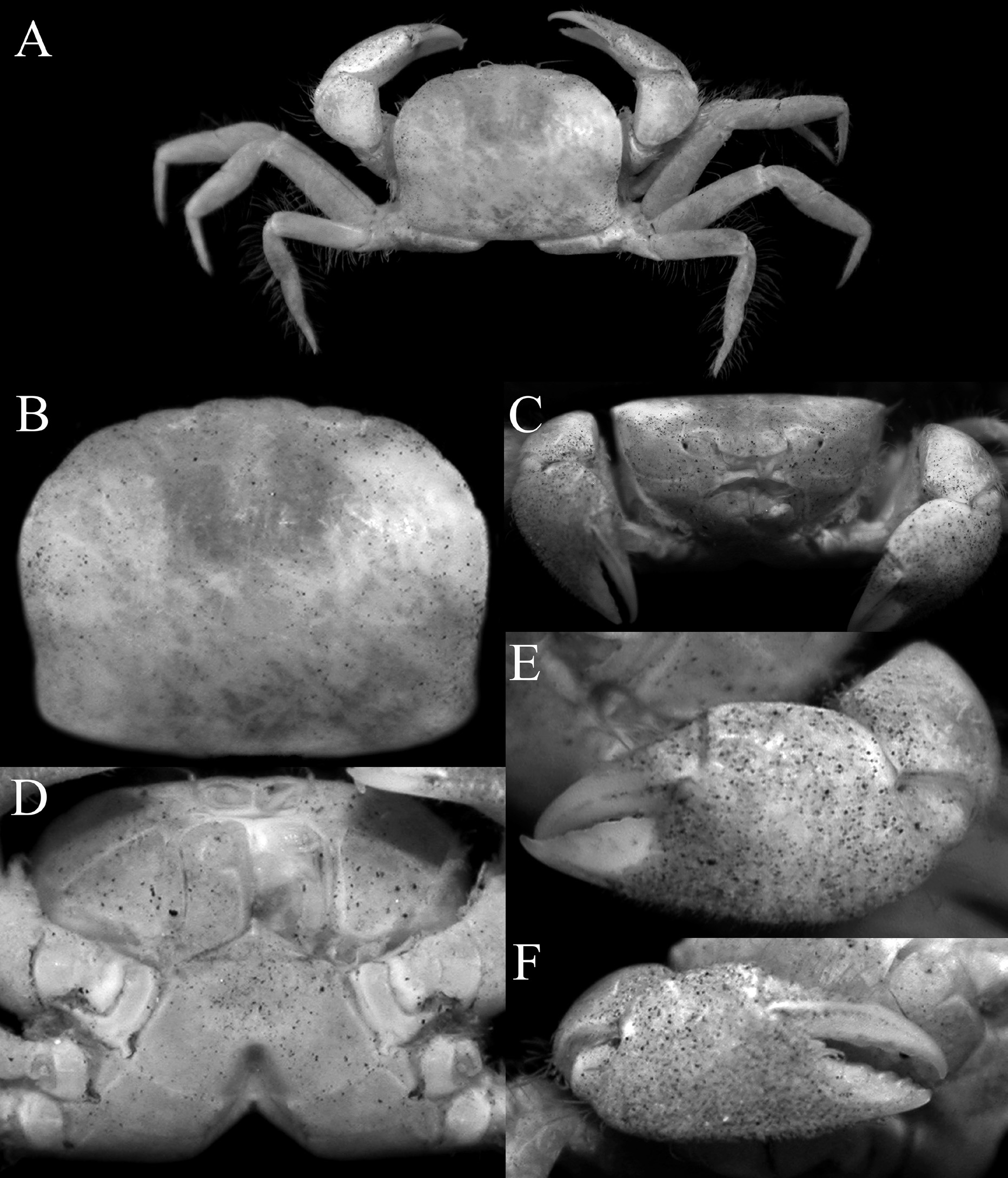
FIGURE 8. Typhlocarcinops canaliculatus Rathbun, 1909, male (5.5 × 4.2 mm) (ZRC 1995.0374), Singapore.A, overall habitus; B, dorsal view of carapace; C, frontal view of cephalothorax; D, anterior thoracic sternum; E, outer view of left chela; F, outer view of right chela.
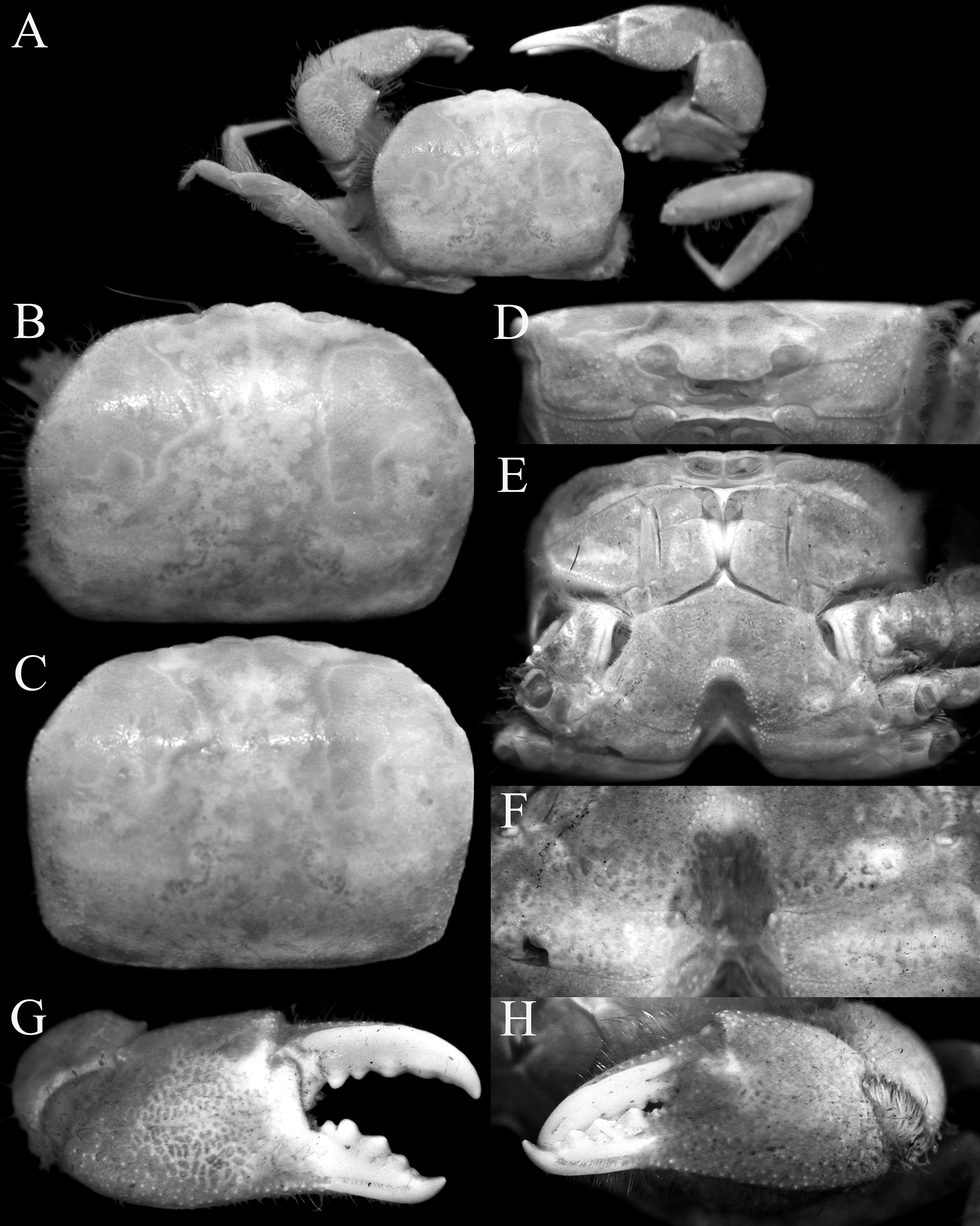
FIGURE 9. Typhlocarcinops canaliculatus Rathbun, 1909, male (8.6 × 6.5 mm) (ZRC 1985.1385), Singapore. A, overall habitus; B, C, dorsal view of carapace; D, frontal view of cephalothorax; E, anterior thoracic sternum; F, sternopleonal cavity showing pleonal locking tubercles; G, outer view of right chela; H, outer view of left chela.
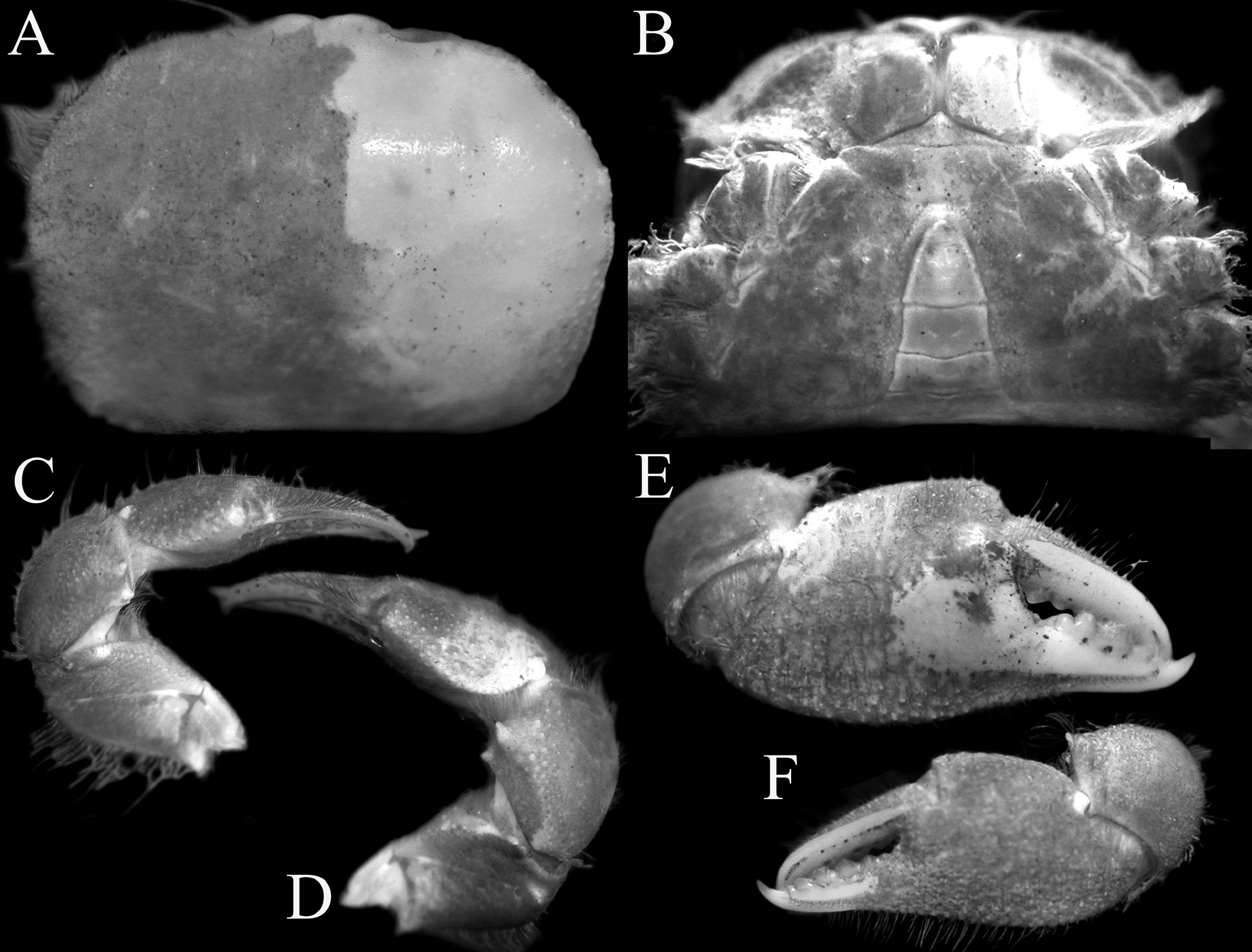
FIGURE 10. Typhlocarcinops canaliculatus Rathbun, 1909, male (8.7 × 6.5 mm) (ZRC 1984.7749), Phuket. A, dorsal view of carapace; B, anterior thoracic sternum and pleon; C, dorsal view of left cheliped; D, dorsal view of right cheliped; E, outer view of right chela; F, outer view of left chela.
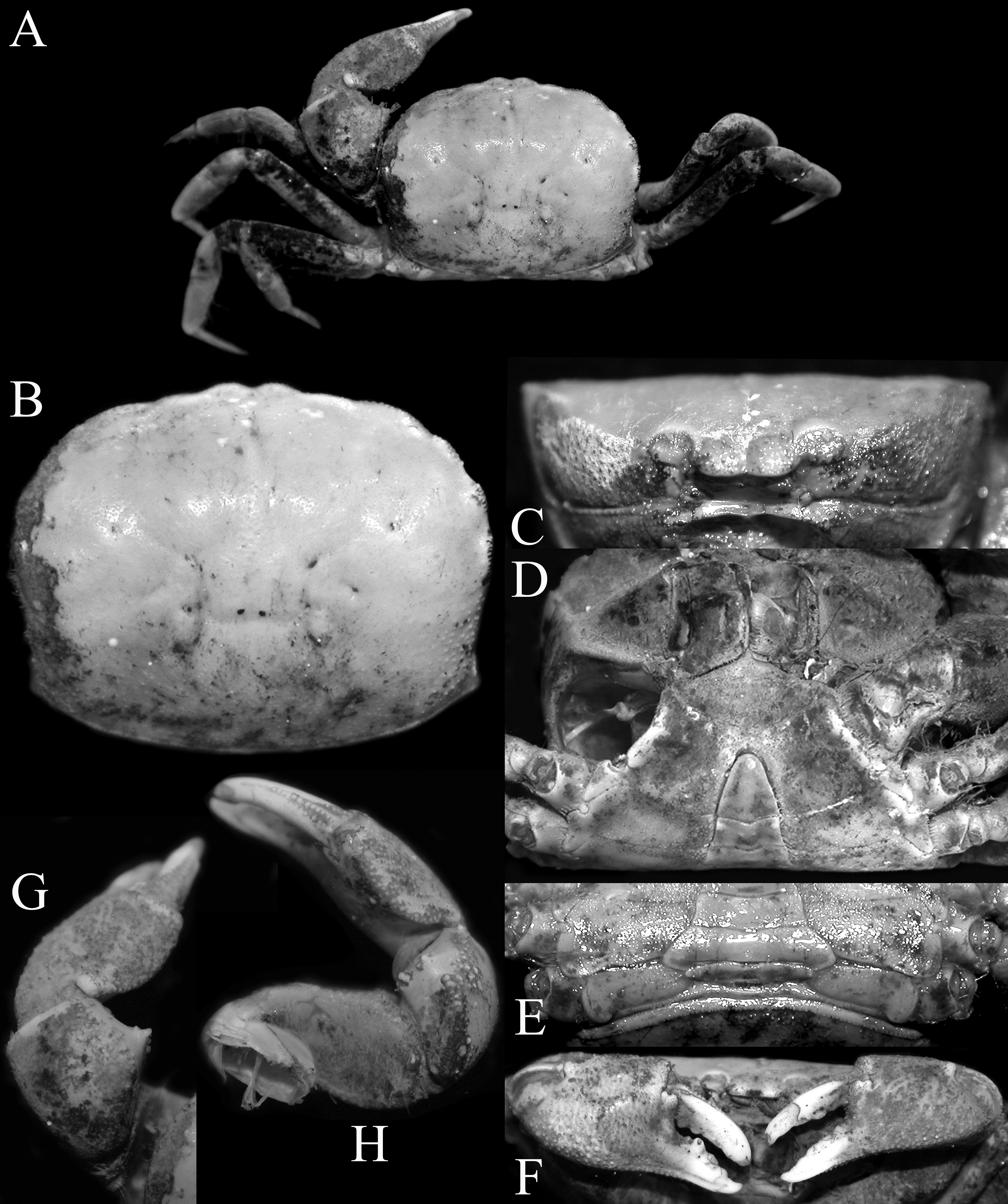
FIGURE 11. Typhlocarcinops canaliculatus Rathbun, 1909, male (13.2 × 10.5 mm) (ZRC 2018.0696), Hong Kong. A, overall habitus; B, dorsal surface of carapace (denuded); C, frontal view of cephalothorax; D, anterior thoracic sternum and pleon; E, posterior thoracic sternum and pleon; F, outer view of chelae; G, dorsal view of left chela; H, dorsal view of right chela.
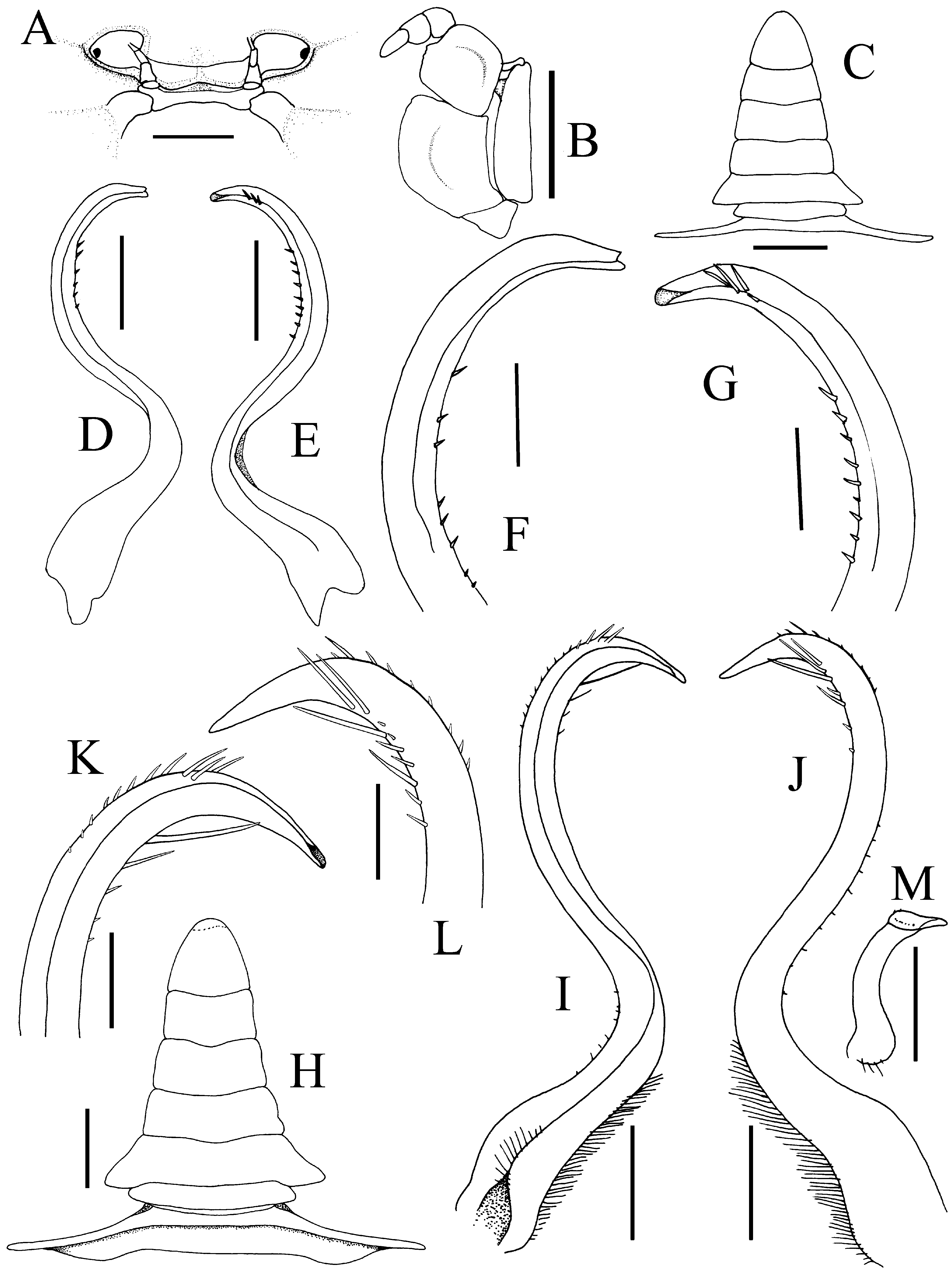
FIGURE 13. Typhlocarcinops canaliculatus Rathbun, 1909. A–G, male (5.5 × 4.2 mm) (ZRC 1995.0374), Singapore; H–M, male (8.6 × 6.5 mm) (ZRC 1985.1385), Singapore.A, frontal view showing orbit, epistome, antenna and antennule; B, left third maxilliped; C, H, male pleon; D, I, left G1 (ventral view); E, J, left G1 (dorsal view); F, K, distal part of left G1 (ventral view); G, L, distal part of left G1 (dorsal view); M, left G2. Scales: A–C, H = 1.0 mm; D, E, I, J, M = 0.5 mm; F, G, K, L = 0.25 mm.
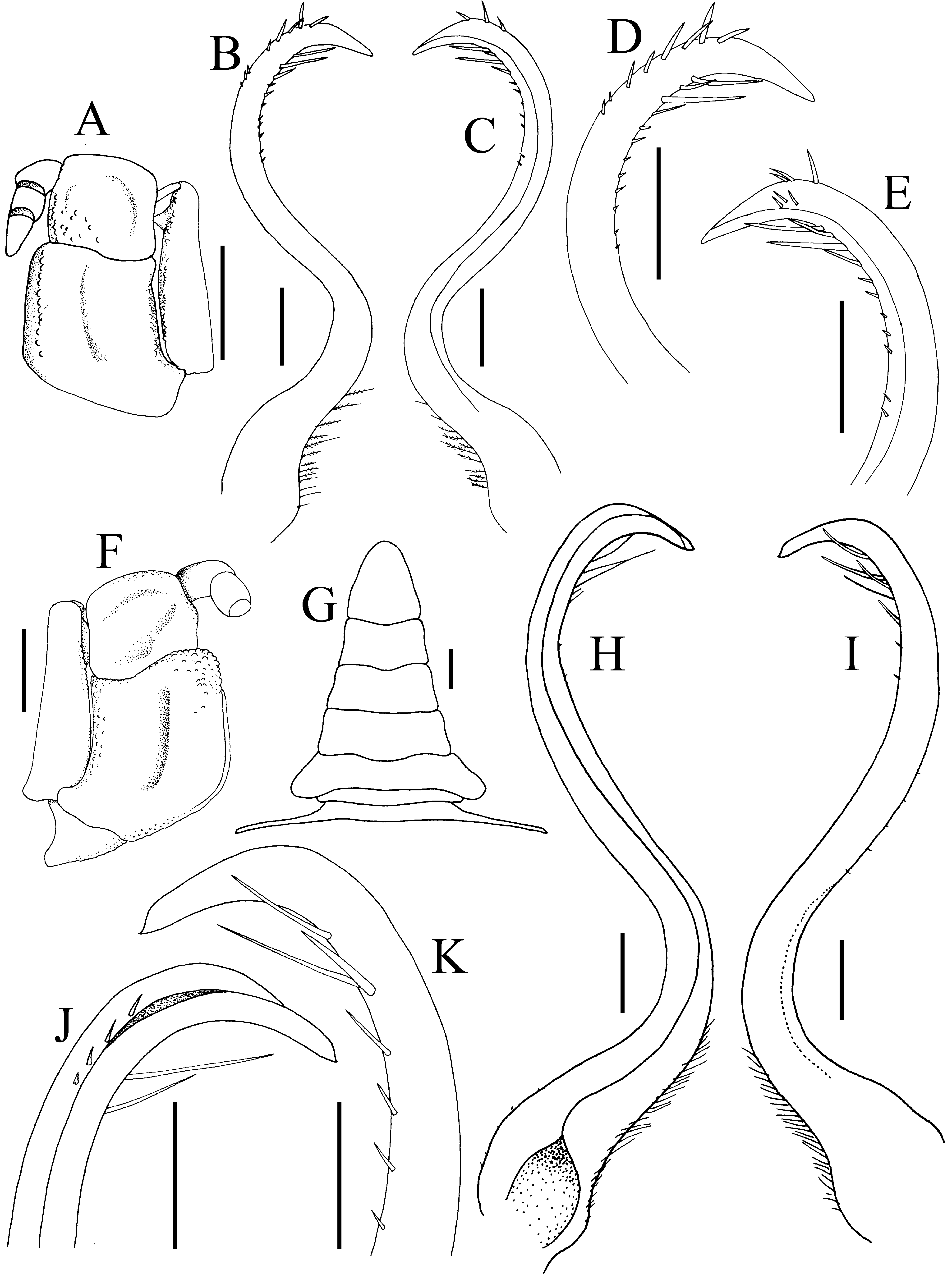
FIGURE 14. Typhlocarcinops canaliculatus Rathbun, 1909. A–E, male (8.7 × 6.6 mm) (ZRC 1984.7749), Phuket; F–J, male (13.2 × 10.5 mm) (ZRC 2018.0696), Hong Kong.A, right third maxilliped; B, right G1 (ventral view); C, right G1 (dorsal view); D, distal part of right G1 (ventral view); E, distal part of right G1 (dorsal view); F, left third maxilliped (dactylus missing); G, male pleon; H, left G1 (ventral view); I, left G1 (dorsal view); J, distal part of left G1 (ventral view); K, distal part of left G1 (dorsal view). Scales: A, B, F = 1.0 mm; B–E, H–K = 0.5 mm.
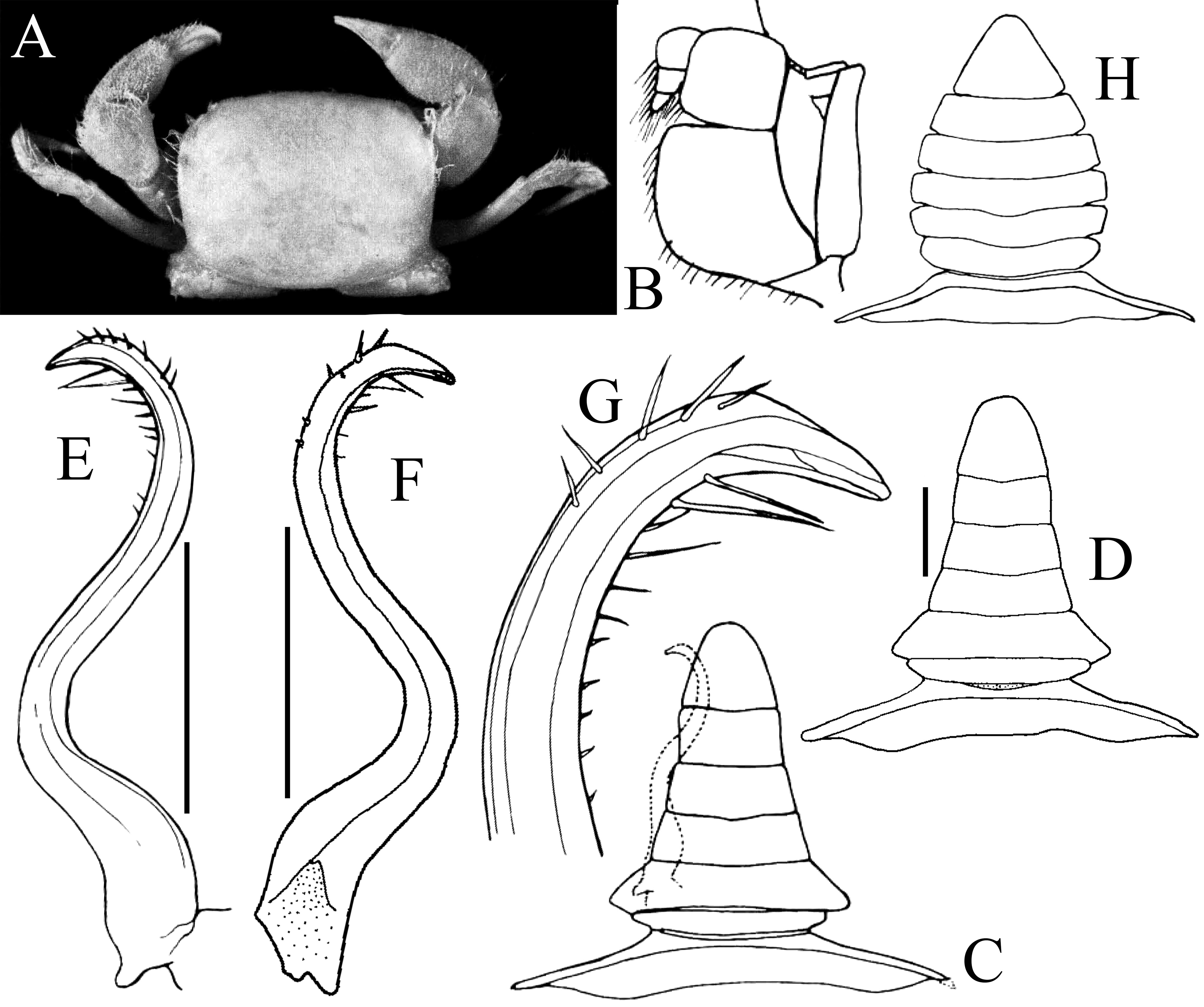
FIGURE 15. Typhlocarcinops canaliculatus Rathbun, 1909, A–D, lectotype male (6.5 × 5.3 mm) of T. gallardoi Serène, 1964 (ZMUC), Java Sea; H, paralectotype female (6.3 × 5.0 mm) of of T. gallardoi Serène, 1964 (ZMUC), Makassar. A, overall habitus; B, left third maxilliped; C, D, male pleon; E, left G1 (dorsal view); F, left G1 (ventral view); DG, distal part of left G1 (ventral view); H, female pleon. Scales: B–F, H = 1.0 mm; G = 0.2 mm. B–E, G, H after Serène (1964: fig. 11); A, D, F after Türkay (1986: text figures 56, 57, pl. 3 fig. 13).
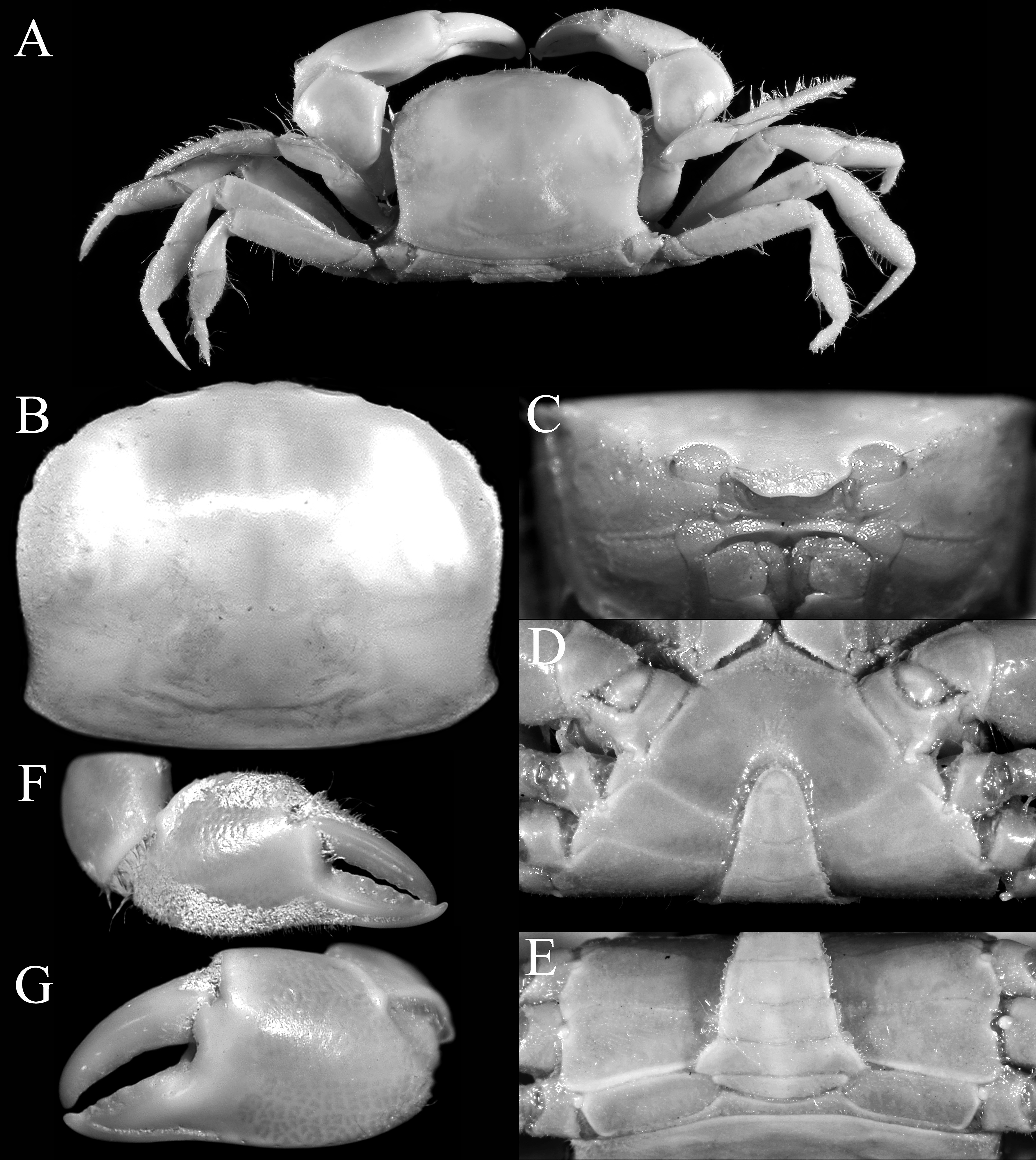
FIGURE 16. Typhlocarcinops decrescens Rathbun, 1914, holotype male (9.7 × 7.6 mm) (USNM 46407), Sulu Archipelago, Philippines. A, overall habitus; B, dorsal view of carapace; C, frontal view of cephalothorax; D, anterior thoracic sternum and pleon; E, posterior thoracic sternum and pleon; F, outer view of right chela; G, outer view of left chela.
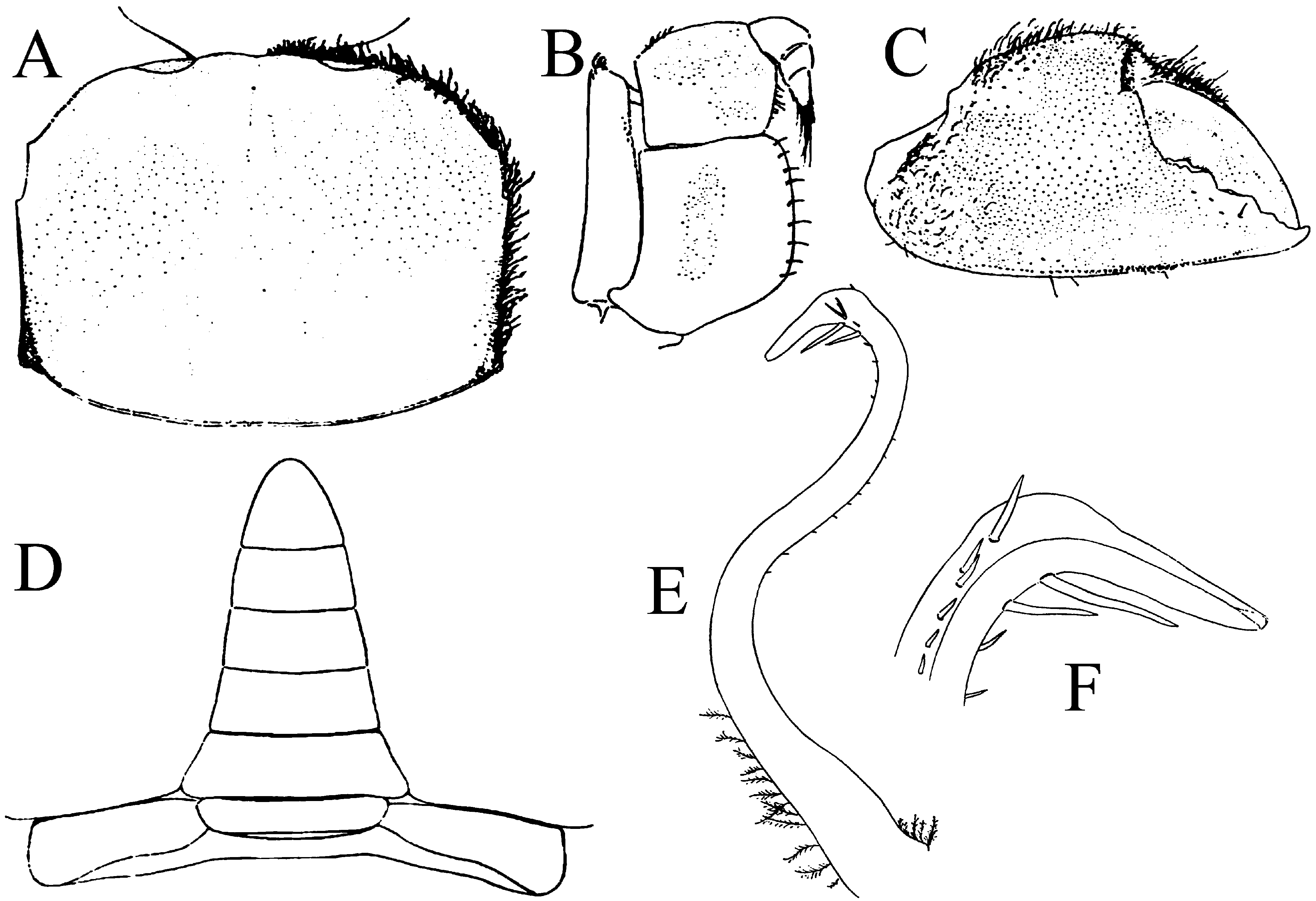
FIGURE 18. Typhlocarcinops decrescens Rathbun, 1914. Holotype male of Typhlocarcinops genkaiae Takeda & Miyake, 1972 (5.4 × 4.0 mm), Japan.A, dorsal view of carapace; B, right third maxilliped; C, outer view of right chela; D, male pleon; E, right G1 (dorsal view); F, distal part of right G1 (ventral view). After Takeda & Miyake (1972: fig. 5).
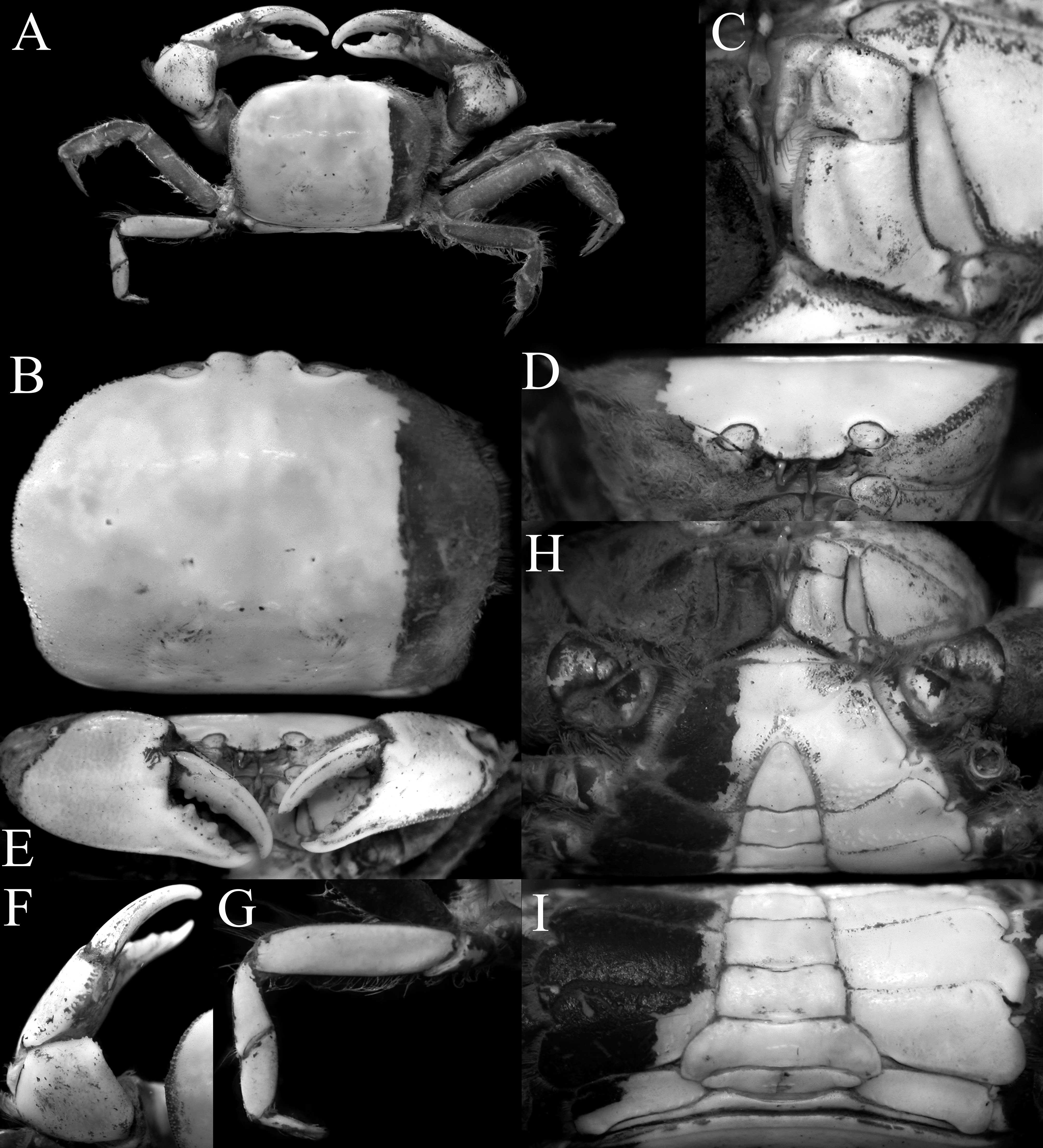
FIGURE 19. Typhlocarcinops decrescens Rathbun, 1914, male (15.2 × 11.9 mm) (ZRC 2018.0699), Hong Kong. A, overall habitus; B, dorsal view of carapace; C, left third maxilliped; D, frontal view of cephalothorax; E, outer view of chelae; F, dorsal view of left cheliped; G, left fourth ambulatory leg; H, anterior thoracic sternum and pleon; I, posterior thoracic sternum and pleon.
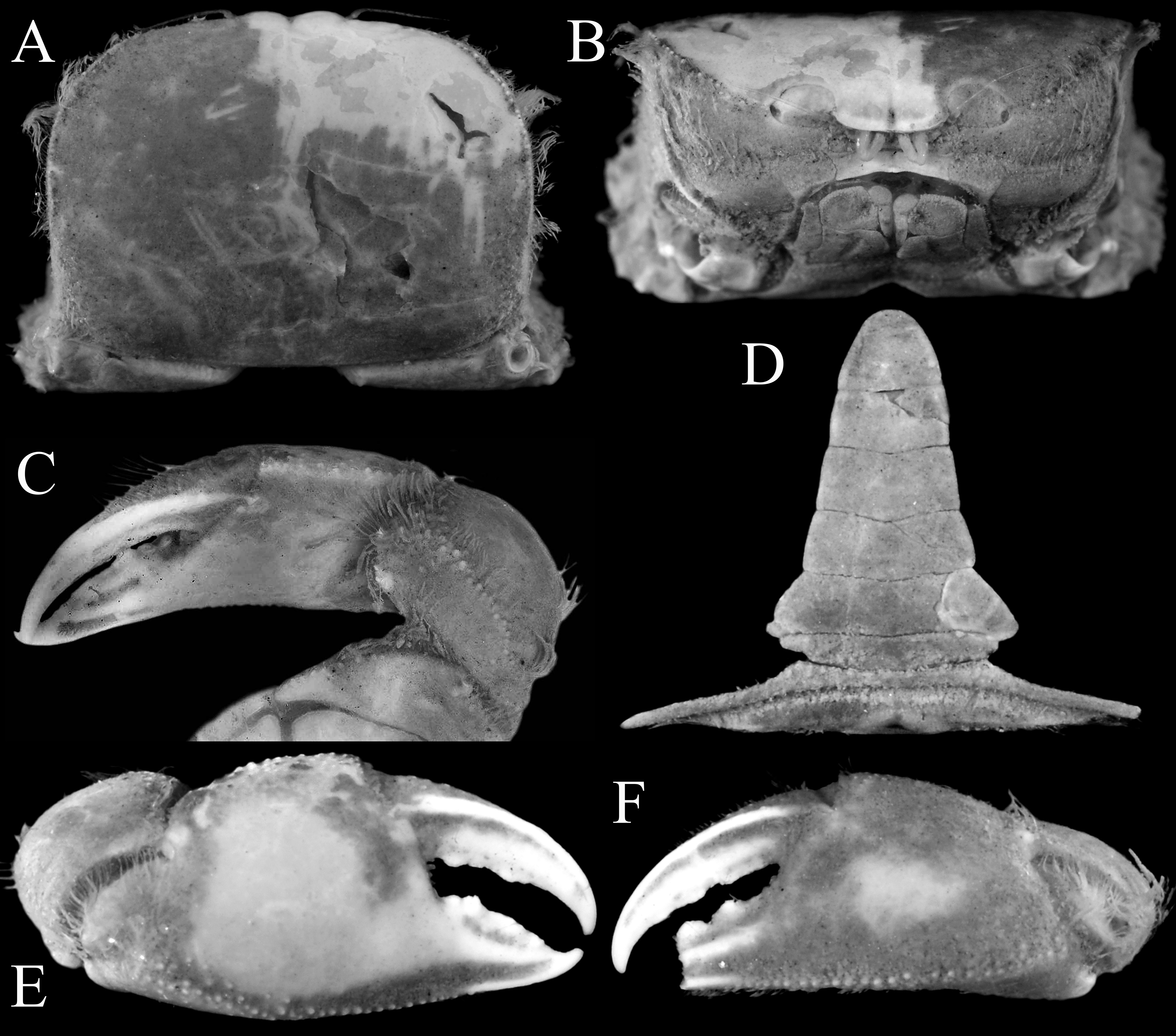
FIGURE 20. Typhlocarcinops decrescens Rathbun, 1914, male (6.1 × 4.5 mm) (AS 2137), China. A, dorsal view of carapace; B, frontal view of cephalothorax; C, dorsal view of left cheliped; D, male pleon; E, outer view of right chela; F, outer view of left chela.
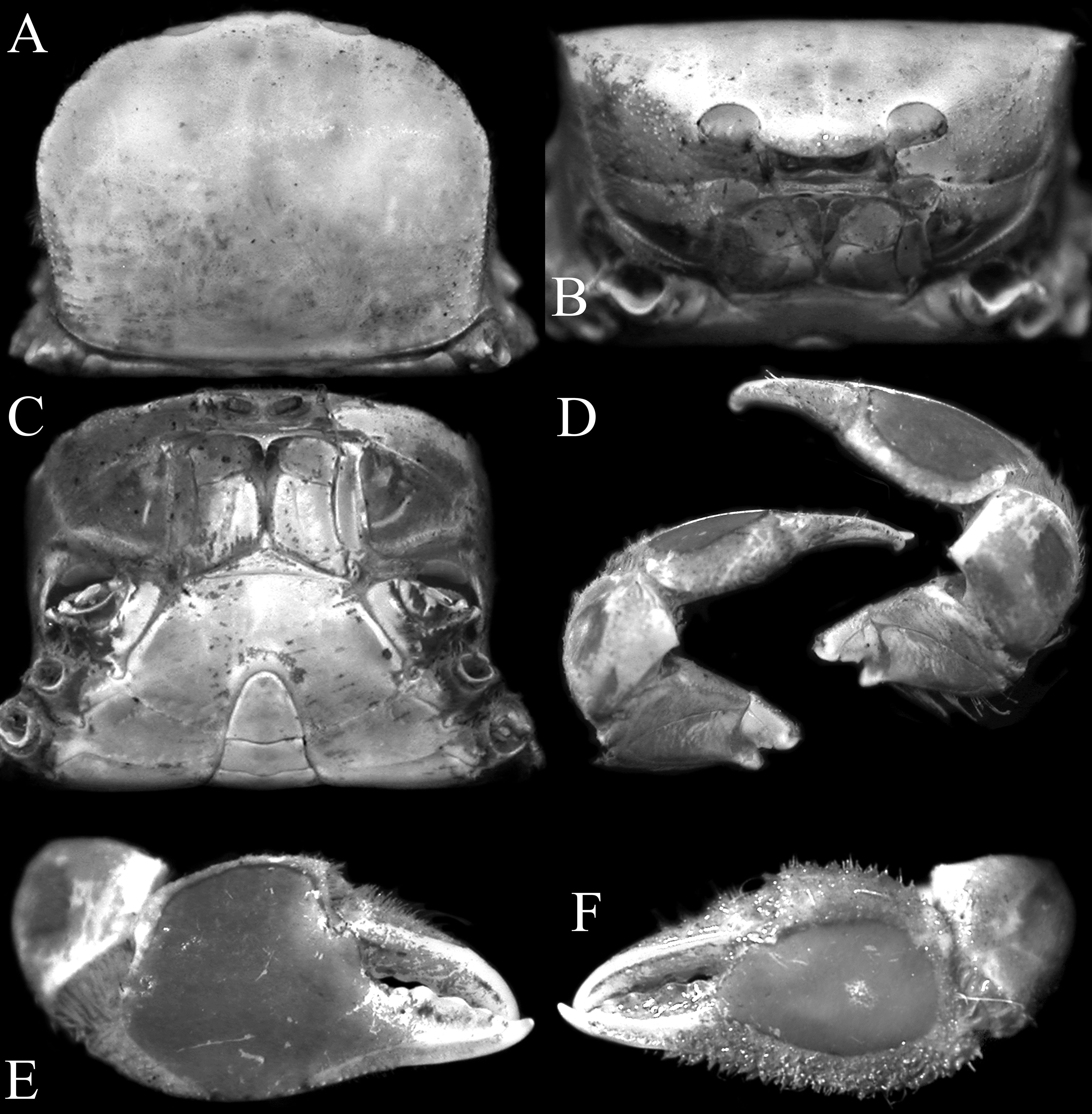
FIGURE 21. Typhlocarcinops decrescens Rathbun, 1914, male (6.8 × 5.3 mm) (ZRC 2018.0269), Papua, Indonesia. A, dorsal view of carapace; B, frontal view of cephalothorax; C, anterior thoracic sternum and pleon; D, dorsal view of left and right chelipeds showing angled inner margin of carpus; E, outer view of right chela; F, outer view of left chela.
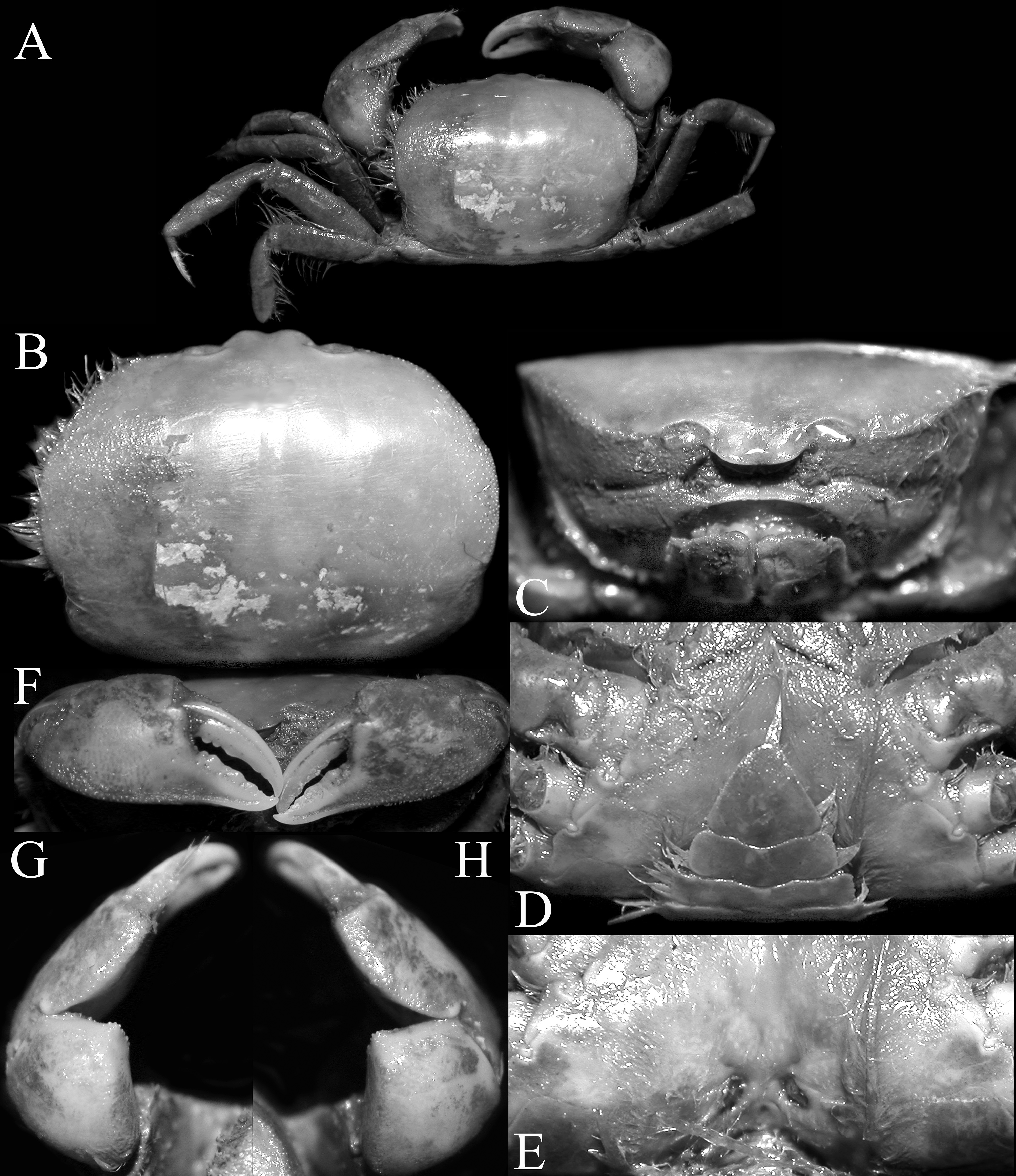
FIGURE 22. Typhlocarcinops decrescens Rathbun, 1914, female (15.3 × 11.6 mm) (ZRC 2018.0695), Hong Kong. A, overall habitus; B, dorsal surface of carapace (denuded); C, frontal view of cephalothorax; D, anterior thoracic sternum and pleon; E, sternopleonal cavity and vulvae; F, outer view of chelae; G, dorsal view of left chela; H, dorsal view of right chela.
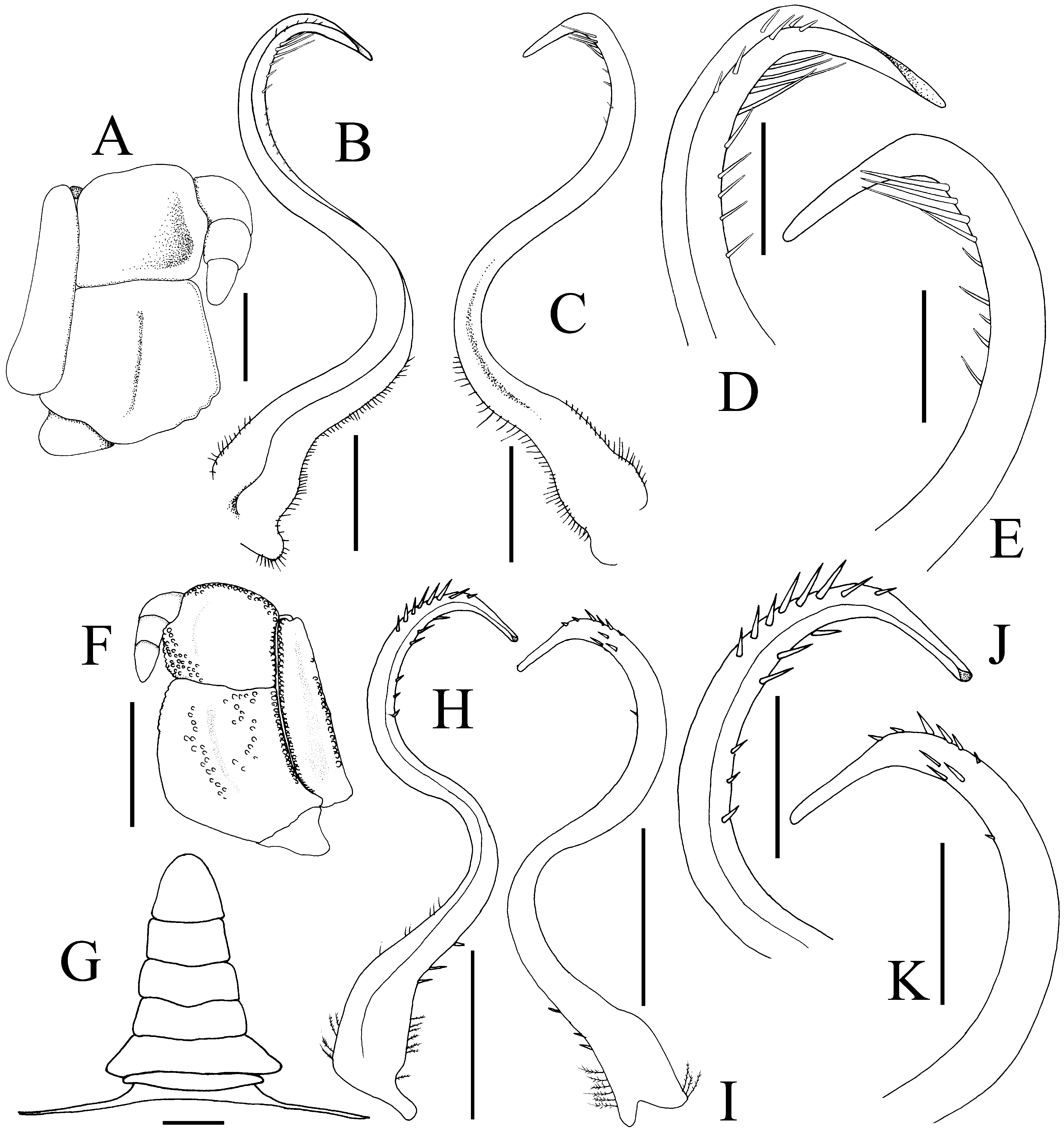
FIGURE 23. Typhlocarcinops decrescens Rathbun, 1914. A–E, holotype male (9.7 × 7.6 mm) (USNM 46407), Sulu Archipelago, Philippines; F–K, male (6.8 × 5.3 mm) (ZRC 2018.0269), Papua, Indonesia. A, right third maxilliped; B, H, left G1 (ventral view); C, I, left G1 (dorsal view); D, J, distal part of left G1 (ventral view); E, K, distal part of left G1 (dorsal view); F, left third maxilliped; G, male pleon. Scales: A–E, J, K = 0.5 mm; F–I = 1.0 mm.
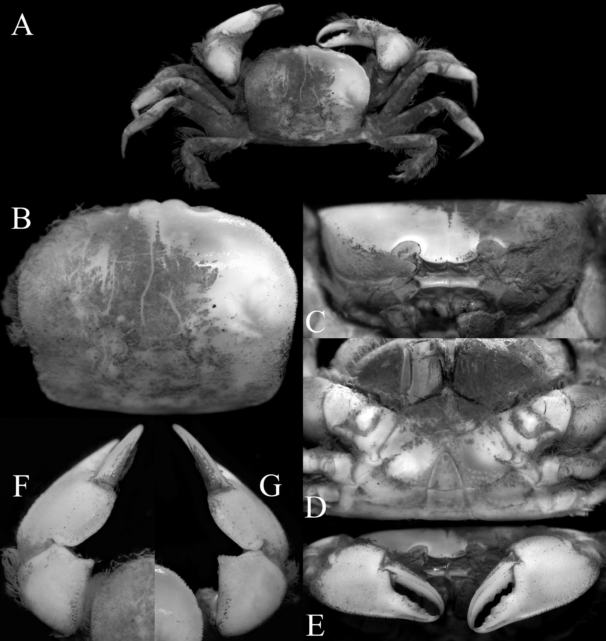
FIGURE 25. Typhlocarcinops denticarpes Dai, Yang, Song & Chen, 1986, male (11.4 × 8.6 mm) (ZRC 2018.0694), Hong Kong.A, overall habitus; B, dorsal surface of carapace (denuded); C, frontal view of cephalothorax; D, anterior thoracic sternum and pleon; E, outer view of chelae; F, dorsal view of left chela; G, dorsal view of right chela.
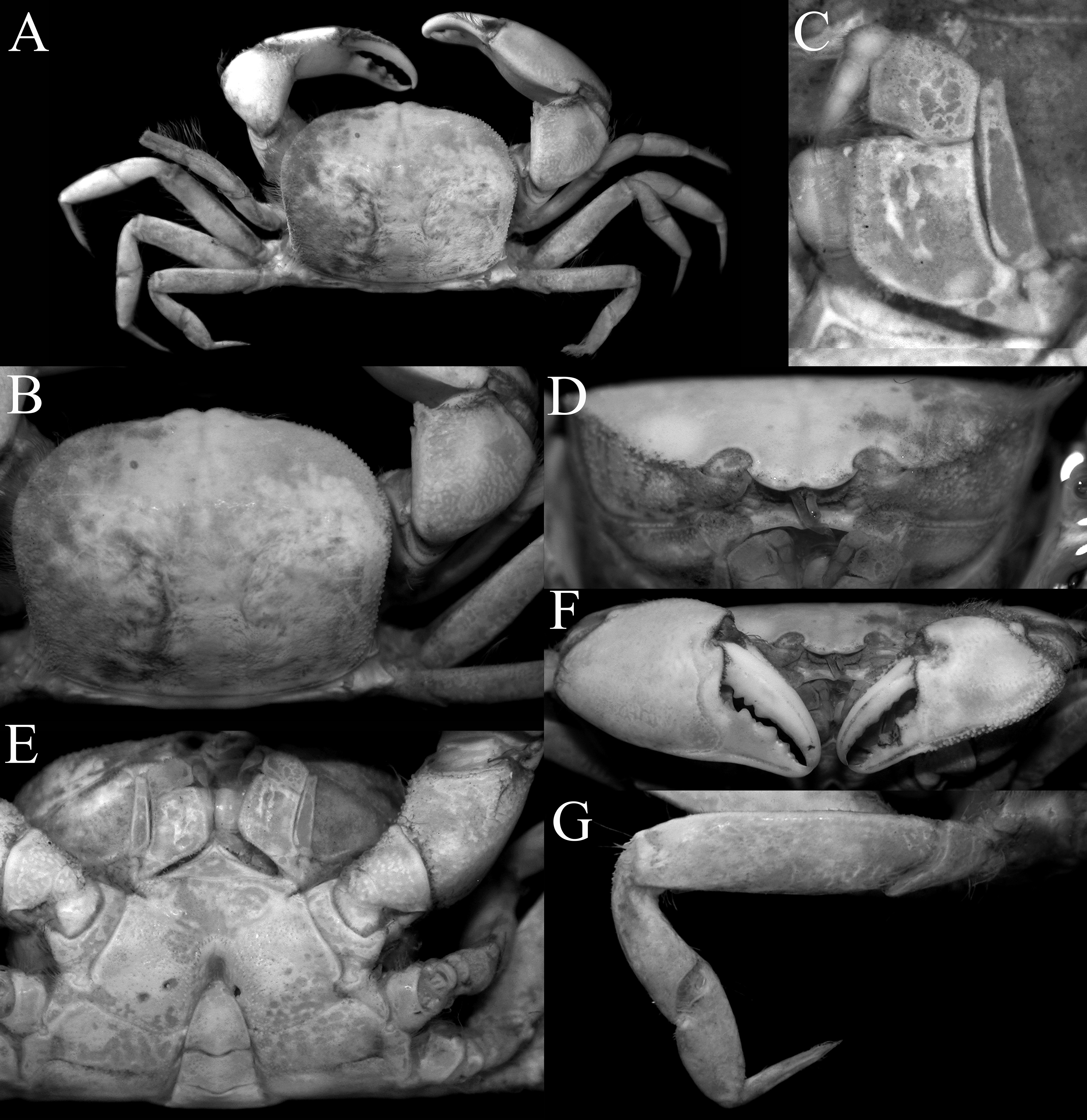
FIGURE 26. Typhlocarcinops denticarpes Dai, Yang, Song & Chen, 1986, male (15.9 × 12.4 mm) (ZRC 2018.0700), Hong Kong. A, overall habitus; B, dorsal view of carapace and right carpus of cheliped; C, left third maxilliped; D, frontal view of cephalothorax; E, anterior thoracic sternum and pleon; F, outer view of chelae; G, left fourth ambulatory leg.
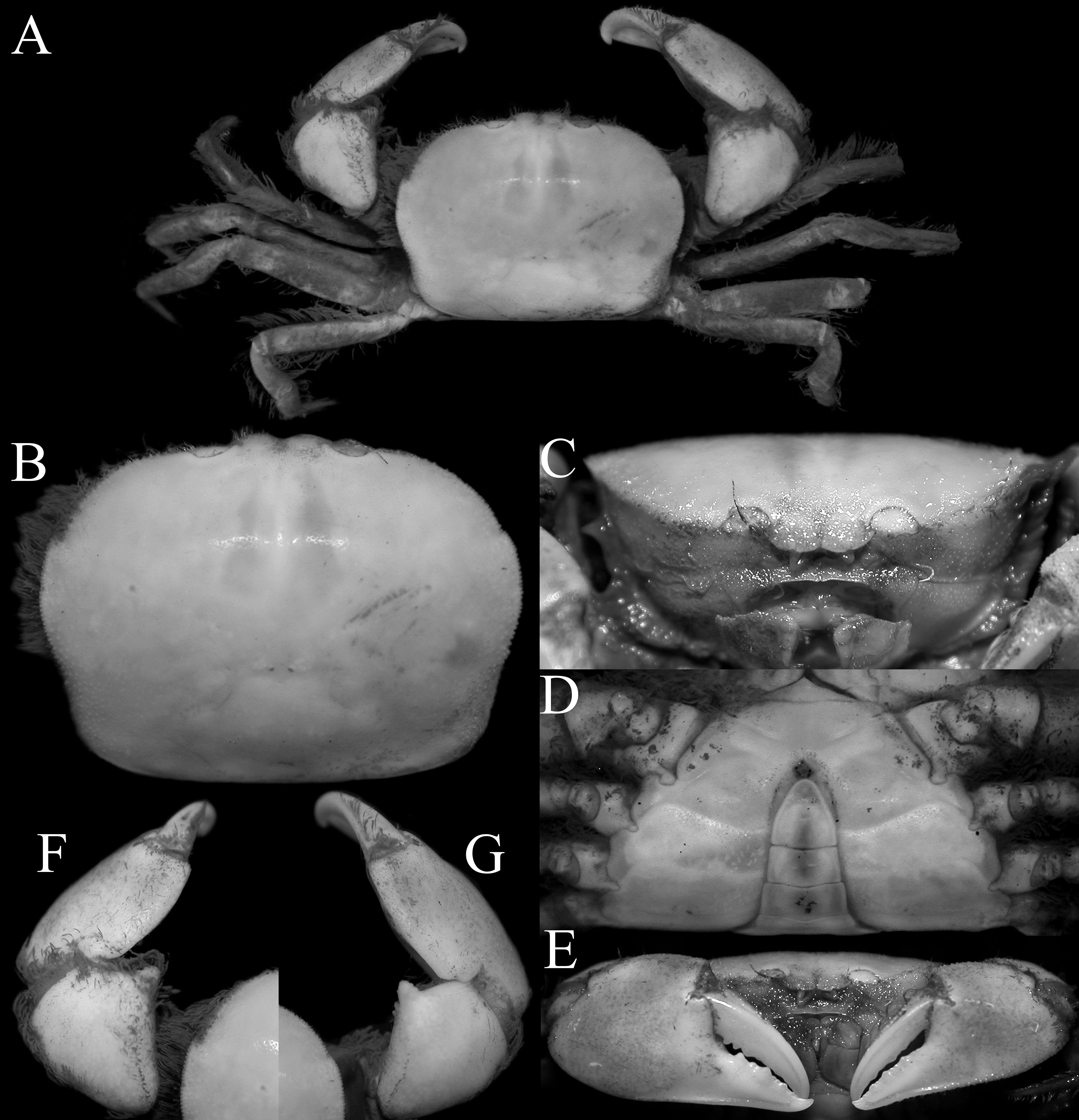
FIGURE 27. Typhlocarcinops denticarpes Dai, Yang, Song & Chen, 1986, male (14.3 × 10.7 mm) (ZRC 2016.0686), Kyushu, Japan. A, overall habitus; B, dorsal view of carapace; C, frontal view of cephalothorax; D, anterior thoracic sternum and pleon; E, outer view of chelae; F, dorsal view of left chela; G, dorsal view of right chela..
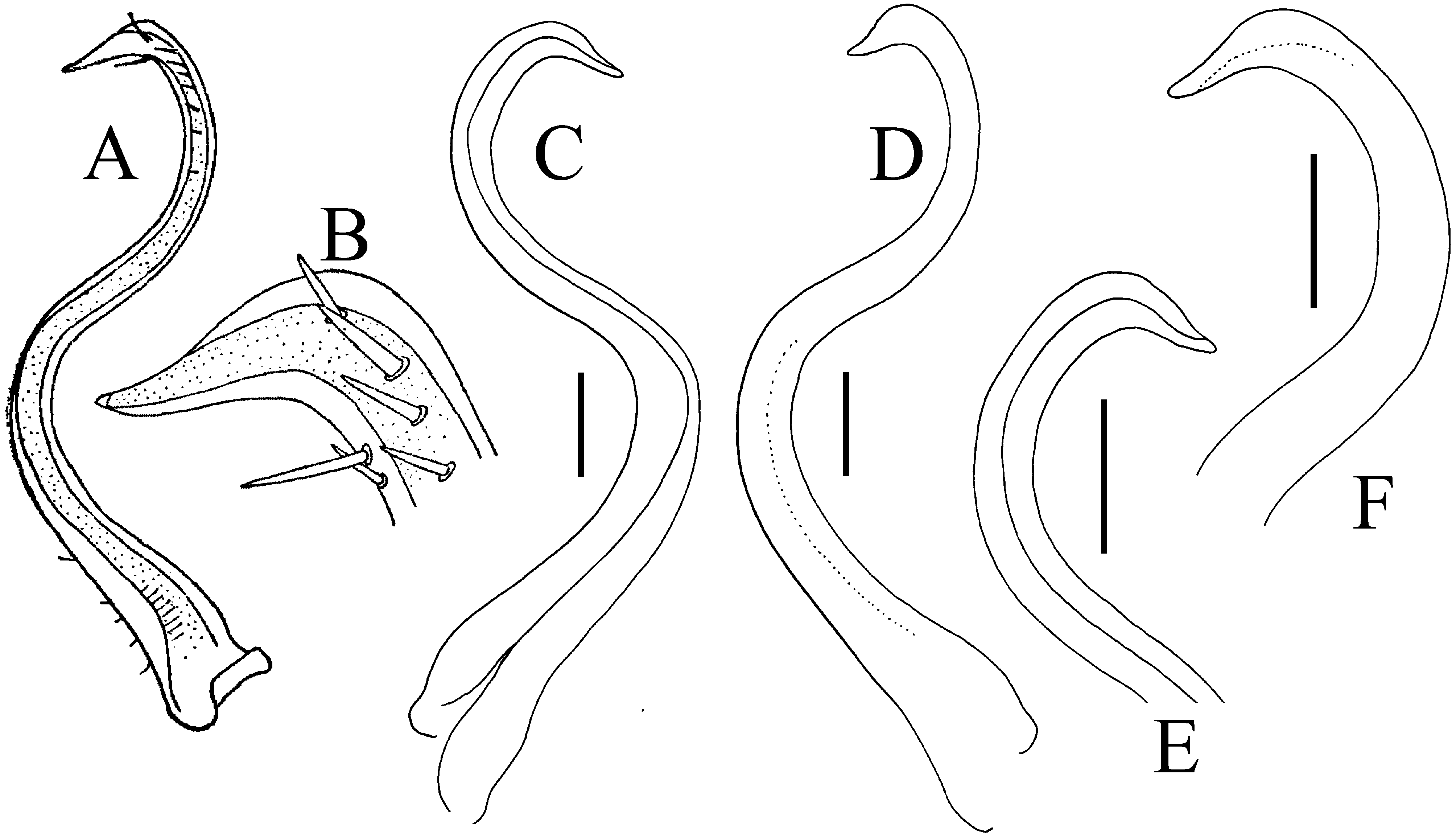
FIGURE 28. Typhlocarcinops denticarpes Dai, Yang, Song & Chen, 1986, holotype male (15.4 × 12.0 mm), Guangdong, China. A, right G1 (ventral view); B, distal part of right G1 (ventral view); C, left G1 (ventral view, setae not drawn); D, left G1 (dorsal view, setae not drawn); E, distal part of left G1 (ventral view, setae not drawn); F, distal part of left G1 (dorsal view, setae not drawn). A, B, after Dai et al. (1986: text fig. 202(1)), incorrectly labelled as “Typhlocarcinops canaliculatus Rathbun, 1909”. Scales = 0.5 mm.
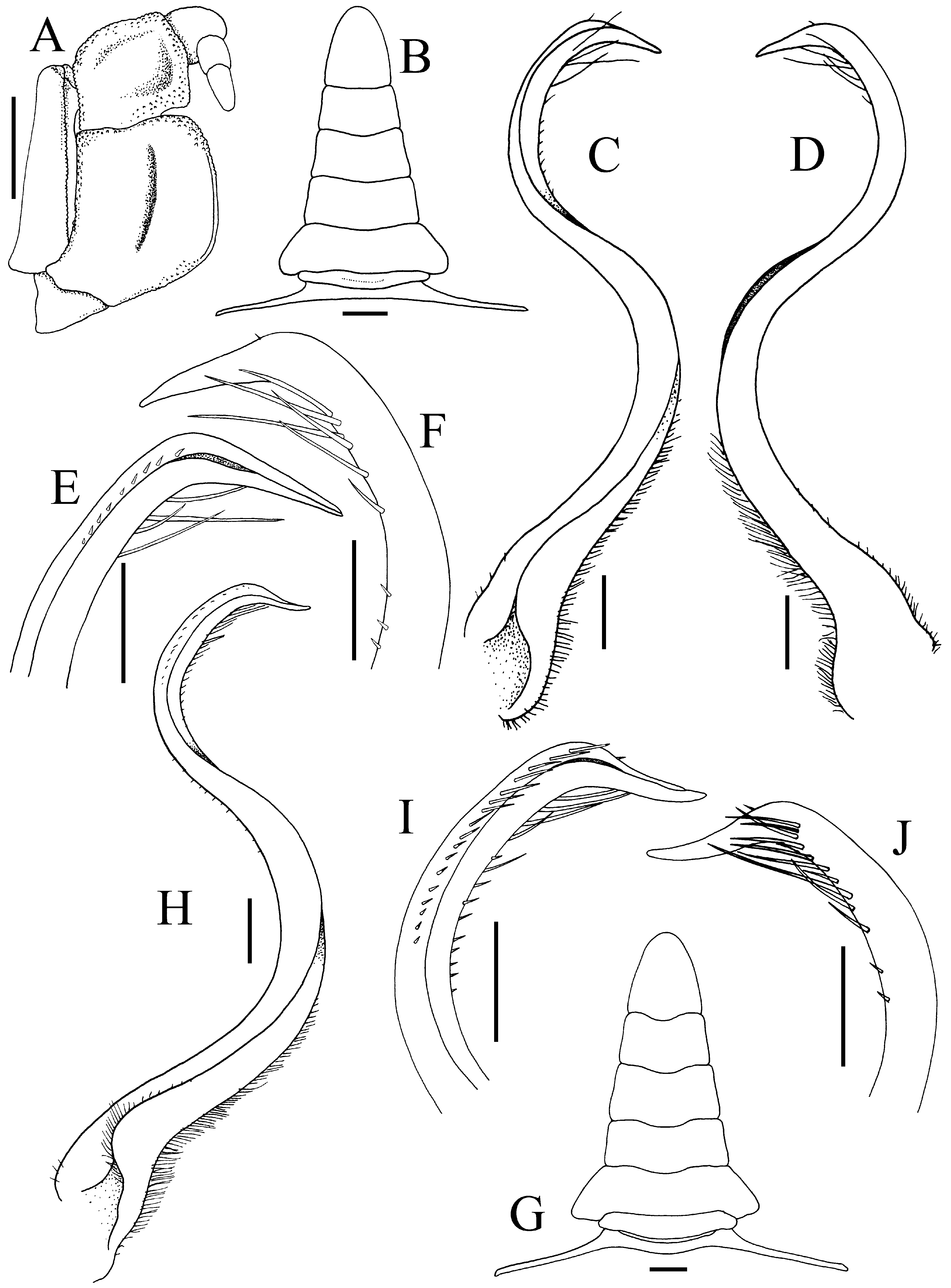
FIGURE 29. Typhlocarcinops denticarpes Dai, Yang, Song & Chen, 1986. A–F, male (11.4 × 8.6 mm) (ZRC 2018.0694), Hong Kong; G–J, male (15.9 × 12.4 mm) (ZRC 2018.0700), Hong Kong. A, left third maxilliped; B, G, male pleon; C, H, left G1 (ventral view); D, left G1 (dorsal view); E, I, distal part of left G1 (ventral view); F, J, distal part of left G1 (dorsal view). Scales: A, B, G = 1.0 mm; C–F, H–J = 0.5 mm.
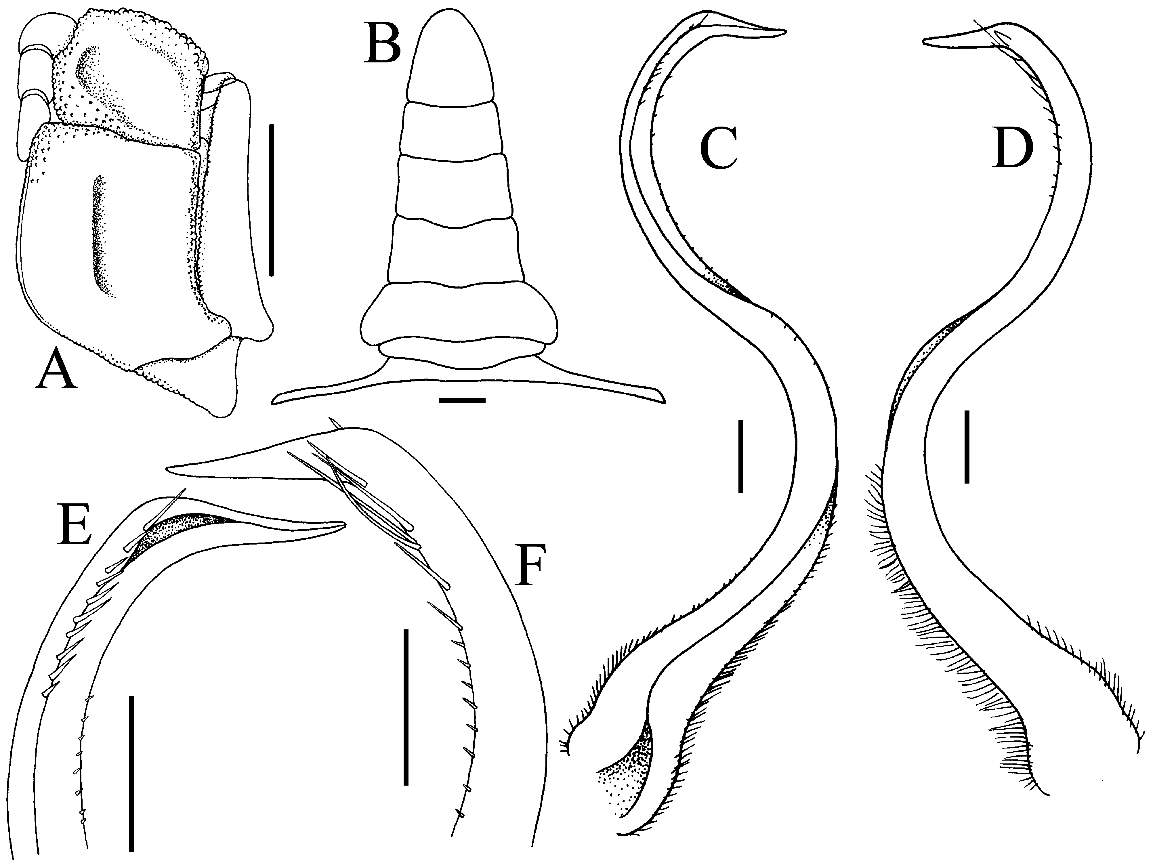
FIGURE 30. Typhlocarcinops denticarpes Dai, Yang, Song & Chen, 1986, male (14.3 × 10.7 mm) (ZRC 2016.0686), Kyushu, Japan.A, left third maxilliped; B, male pleon; C, left G1 (ventral view); D, left G1 (dorsal view); E, distal part of left G1 (ventral view); F, distal part of left G1 (dorsal view). Scales: A, B = 1.0 mm; C–F = 0.5 mm.
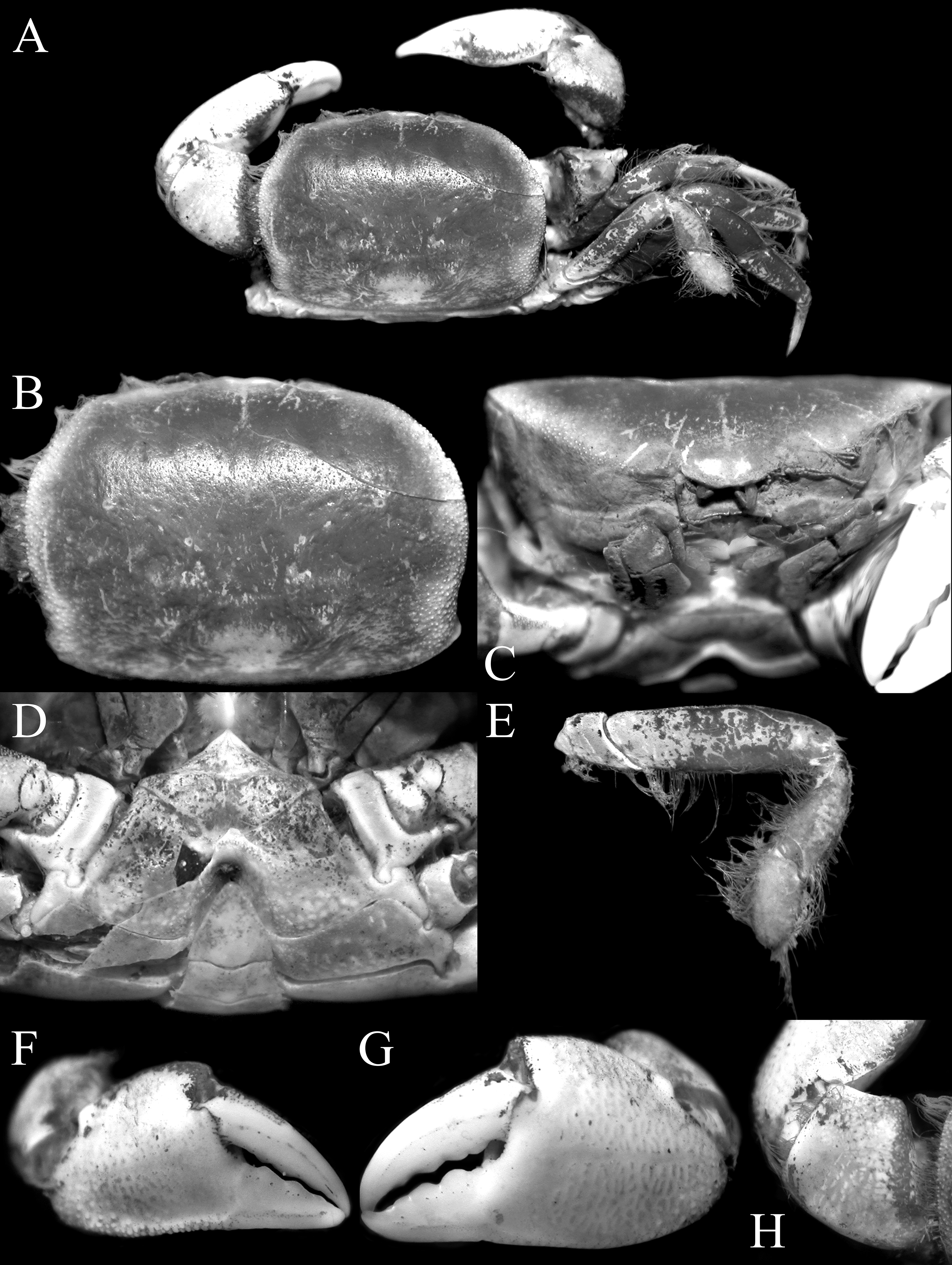
FIGURE 31. Typhlocarcinops yui Ng & Ho, 2003, male (10.7 × 8.1 mm) (SMF 37555), Japan. A, overall habitus; B, dorsal view of carapace; C, frontal view of cephalothorax; D, anterior thoracic sternum and pleon; E, right fourth ambulatory leg; F, outer view of left chela; G, outer view of right chela; H, right carpus of cheliped.
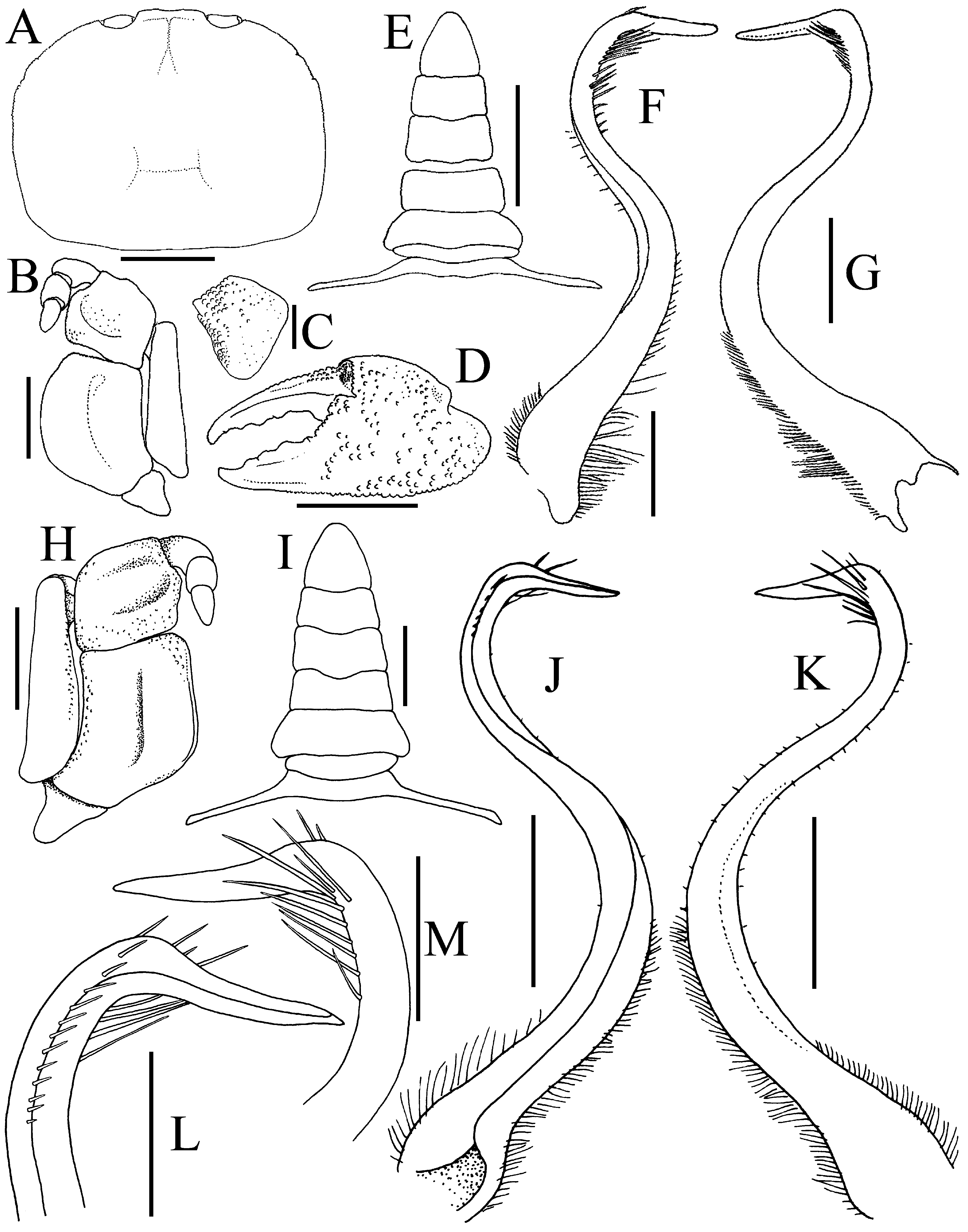
FIGURE 32. Typhlocarcinops yui Ng & Ho, 2003.A–F, holotype male (6.5 × 4.7 mm) (NTOU), Taiwan; G–L, male (10.7 × 8.1 mm) (SMF 37555), Japan.A, carapace; B, right third maxilliped; C, outer view of carpus of right cheliped; D, outer view of left chela; E, male pleon; H, left third maxilliped; F, J, left G1 (ventral view); G, K, left G1 (dorsal view); L, distal part of left G1 (ventral view); M, distal part of left G1 (dorsal view). Scales: B, D, E = 2.0 mm; C, H, J, K = 1.0 mm; I = 2.0 mm; F, G, L, M = 0.5 mm. A–G after Ng & Ho (2003: fig. 3).
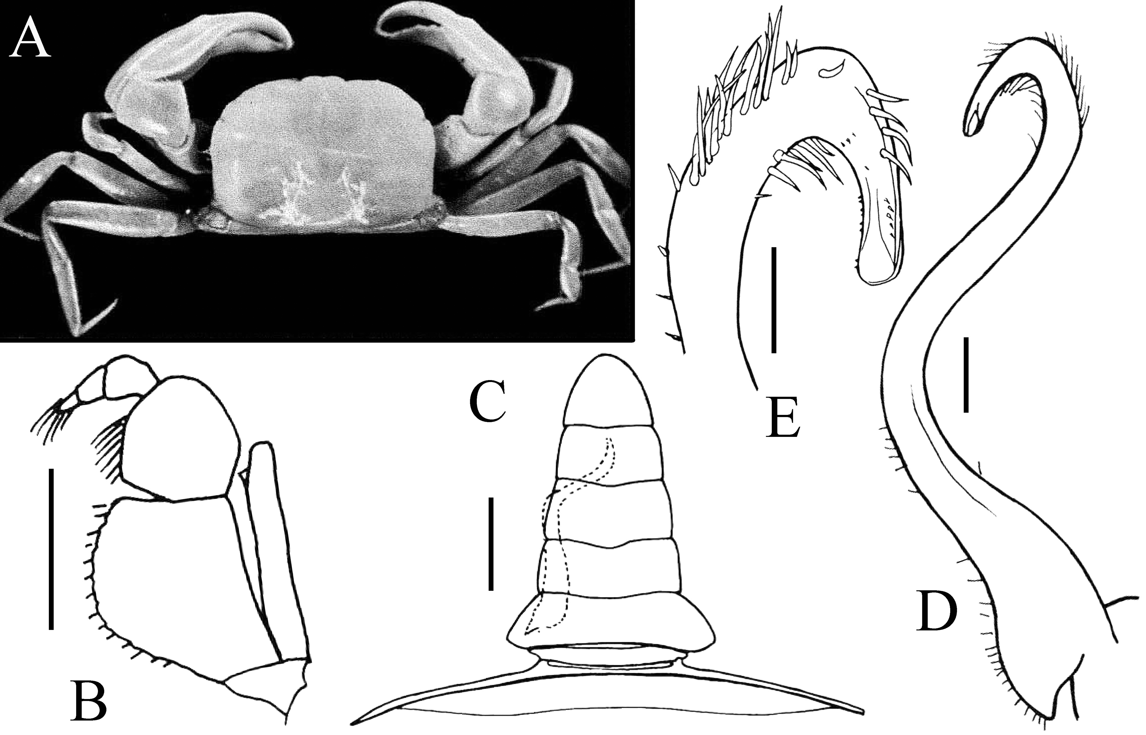
FIGURE 33. Typhlocarcinops stephenseni Serène, 1964, lectotype male (7.5 × 5.3 mm) (Zoological Survey of India no. 4.991), Indian Ocean. A, overall habitus; B, left third maxilliped; C, pleon; D, left G1 (dorsal view); E, distal part of left G1 (ventral view). After Serène (1964: text-fig. 8, pl. 19 fig. B). Scales: B, C = 1.0 mm; D = 0.3 mm; E = 0.2 mm.
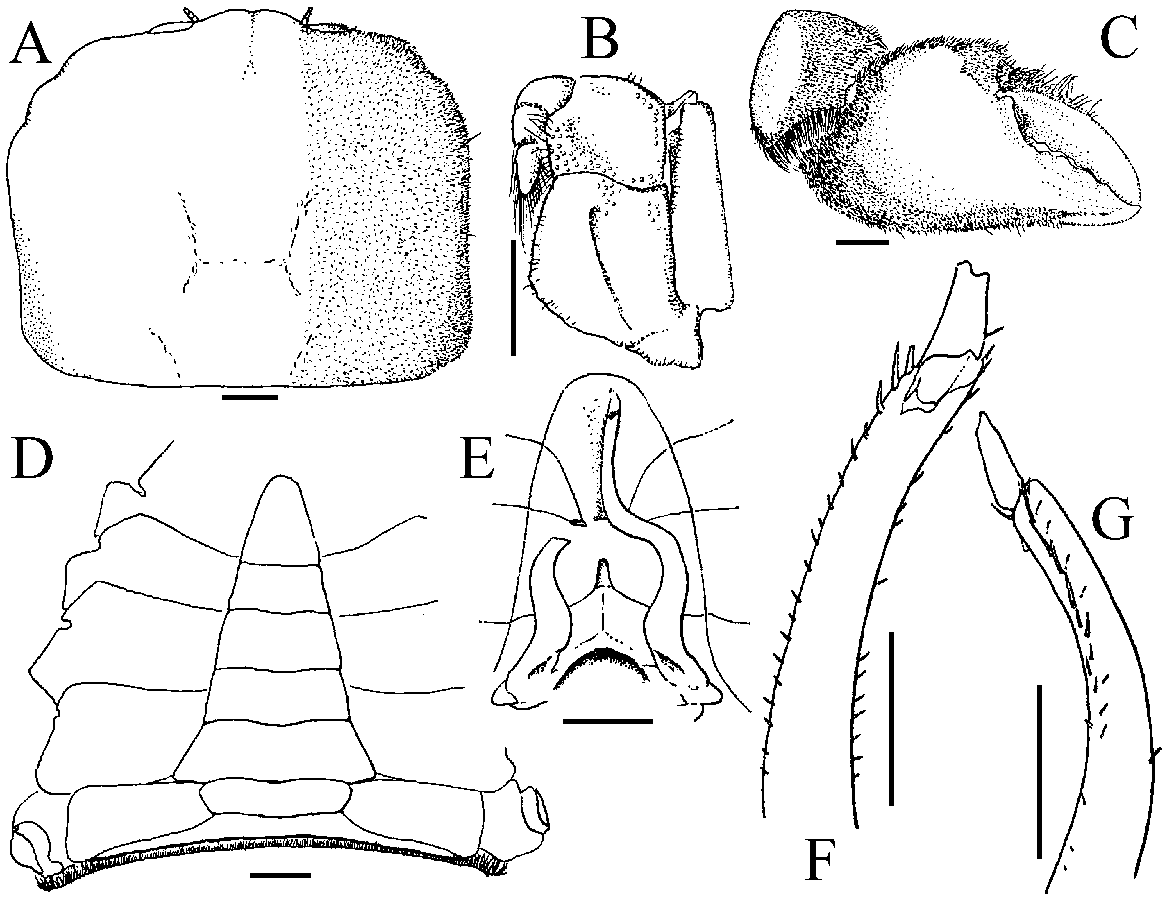
FIGURE 34. Typhlocarcinops tonsuratus Griffin & Campbell, 1969, holotype male (8.8 × 7.2 mm) (Queensland Museum W2911), Queensland, Australia.A, dorsal view of carapace; B, left third maxilliped; C, outer view of right cheliped; D, thoracic sternum and pleon; E, sternopleonal cavity with G1 (right one broken); F, distal part of left G1 (ventral view); G, distal part of left G1 (dorsal view). After Griffin & Campbell (1969: figs. 3, 6B). Scales = 1.0 mm.
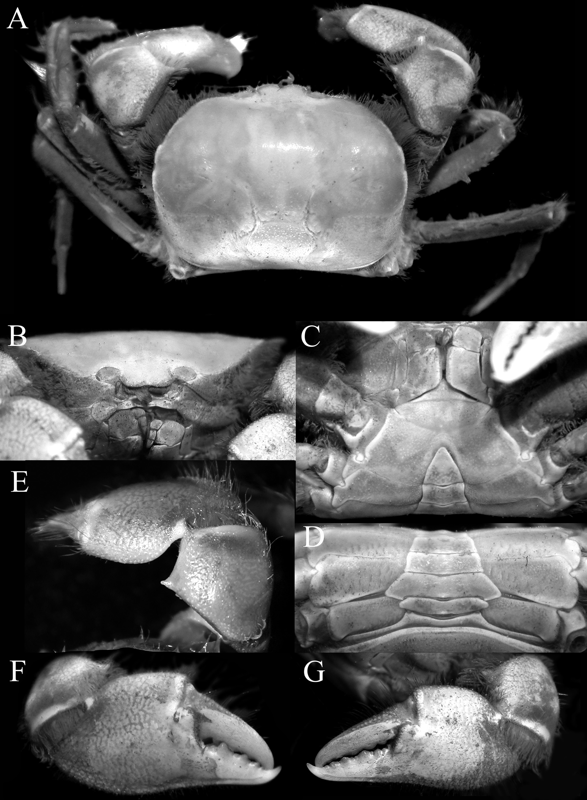
FIGURE 35. Typhlocarcinops robustus n. sp., holotype male (15.2 × 11.3 mm) (MZB Cru 4804), Papua, Indonesia. A, overall habitus; B, frontal view of cephalothorax; C, anterior thoracic sternum and pleon; D, posterior thoracic sternum and pleon; E, carpus of right cheliped; F, outer view of right chela; G, outer view of left chela.
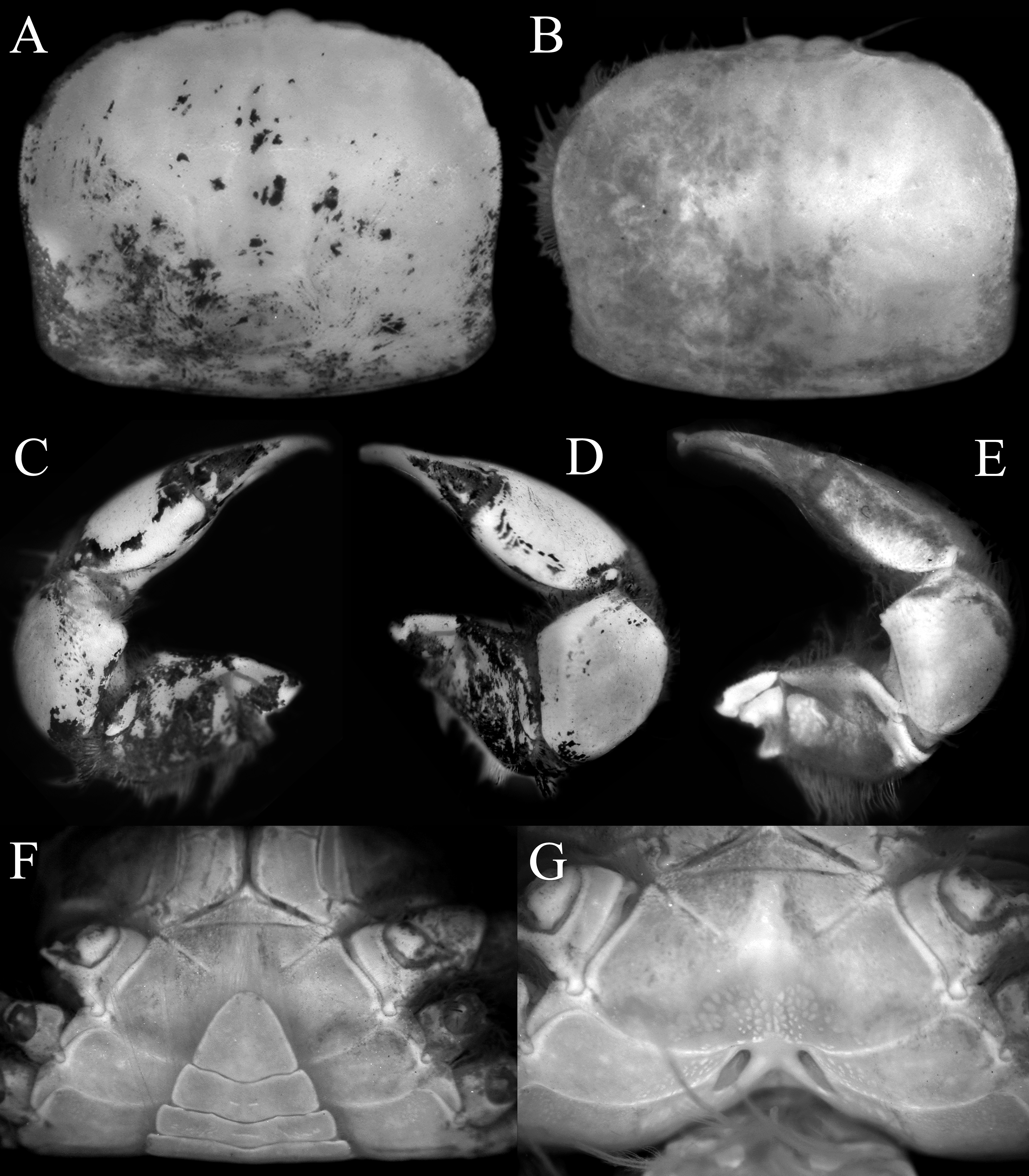
FIGURE 36. Typhlocarcinops robustus n. sp., A, C, D, paratype male (8.5 × 6.8 mm) (ZRC 2018.0275), Indonesia; B, E–G, paratype female (8.5 × 6.5 mm) (ZRC 2018.0275), Indonesia. A, B, dorsal view of carapace; C, dorsal view of left cheliped; D, E, dorsal view of right cheliped; F, anterior thoracic sternum and pleon; G, sternopleonal cavity and vulvae.
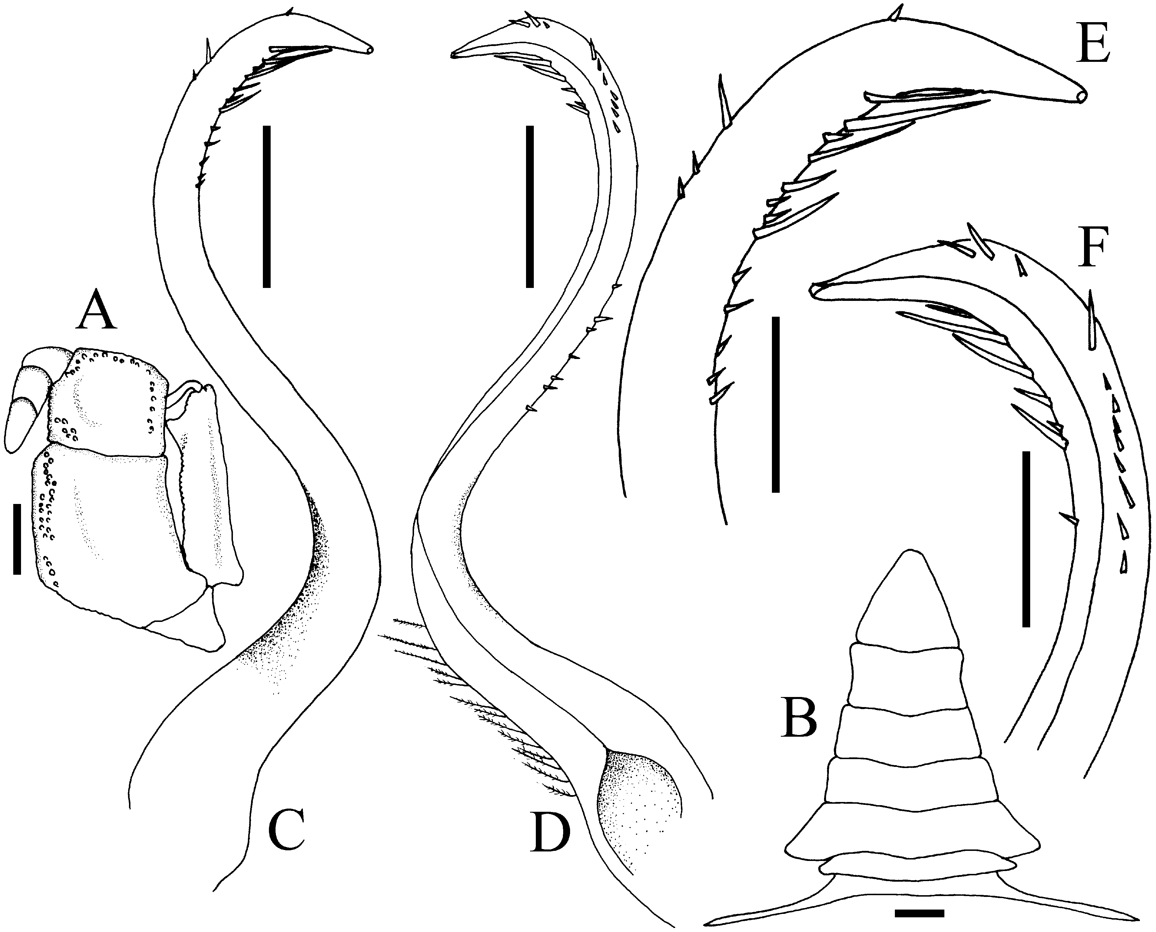
FIGURE 37. Typhlocarcinops robustus n. sp., holotype male (15.2 × 11.3 mm) (MZB Cru 4804), Papua, Indonesia.A, left third maxilliped; B, male pleon; C, right G1 (dorsal view); D, right G1 (ventral view); E, distal part of right G1 (dorsal view); F, distal part of right G1 (ventral view). Scales: A–D = 1.0 mm; E, F = 0.5 mm.

FIGURE 38. Typhlocarcinops hamus n. sp., holotype male (5.0 × 3.5 mm) (MNHN-IU-2013-1441a), Papua New Guinea. A, overall habitus; B, dorsal view of carapace; C, frontal view of cephalothorax; D, anterior thoracic sternum and pleon; E, carpus of left cheliped; F, carpus of right cheliped; G, outer view of right chela; H, outer view of left chela.
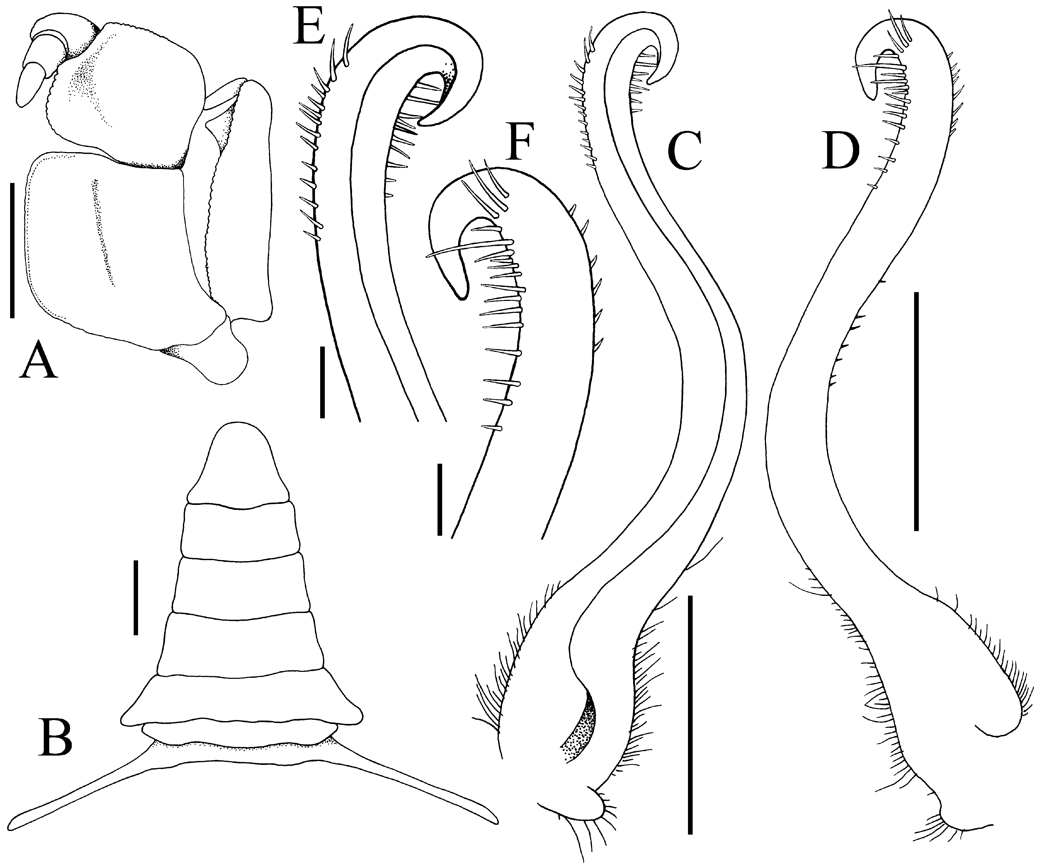
FIGURE 39. Typhlocarcinops hamus n. sp., holotype male (5.0 × 3.5 mm) (MNHN-IU-2013-1441a), Papua New Guinea. A, left third maxilliped; B, male pleon; C, left G1 (ventral view); D, left G1 (dorsal view); E, distal part of left G1 (ventral view); F, distal part of left G1 (dorsal view). Scales: A–D = 0.5 mm; E, F = 0.1 mm.
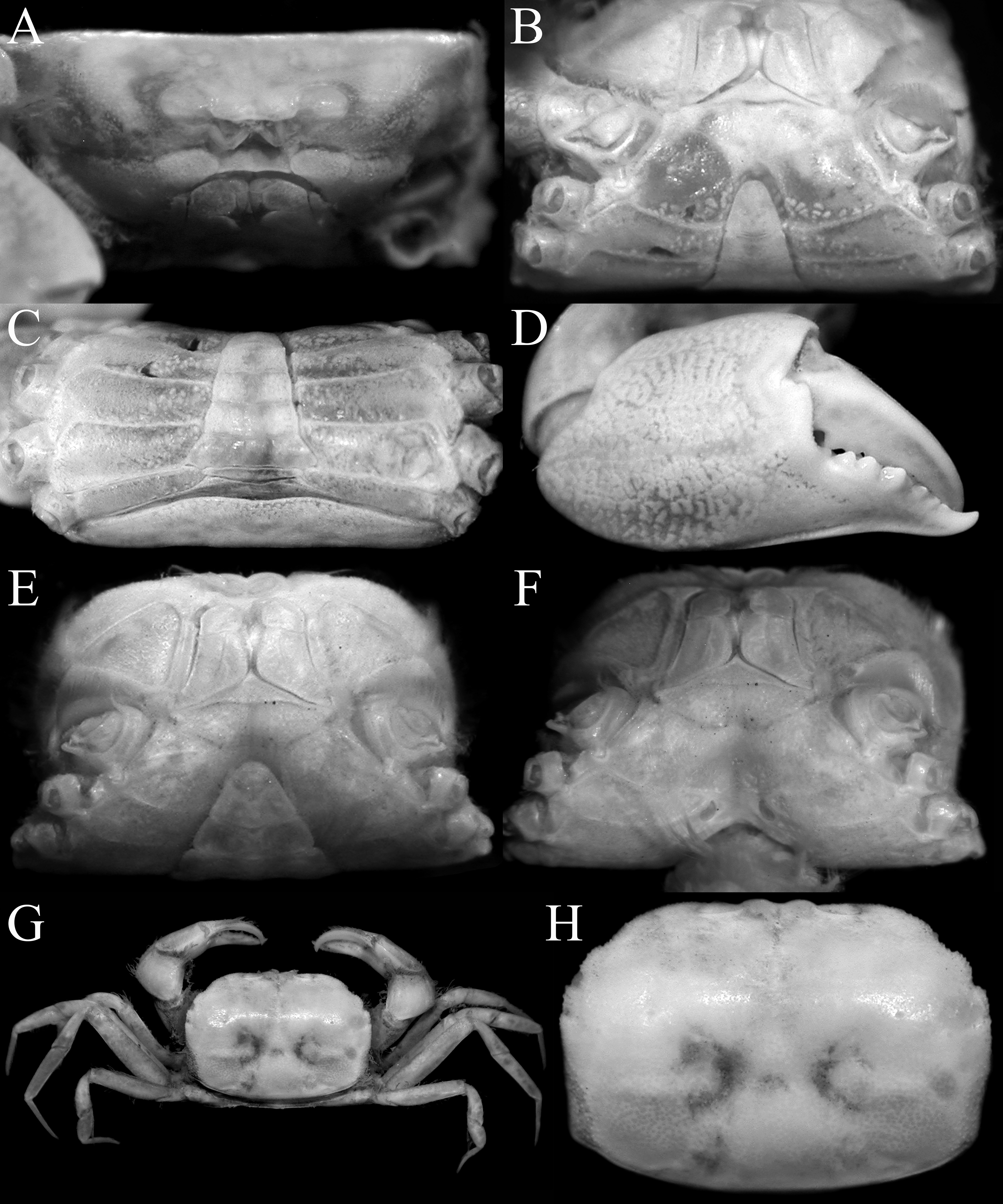
FIGURE 42. Typhlocarcinops raouli n. sp. A–D, holotype male (8.0 × 5.7 mm) (MZB Cru 4806), Papua, Indonesia; E, F, paratype female (5.3 × 3.6 mm) (ZRC 2018.0262), Papua, Indonesia; G, H, female (8.7 × 6.1 mm) (ZRC 2018.0701), Hong Kong. A, frontal view of cephalothorax; B, C, posterior thoracic sternum and pleon; D, outer view of right chela; E, anterior thoracic sternum and pleon; F, sternopleonal cavity and vulvae; G, overall habitus; H, dorsal view of carapace.
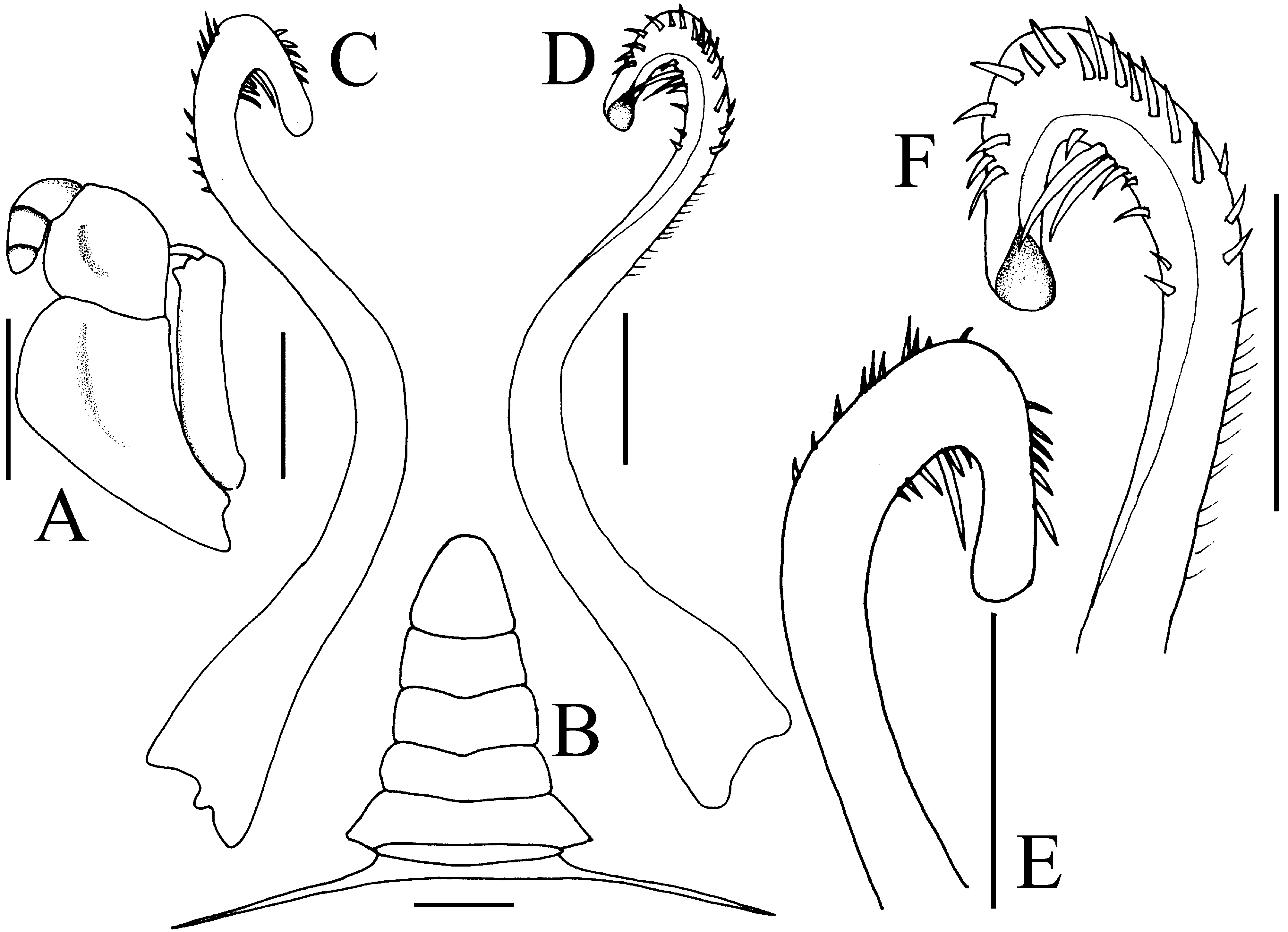
FIGURE 44. Typhlocarcinops raouli n. sp., holotype male (8.0 × 5.7 mm) (MZB Cru 4806), Papua, Indonesia. A, left third maxilliped; B, male pleon; C, left G1 (ventral view); D, left G1 (dorsal view); E, distal part of left G1 (ventral view); F, distal part of left G1 (dorsal view). Scales: A–D = 1.0 mm; E, F = 0.5 mm.
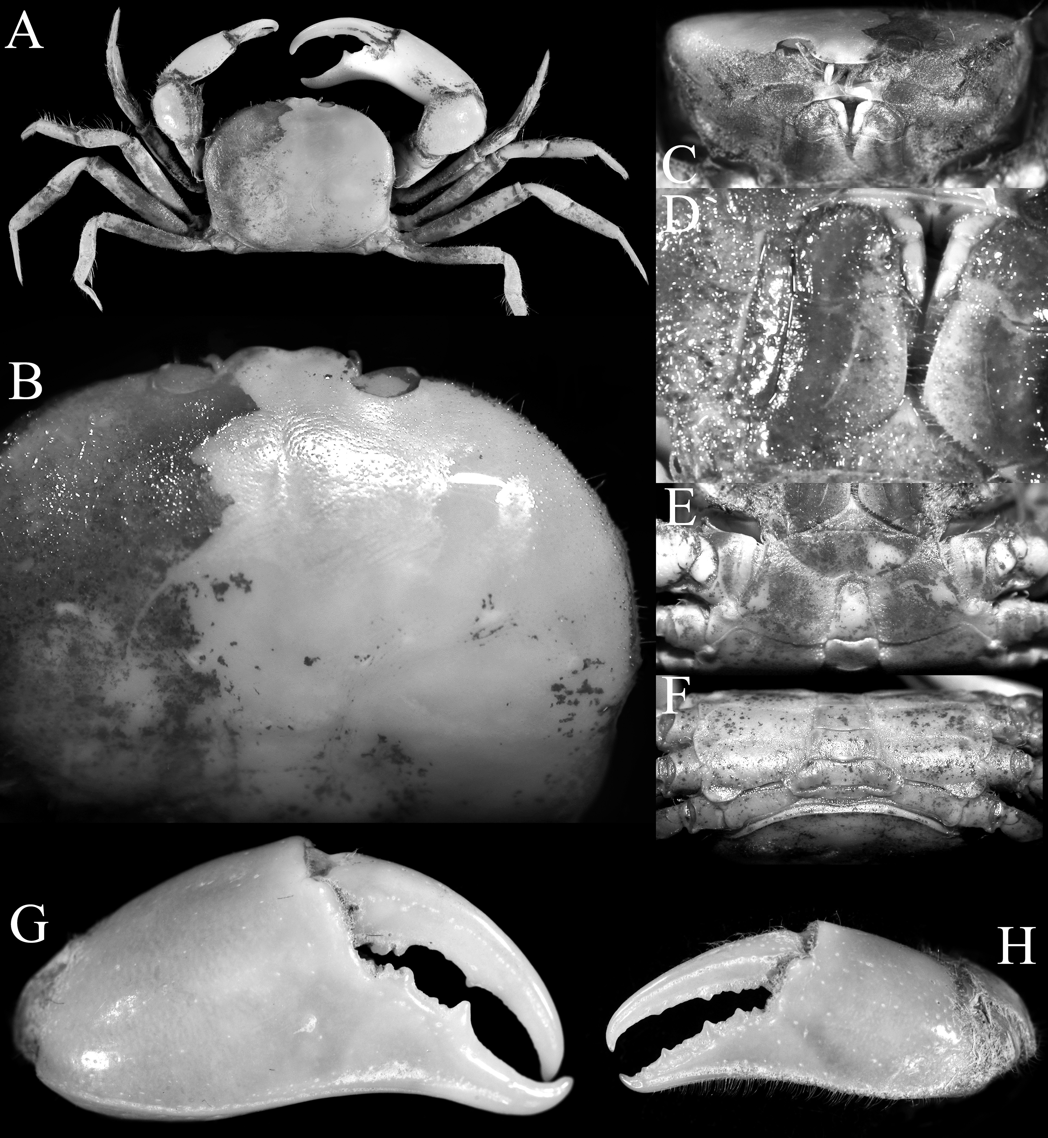
FIGURE 45. Typhlocarcinops ocularius Rathbun, 1914, holotype male (17.0 × 13.5 mm) (USNM 46408), between Samar and Masbate, Philippines. A, overall habitus; B, dorsal view of carapace (right side denuded); C, frontal view of cephalothorax; D, right third maxilliped; E, anterior thoracic sternum and pleon; F, posterior thoracic sternum and pleon; G, outer view of right chela; H, outer view of left chela.
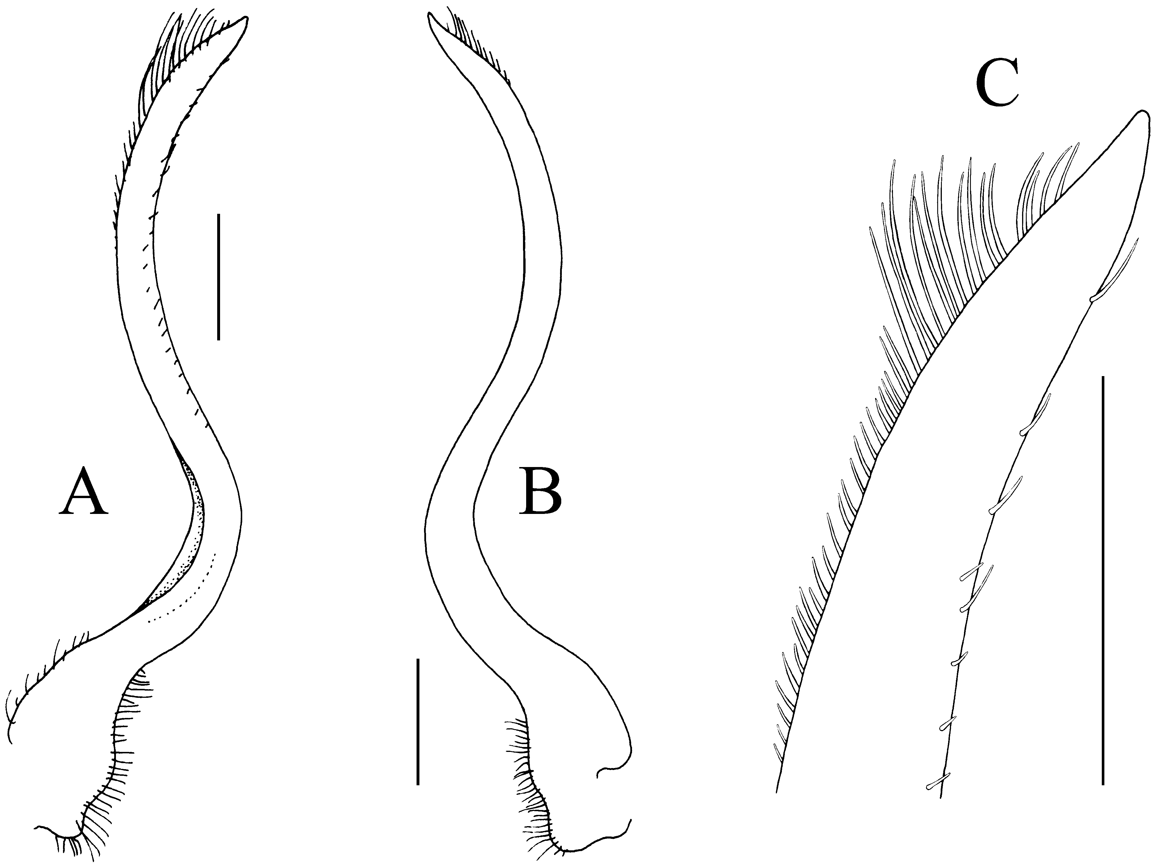
FIGURE 46. Typhlocarcinops ocularius Rathbun, 1914, holotype male (17.0 × 13.5 mm) (USNM 46408), between Samar and Masbate, Philippines. A, left G1 (ventral view); B, left G1 (dorsal view); C, distal part of left G1 (ventral view). Scales = 0.5 mm.

FIGURE 47. Typhlocarcinops atimovatae n. sp., holotype male (15.9 × 11.9 mm) (MNHN-IU-200-4369a), Madagascar. A, overall habitus; B, dorsal view of carapace (right side denuded); C, frontal view of cephalothorax; D, anterior thoracic sternum and pleon; E, carpus of right cheliped; F, left fourth ambulatory leg; G, outer view of chelae.
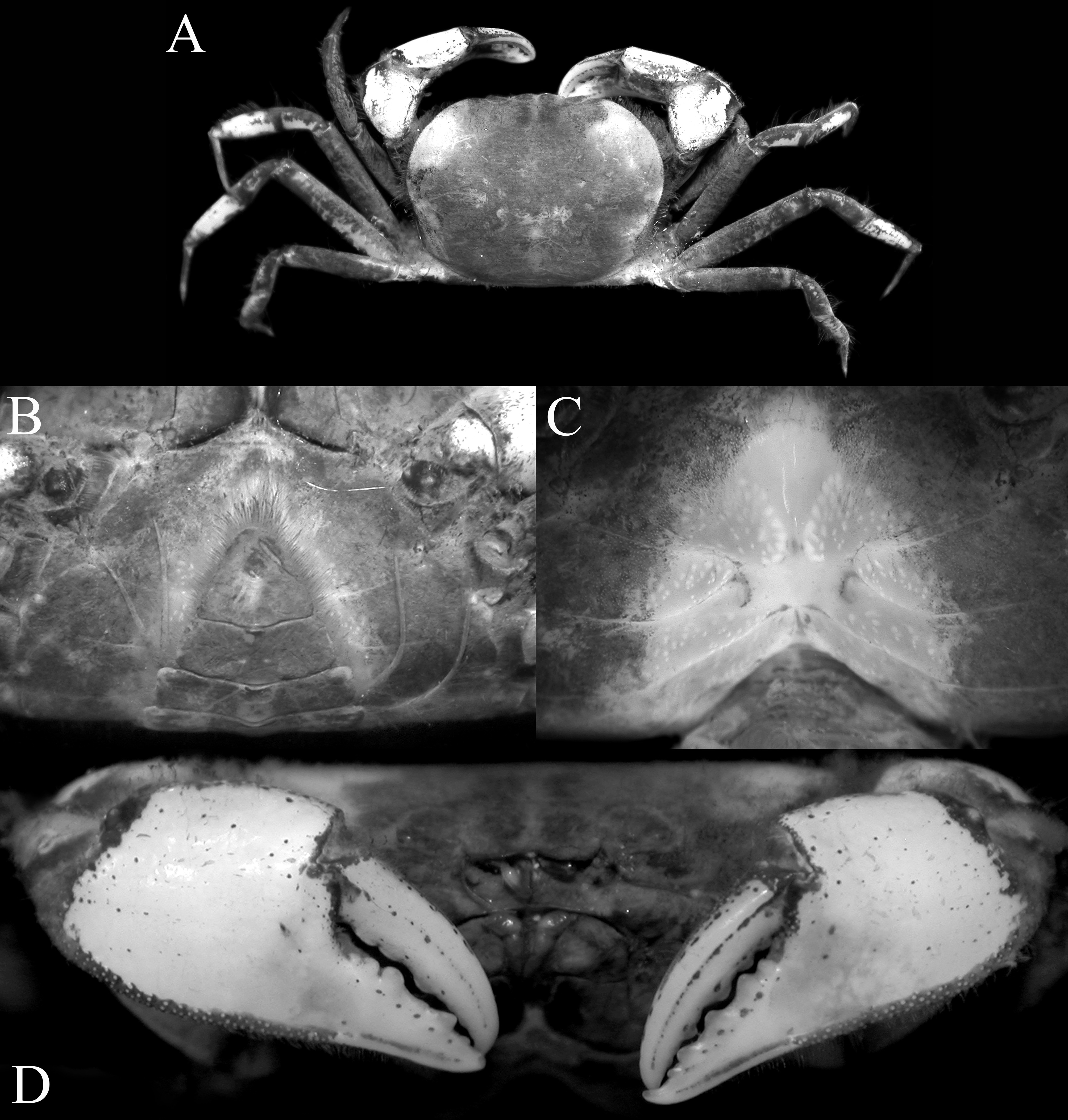
FIGURE 48. Typhlocarcinops atimovatae n. sp., paratype female (13.9 × 10.4 mm) (MNHN-IU-200-4369b), A, overall habitus; B, anterior thoracic sternum and pleon; C, sternopleonal cavity and vulvae; D, outer view of chelae.

FIGURE 49. Typhlocarcinops atimovatae n. sp., holotype male (15.9 × 11.9 mm mm) (MNHN-IU-200-4369a), Madagascar. A, left third maxilliped; B, pleon; C, left G1 (ventral view); D, left G1 (dorsal view); E, distal part of left G1 (ventral view); F, dista part of left G1 (dorsal view). Scales: A–D = 1.0 mm; E, F = 0.5 mm.

FIGURE 50. Typhlocarcinops hadrotes n. sp., holotype male (17.5 × 14.5 mm) (MZB Cru 4813), southern Java. A, overall habitus; B, dorsal view of carapace; C, frontal view of cephalothorax; D, anterior thoracic sternum and pleon; E, outer view of chelae.
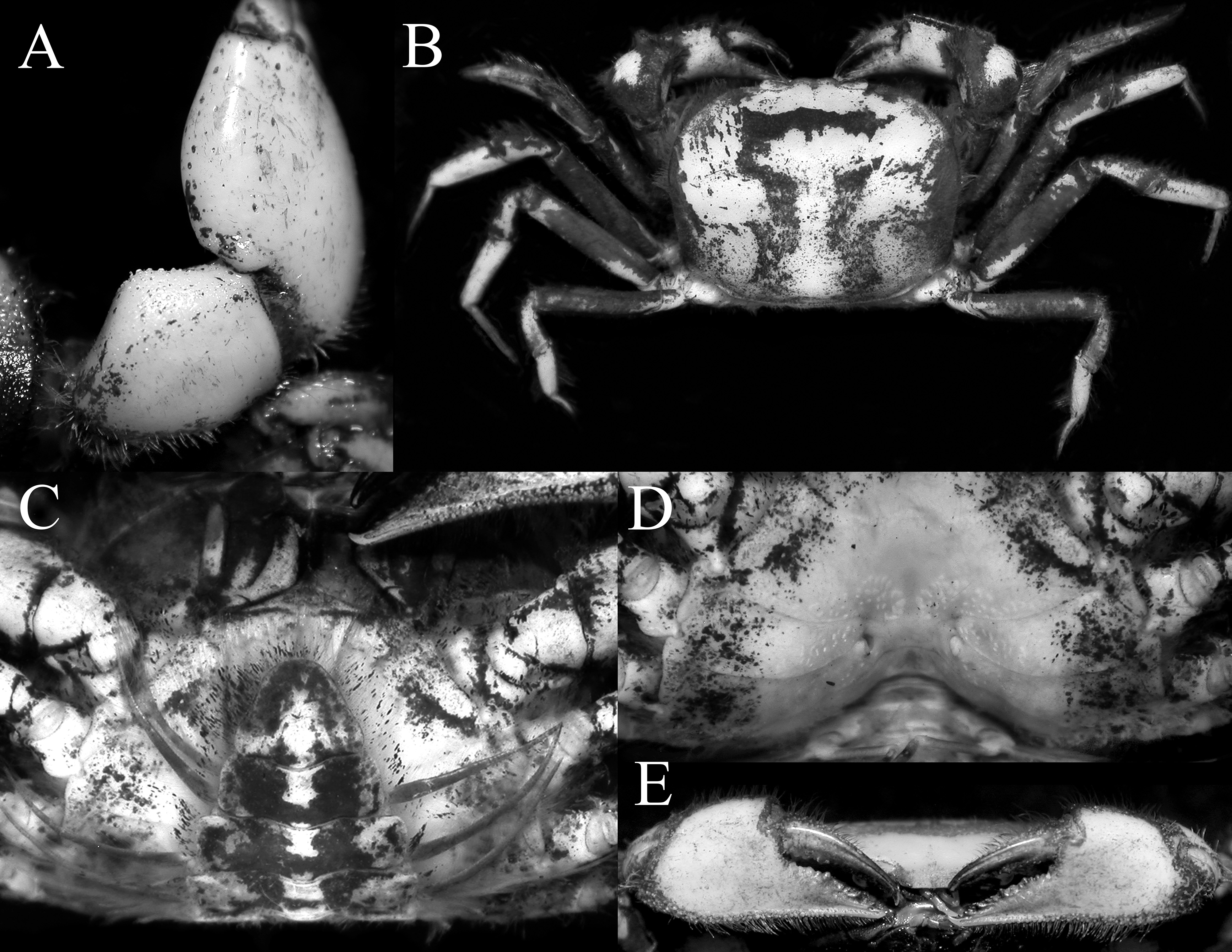
FIGURE 51. Typhlocarcinops hadrotes n. sp. A, paratype male (16.2 × 12.8 mm) (ZRC 2018.278), Indonesia; B–E, paratype female (18.3 × 14.3 mm) (ZRC 2018.0278), southern Java. A, carpus of right cheliped; B, overall habitus; C, anterior thoracic sternum and pleon; D, sternopleonal cavity and vulvae; E, outer view of chelae.

FIGURE 52. Typhlocarcinops hadrotes n. sp., holotype male (17.5 × 14.5 mm) (MZB Cru 4813), southern Java. A, left third maxilliped; B, male pleon; C, left G1 (ventral view); D, left G1 (marginal view); E, left G1 (dorsal view); F, distal part of left G1 (ventral view); D, distal part of left G1 (marginal view); E, distal part of left G1 (dorsal view). Scales: A–E = 1.0 mm; F–H = 0.5 mm.
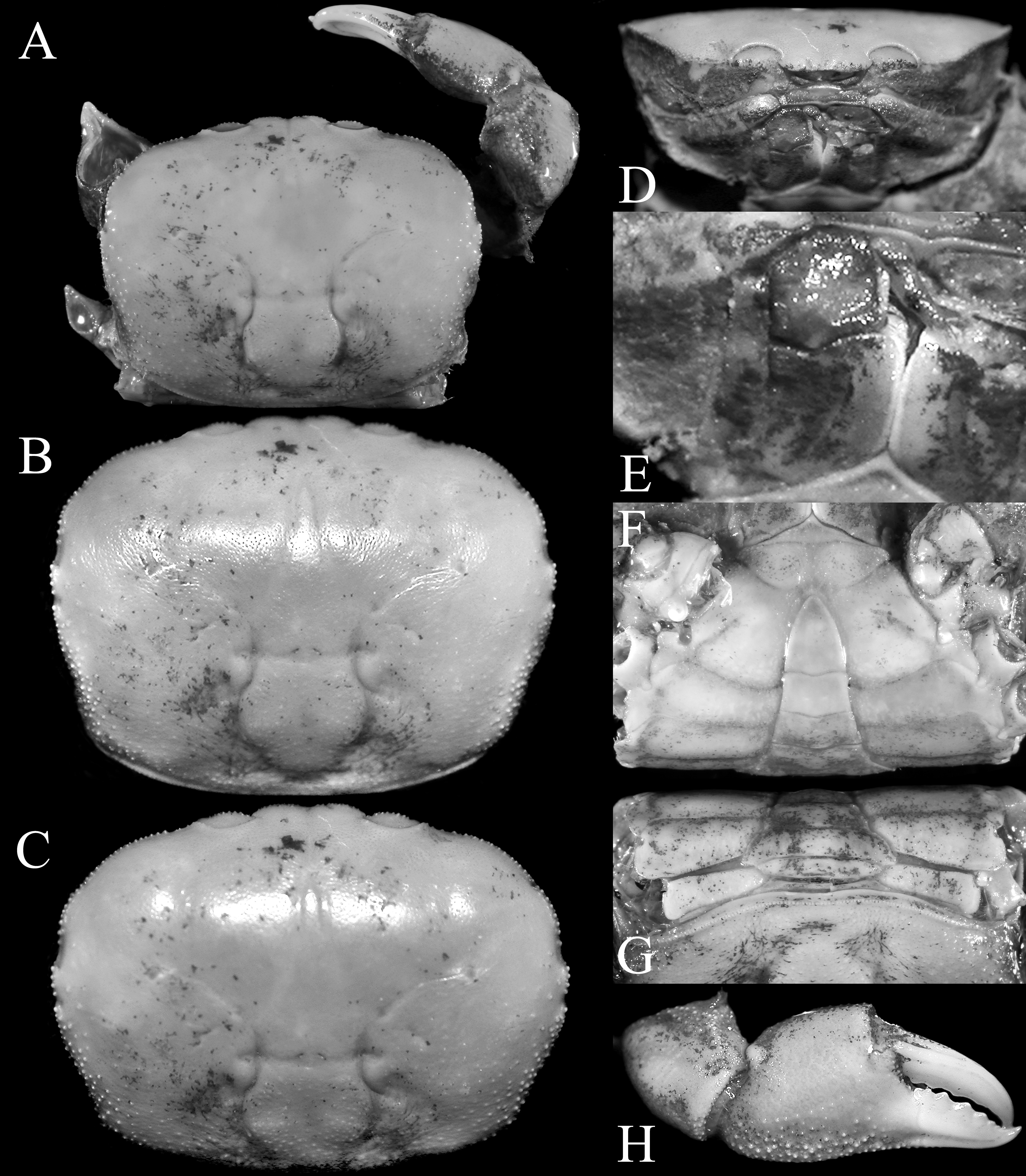
FIGURE 53. Typhlocarcinops angustifrons Rathbun, 1914, holotype male (14.9 × 11.3 mm) (USNM 46409), between Marinduque and Luzon, Philippines. A, overall habitus; B, C, doral view of carapace; D, frontal view of cephalothorax; E, right third maxilliped; F, anterior thoracic sternum and pleon; G, posterior thoracic sternum and pleon; H, outer view of right chela.
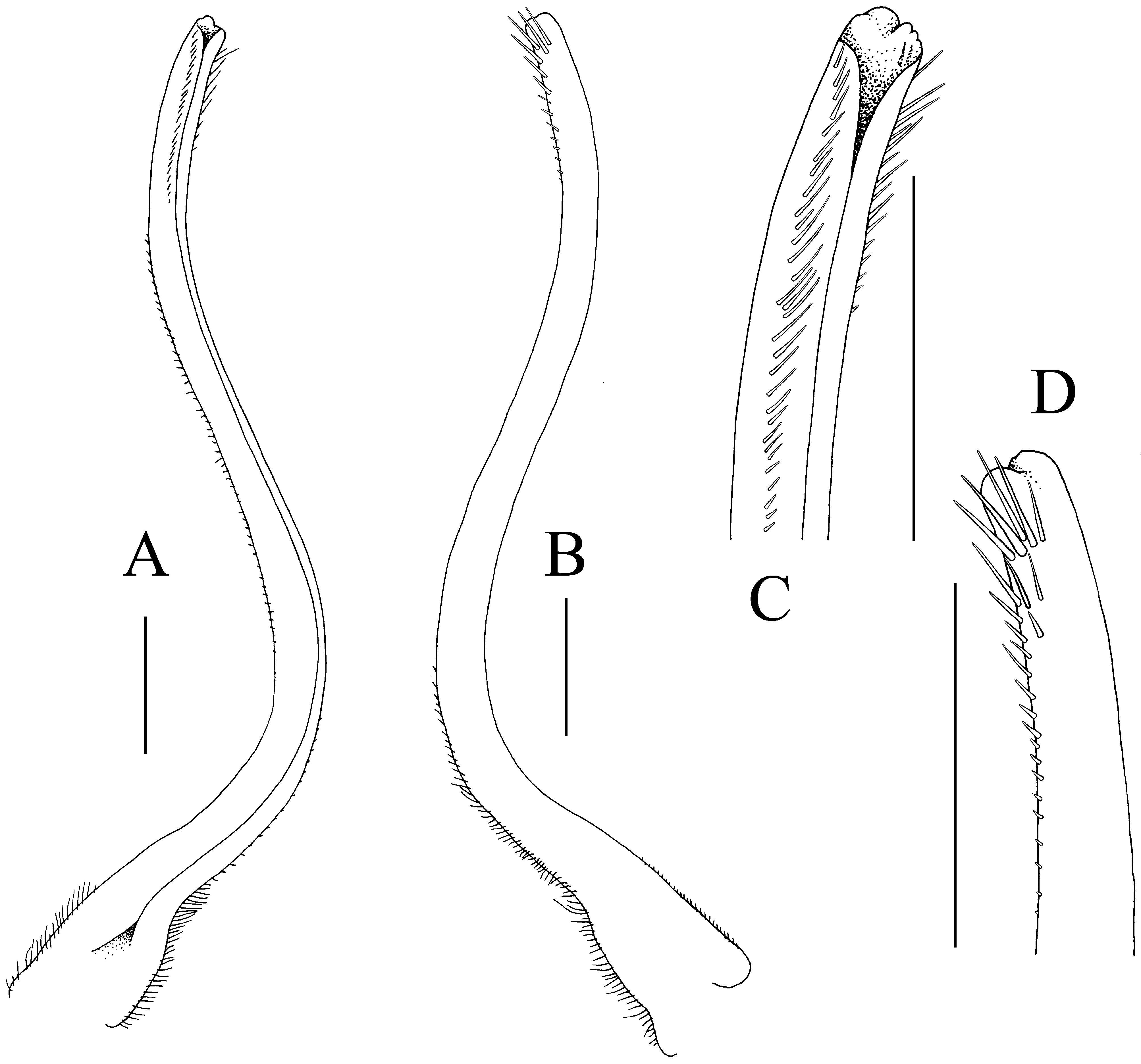
FIGURE 54. Typhlocarcinops angustifrons Rathbun, 1914, holotype male (14.9 × 11.3 mm) (USNM 46409), between Marinduque and Luzon, Philippines. A, left G1 (ventral view); B, left G1 (dorsal view); C, distal part of left G1 (ventral view); D, distal part of left G1 (dorsal view). Scales = 0.5 mm.
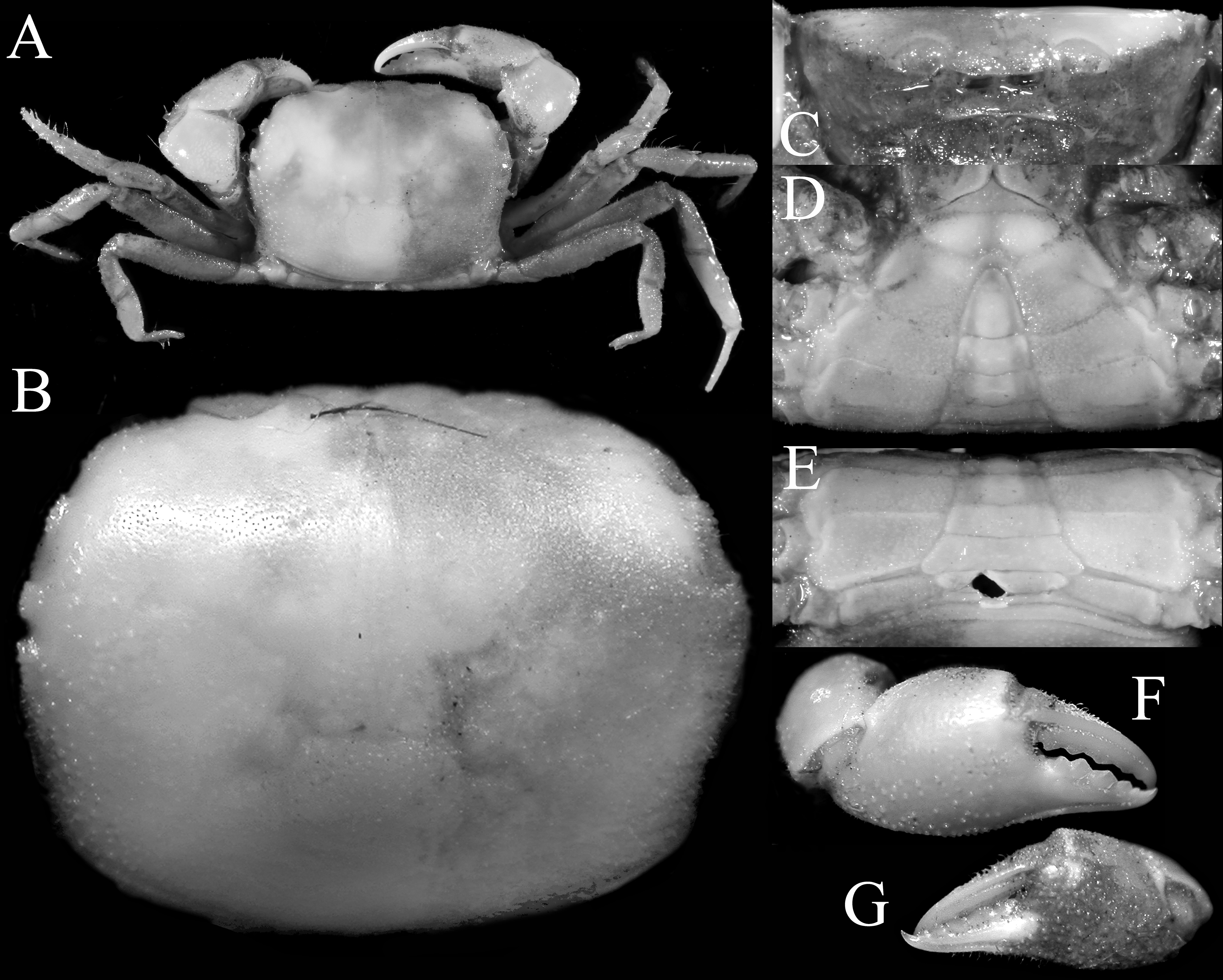
FIGURE 55. Typhlocarcinops marginatus Rathbun, 1914, holotype male (8.1 × 6.3 mm) (USNM 46395), Palawan, Philippines. A, overall habitus; B, dorsal view of carapace; C, frontal view of cephalothorax; D, anterior thoracic sternum and pleon; E, posterior thoracic sternum and pleon; F, outer view of right chela; G, outer view of left chela.
FIGURE 56. Typhlocarcinops marginatus Rathbun, 1914, holotype male (8.1 × 6.3 mm) (USNM 46345), Palawan, Philippines. A, left third maxilliped; B, left G1 (ventral view); C, left G1 (dorsal view). Scales = 0.5 mm.
FIGURE 60. Typhlocarcinops transversus Tesch, 1918. A, B, D–G, male (7.5 × 5.5 mm) (ZRC 2018.0272), Philippines; C, holotype male (8.0 × 4.1 mm) (NNM-ZMA De 103.003), Sumbawa, Indonesia. A, left third maxilliped; B, male pleon; C, right G1 (ventral view); D, left G1 (ventral view); E, left G1 (dorsal view); F, distal part of left G1 (ventral view); G, distal part of left G1 (dorsal view). Scales: A, D, E = 0.5 mm; B, C = 1.0 mm; F, G = 0.25 mm. C after Türkay (1986: text fig 59).

FIGURE 61. Typhlocarcinops serenei Türkay, 1986. A–C, I–L, paratype male (4.2 × 3.4 mm) (SMF 13559), Red Sea; D–H, holotype male (7.1 × 5.0 mm) (SMF 13559), Red Sea. A, dorsal view of carapace; B, frontal view of cephalothorax; C, anterior thoracic sternum and pleon; D, dorsal view of left cheliped; E, outer view of left chela; F, left first ambulatory leg; G, male pleon; H, J, right G1 (ventral view); I, right G1 (dorsal view); K, distal part of right G1 (dorsal view); L, distal part of right G1 (ventral view). D–H after Türkay (1986: text figs. 51–55). Scales: D–H = 1.0 mm; I, J = 0.25 mm; K, L = 0.12 mm.

FIGURE 62. Typhlocarcinops hirtus n. sp., holotype male (10.6 × 8.1 mm) (MZB Cru 4810), Lombok, Indonesia. A, overall habitus; B, dorsal view of carapace; C, frontal view of cephalothorax; D, anterior thoracic sternum and pleon; E, posterior thoracic sternum and pleon; F, outer view of right chela; G, outer view of left chela.
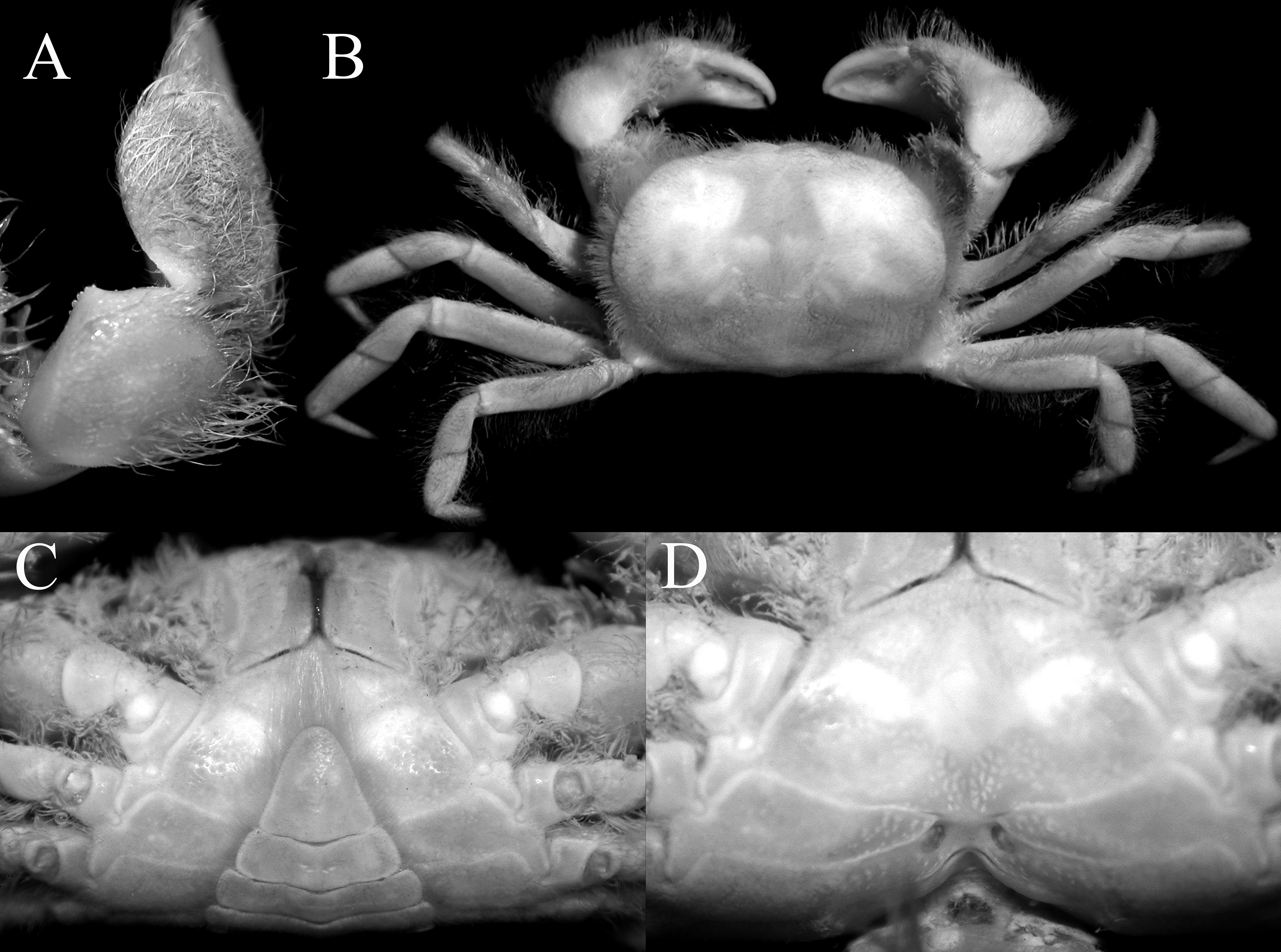
FIGURE 63. Typhlocarcinops hirtus n. sp. A, paratype male (15.0 × 11.0 mm) (ZRC 2015.0486), Lombok, Indonesia; B–D, paratype female (11.4 × 8.3 mm) (MZB Cru 4811), Lombok, Indonesia.A, carpus of right cheliped; B, overall habitus; C, anterior thoracic sternum and pleon; D, sternopleonal cavity and vulvae.
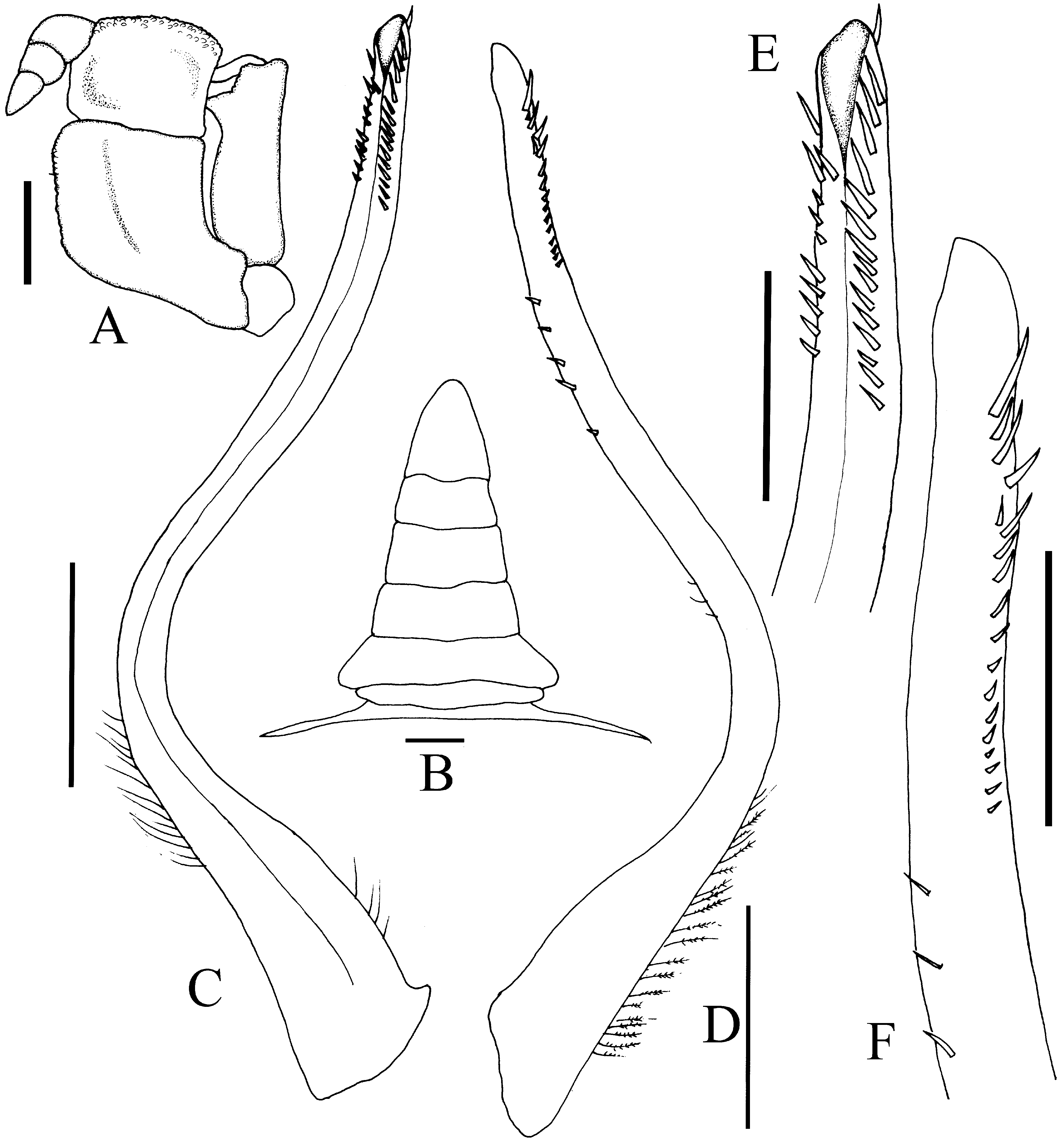
FIGURE 64. Typhlocarcinops hirtus n. sp., holotype male (10.6 × 8.1 mm) (MZB Cru 4810), Lombok, Indonesia.A, left third maxilliped; B, male pleon; C, left G1 (ventral view); D, left G1 (dorsal view); E, distal part of left G1 (ventral view); F, distal part of left G1 (dorsal view). Scales: A–D = 1.0 mm; E, F = 0.5 mm.
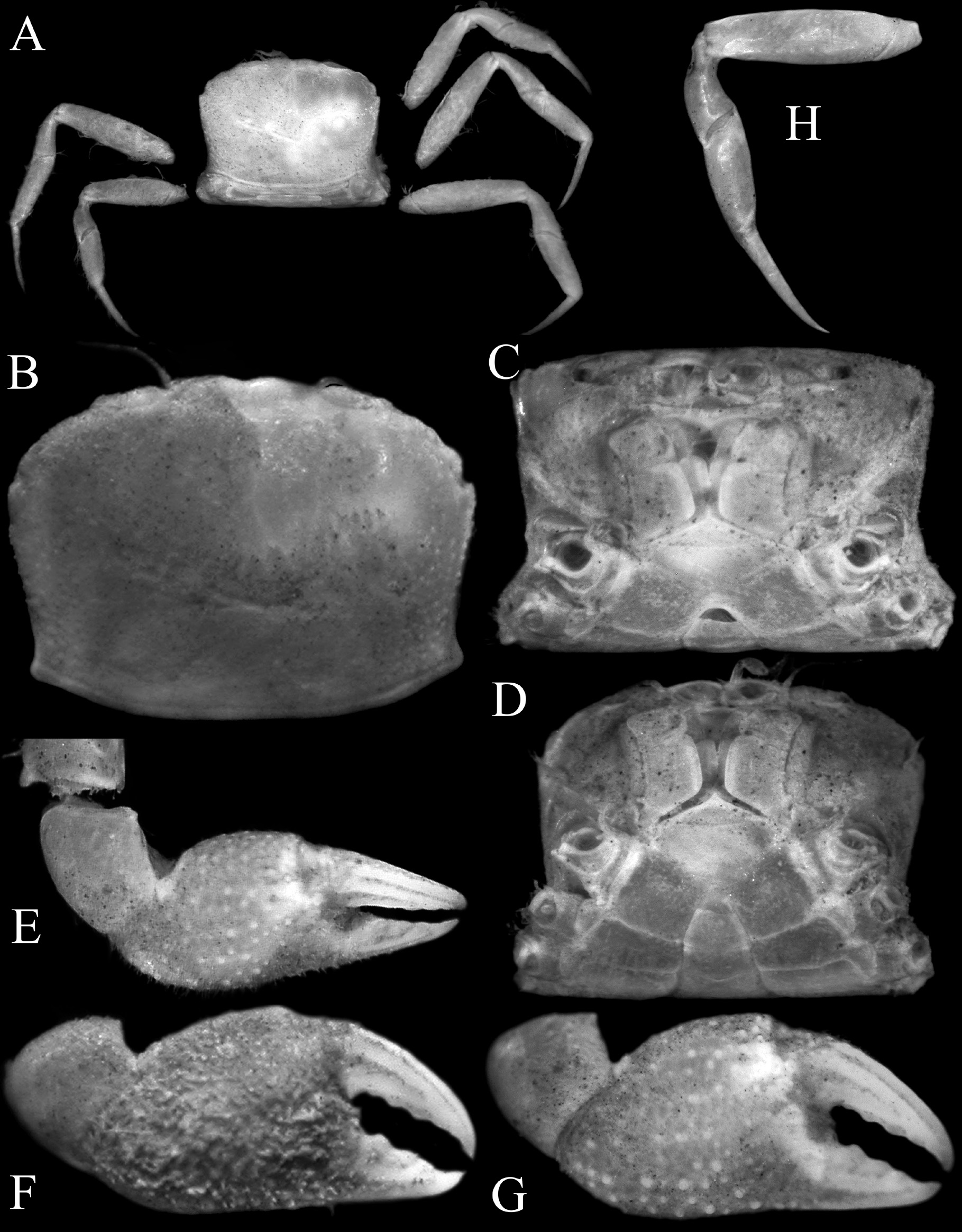
FIGURE 67. Typhlocarcinops diminutus n. sp., male (3.5 × 2.5 mm) (ZRC 2018.0295), Singapore.A, overall habitus; B, dorsal view of carapace; C, frontal view of cephalothorax; D, anterior thoracic sternum and telson; E, subdorsal view of right cheliped (denuded); F, outer view of right chela; G, outer view of right chela (denuded); H, left fourth ambulatory leg (denuded).
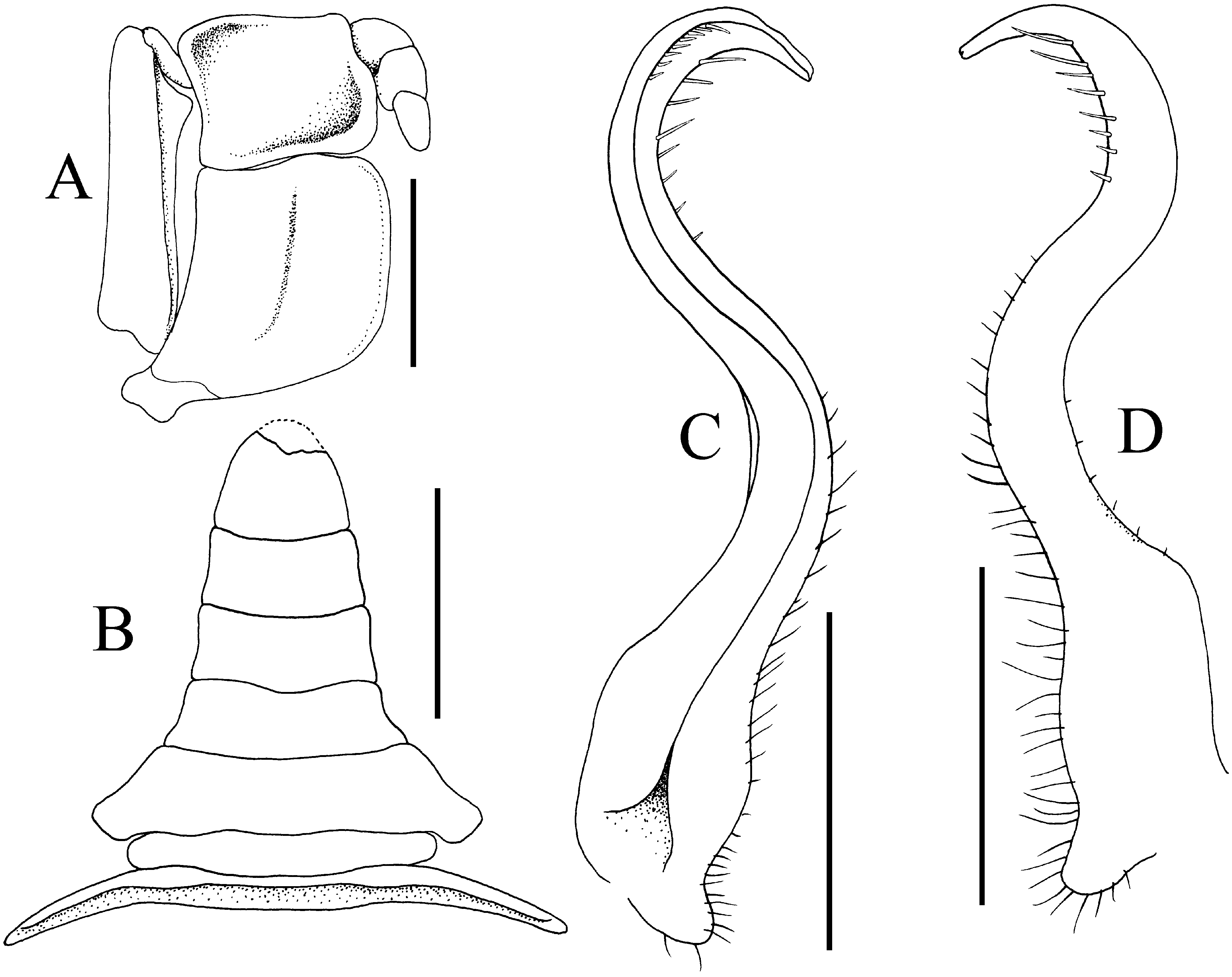
FIGURE 69. Typhlocarcinops diminutus n. sp., male (3.5 × 2.5 mm) (ZRC 2018.0295), Singapore.A, right third maxilliped; B, pleon; C, left G1 (ventral view); D, left G1 (dorsal view). Scales: A, C, D = 0.5 mm; B = 1.0 mm.
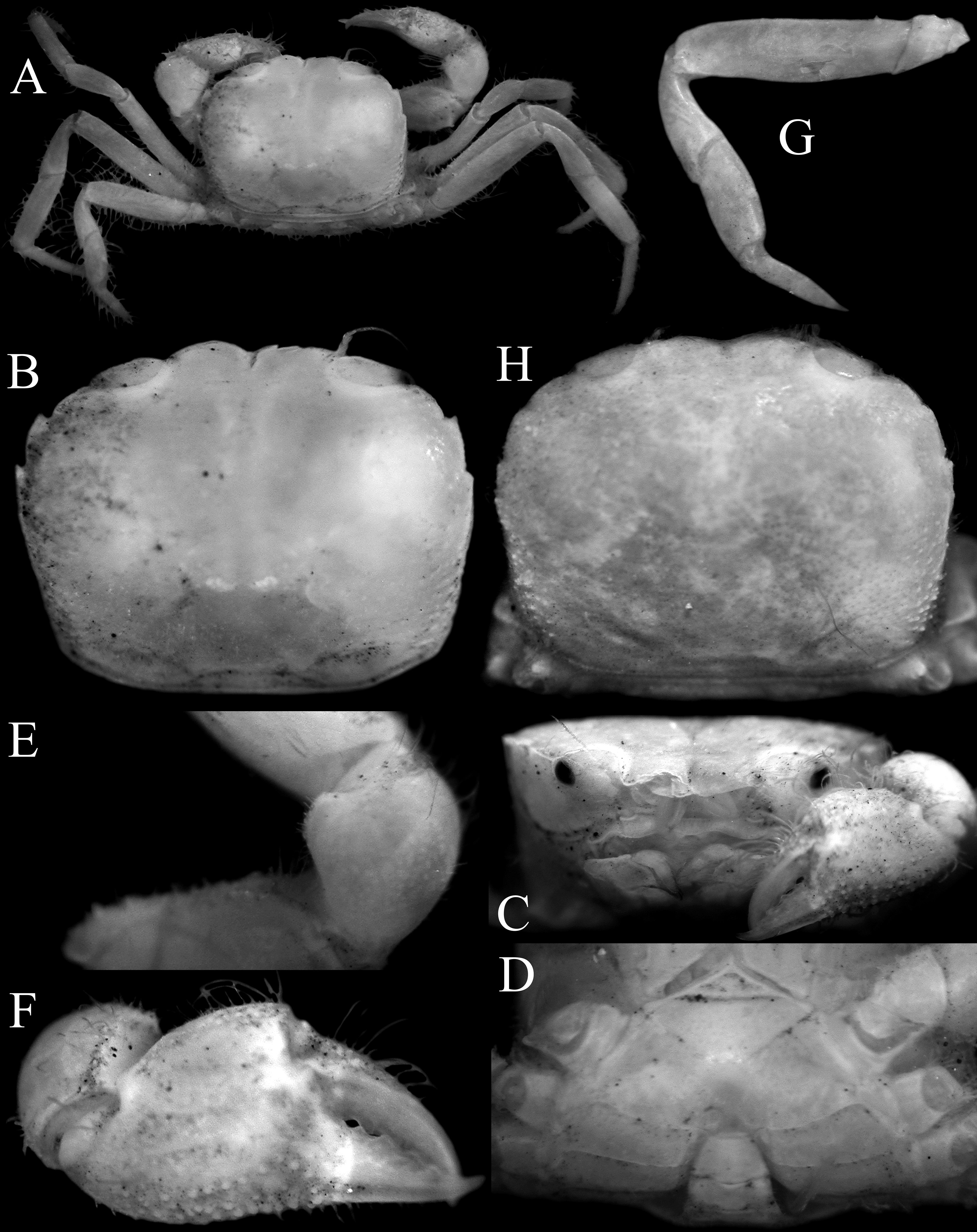
FIGURE 70. Typhlocarcinops kanashi n. sp. A–G, holotype male (3.9 × 3.0 mm) (NSMT-Cr 11575a), Japan; H, paratype male (4.5 × 3.5 mm) (NSMT-Cr 1212), Japan.A, overall habitus; B, H, dorsal view of carapace; C, frontal view of cephalothorax; D, anterior thoracic sternum and telson; E, subdorsal view of carpus of right cheliped (denuded); F, outer view of right chela; G, left fourth ambulatory leg (denuded).
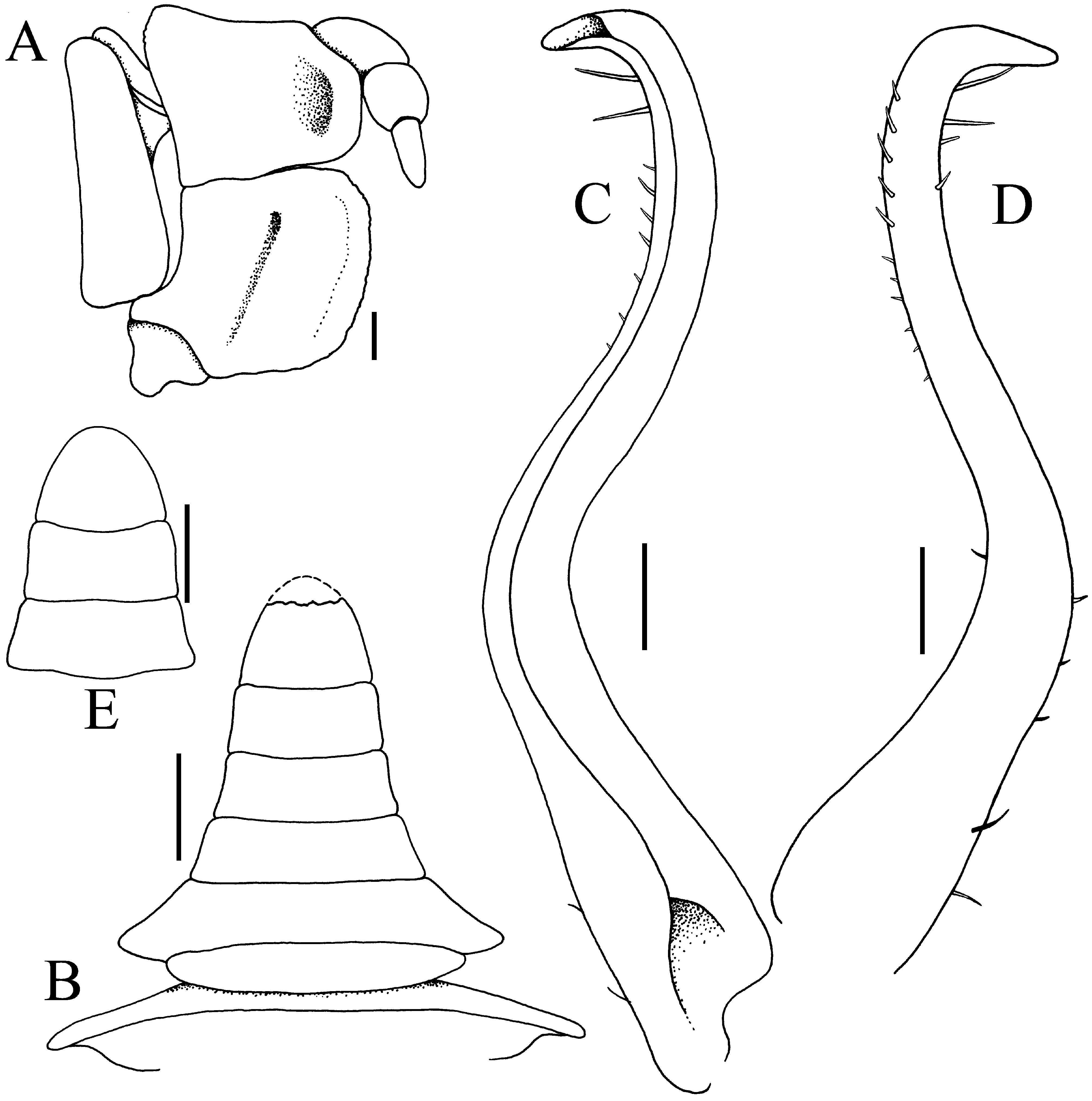
FIGURE 71. Typhlocarcinops kanashi n. sp. A–D, holotype male (3.9 × 3.0 mm) (NSMT-Cr 11575a), Japan; E, paratype male (4.5 × 3.5 mm) (NSMT-Cr 1212), Japan. A, right third maxilliped; B, pleon (telson tip damaged); C, right G1 (ventral view); D, right G1 (dorsal view); E, pleonal somites 5, 6 and telson. Scales: A, C, D = 0.1 mm; B, E = 0.5 mm.
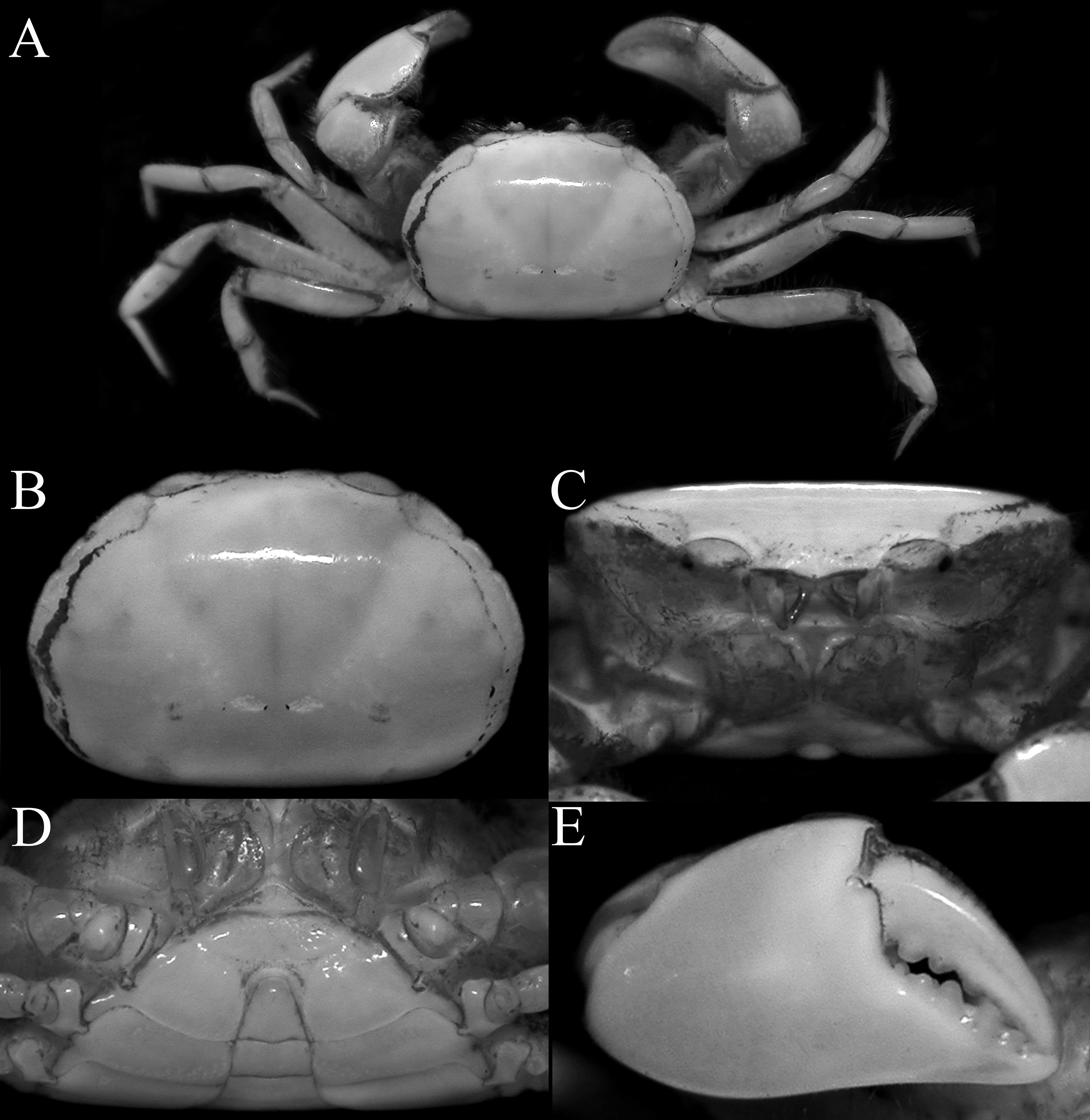
FIGURE 72. Typhlocarcinops lapillus n. sp. A–E, holotype male (6.0 × 4.2 mm) (NMCR), Bohol, Philippines. A, overall habitus; B, dorsal view of carapace; C, frontal view of cephalothorax; D, anterior thoracic sternum and pleon; E, outer view of right chela.
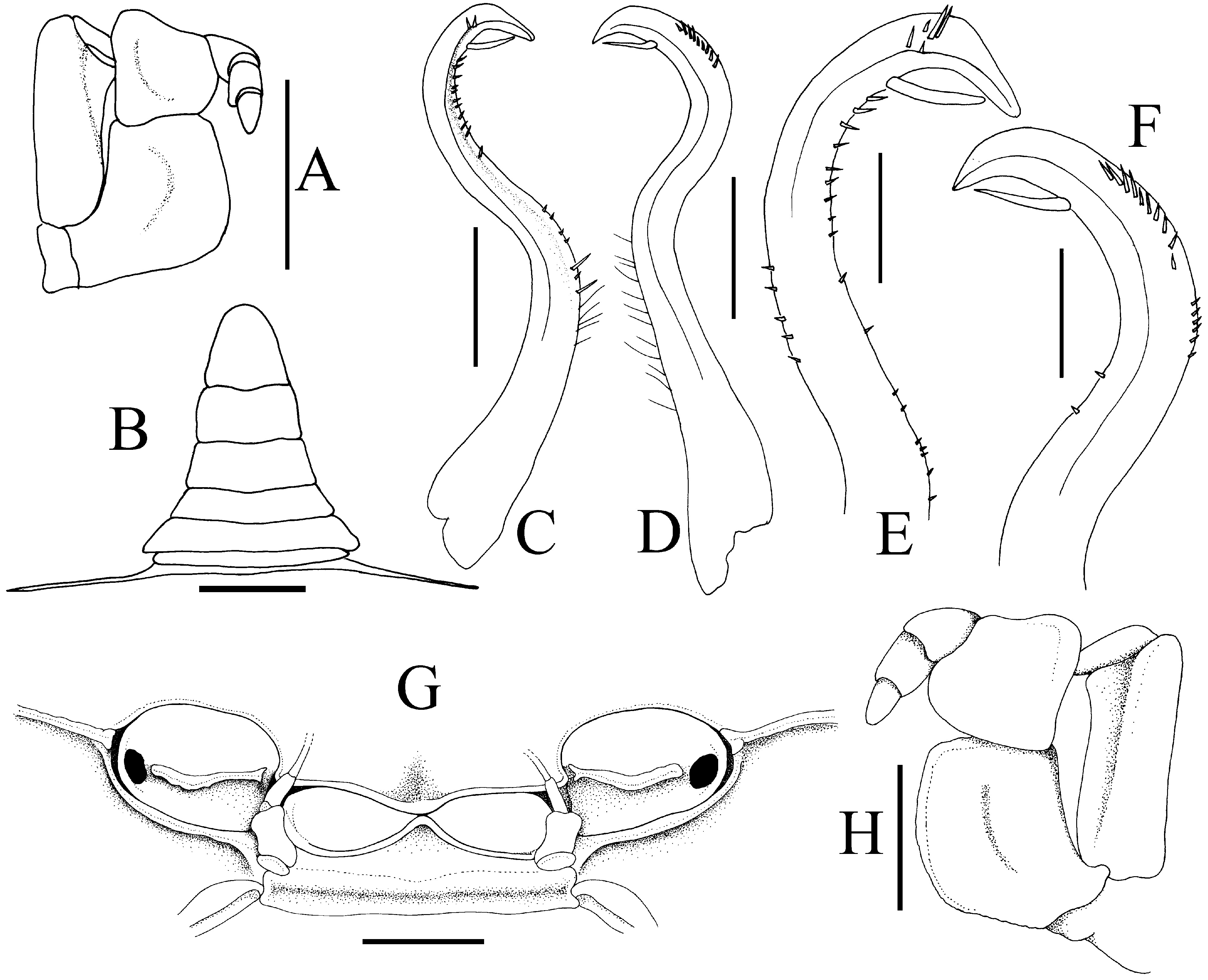
FIGURE 73. Typhlocarcinops lapillus n. sp. A–F, holotype male (6.0 × 4.2 mm) (NMCR), Bohol, Philippines; G, H, female (4.5 × 2.8 mm) (ZRC 2018.0281), Tanimbar Islands. A, right third maxilliped; B, male pleon; C, left G1 (ventral view); D, left G1 (dorsal view); E, distal part of left G1 (ventral view); F, distal part of left G1 (dorsal view); G, frontal view showing epistome, antennae and orbits (antennules not drawn); H, left third maxilliped. Scales: A, B = 1.0 mm; C, D, G, H = 0.5 mm; E, F = 0.25 mm.
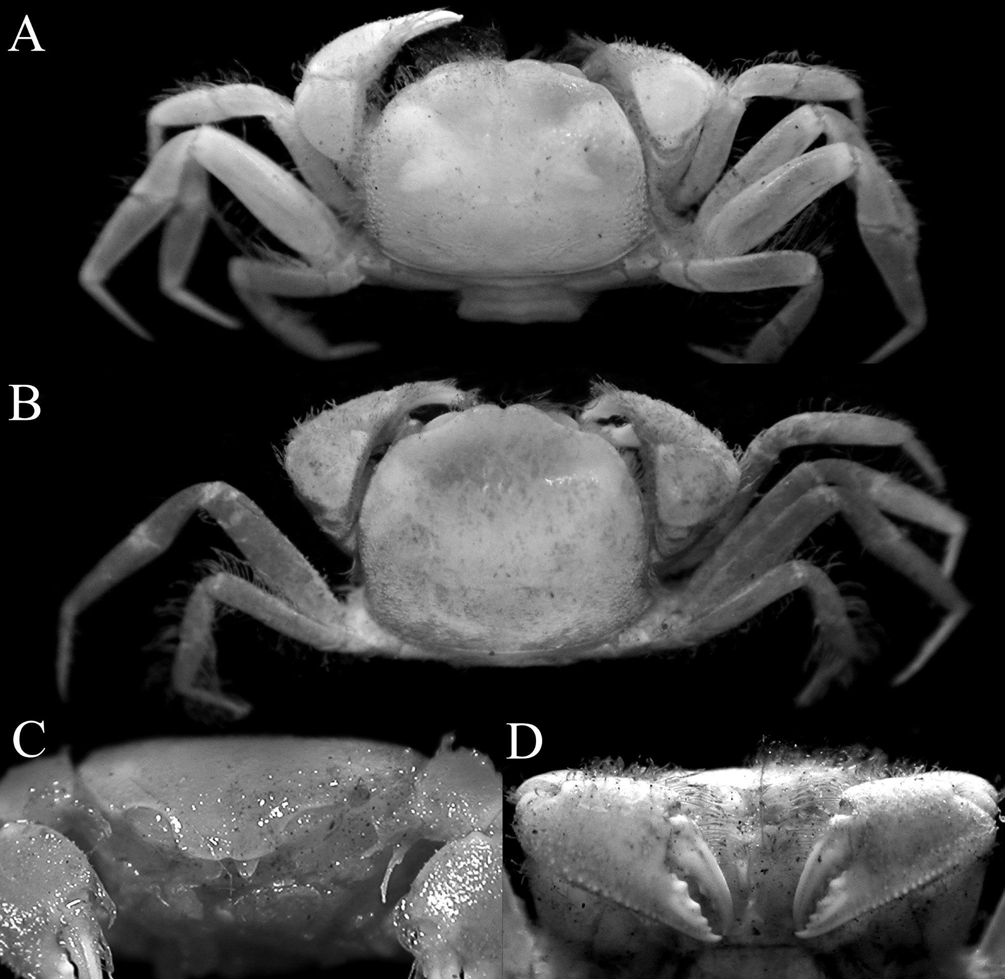
FIGURE 76. Typhlocarcinops angustipes Tesch, 1918. A, C, D, ovigerous syntype female (4.5 × 3.2 mm) (NNM-ZMA Crust De 103.004), Talaut Islands, Indonesia; B, syntype female (5.5 × 4.7 mm) (NNM-ZMA Crust De 103.005), Aru Islands, Indonesia.A, B, overall habitus; C, frontal view of cephalothorax; D, outer view of chelae.
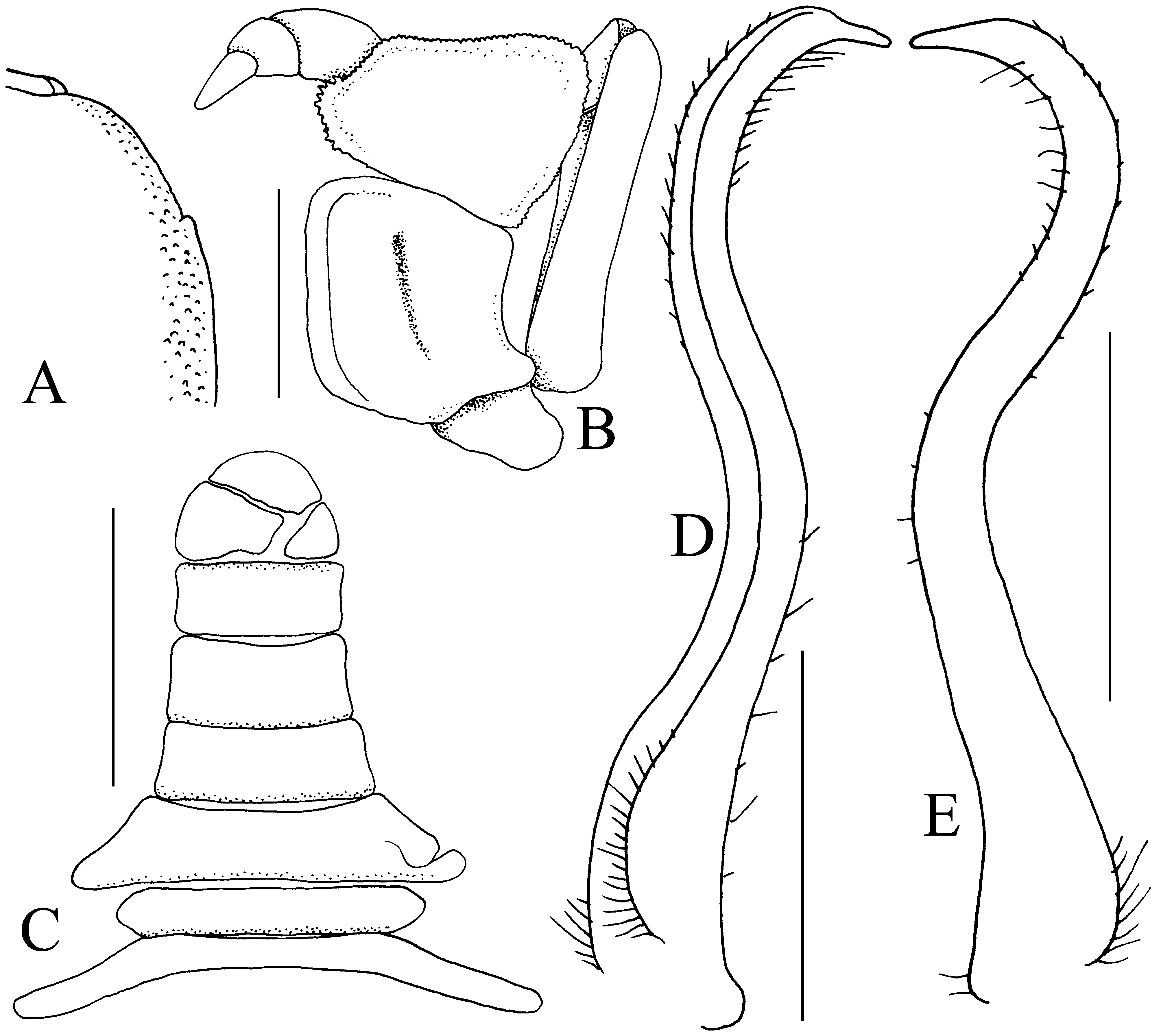
FIGURE 77. Typhlocarcinops angustipes Tesch, 1918. 1 male (3.7 × 2.8 mm, in bad condition) (NNM-ZMA Crust De 103.005), Aru Islands, Indonesia. A, right side of carapace (denuded); B, left third maxilliped; C, male pleon (telson broken); D, left G1 (ventral view); E, left G1 (dorsal view). Scales: A, C = 0.1 mm; B, D, E = 0.05 mm.
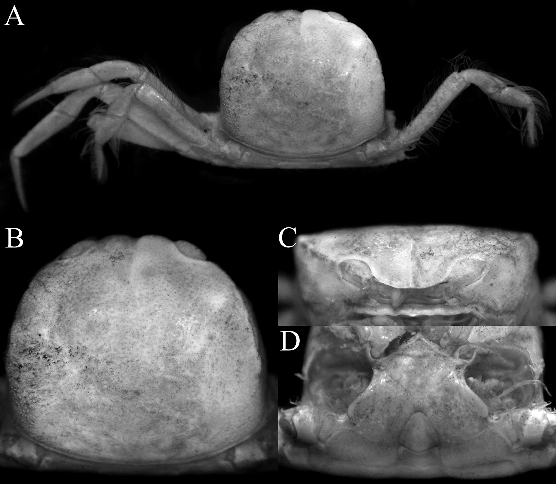
FIGURE 78. Typhlocarcinops arcuatus (Miers, 1884), holotype male (6.5 × 6.0 mm) (NNM 1882.7), Australia.A, overall habitus; B, dorsal view of carapace; C, frontal view of cephalothorax; D, anterior thoracic sternum and pleon.
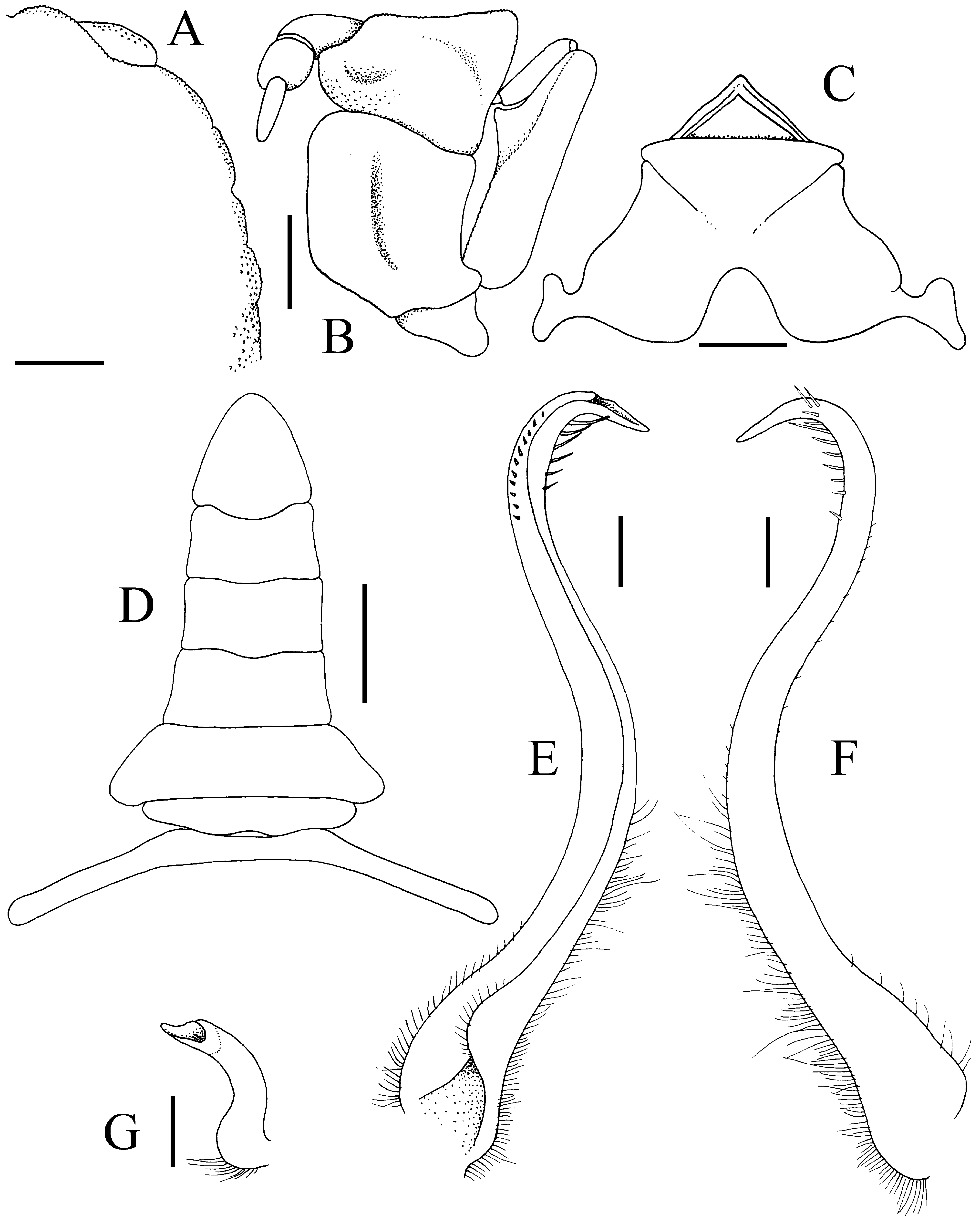
FIGURE 80. Typhlocarcinops arcuatus (Miers, 1884), holotype male (6.5 × 6.0 mm) (NNM 1882.7), Australia.A, right side of carapace (denuded); B, left third maxilliped; C, anterior thoracic sternum; D, pleon; E, left G1 (ventral view); F, left G1 (dorsal view); G, right G2. Scales: A, C, D = 1.0 mm; B = 0.5 mm; E–G = 0.2 mm.
No known copyright restrictions apply. See Agosti, D., Egloff, W., 2009. Taxonomic information exchange and copyright: the Plazi approach. BMC Research Notes 2009, 2:53 for further explanation.
|
Kingdom |
|
|
Phylum |
|
|
Class |
|
|
Order |
|
|
Family |