
Argentinomyia, Lynch-Arribalzaga, 1891
|
publication ID |
https://doi.org/10.11646/zootaxa.5234.1.1 |
|
publication LSID |
lsid:zoobank.org:pub:A540F250-BDE2-43F7-83A1-DA261F914B41 |
|
DOI |
https://doi.org/10.5281/zenodo.7621112 |
|
persistent identifier |
https://treatment.plazi.org/id/03C03256-AC55-FFF9-0FF9-FBD8F7BEF84B |
|
treatment provided by |
Plazi (2023-02-06 09:25:52, last updated 2024-11-28 20:14:13) |
|
scientific name |
Argentinomyia |
| status |
|
Identification key to the species of Argentinomyia View in CoL View at ENA
The last key of Fluke (1945) included only 19 valid species. A new key to the known species of Argentinomyia is provided based on characters published by Fluke (1945, 1957) and Thompson (1999a; 2006), including some modifications and couplet´s order to distinguish them from the sixteen new species. Argentinomyia scitula is not included in the key due to its doubtful validity, since the only known specimen is a teneral, headless, with no additional males found in good condition for dissection and deep examination.
1. Basoflagellomere large, slightly oval and apically rounded (SNLSA Figs 1C, F View FIGURE 1 ); face with a well-rounded tubercle, never with transversal grooves dorsally along tubercle or broadly punctuate (SNLSA Figs 1A, D View FIGURE 1 ); scutellum with a deep groove next to the rim (emarginate) (SNLSA Figs 5F View FIGURE 5 , 1B, 1E View FIGURE 1 , 12D View FIGURE 12 ); metacoxa with pile posteromedial on the apical angle (SNLSA Fig. 12C View FIGURE 12 ); abdomen elongated, with large markings on terga, sometimes with a transverse fascia on 3 rd or with a pair of small maculae in the basal corners of the 5 th tergum (SNLSA Figs 1B, E View FIGURE 1 ).............................................................................................. larger species of Argentinomyia View in CoL (in Montoya & Wolff 2020, not treated here)
- Basoflagellomere oval or slightly elongated (MCAD Fig. 22 View FIGURE 22 ); face usually with transversal grooves dorsally along tubercle (MCAD Fig. 23 View FIGURE 23 ) and shine (bare) punctuate maculae laterally; scutellum without a deep groove next to the rim; metacoxa without a pile tuft at the posteromedial apical angle; abdomen slightly spatulate, oval or with parallel sides, with triangular to quadrate or oval markings.............................................................................. 2
2. Antenna elongated, scape about as long as or longer than pedicel and basoflagellomere combined, scape always twice as long as pedicel ( Figs 1A–B View FIGURE 1 , 4J, K, L View FIGURE 4 , 6B, C View FIGURE 6 , 26A View FIGURE 26 , 55A, D View FIGURE 55 , 81A, D View FIGURE 81 )................................................. 3
- Antenna short, scape shorter than pedicel and basoflagellomere combined ( Figs 2A View FIGURE 2 , 4D, F, G, I View FIGURE 4 )....................... 7
3. Face with seven or eight shallows, transverse grooves above tubercle; sides of the face with a thin white line of pollen; pedicel and basoflagellomere about equal in length; wings with two short, narrow, transverse brown bands near the middle of the anterior half ( Fig. 8A View FIGURE 8 ); metafemur and protibia black ( Figs 7A View FIGURE 7 , 26A–F View FIGURE 26 ); male genitalia: surstylus in lateral view ( Fig. 27A View FIGURE 27 ) with dorsal and ventral margins approximately of the same width in the whole length; hypandrium in ventral view ( Fig. 27C View FIGURE 27 ) narrowed laterally towards the apex; aedeagal lobe in ventral view ( Fig. 27C View FIGURE 27 ) circular, apex rounded [ Bolivia, Colombia, Costa Rica, Guatemala, Guatemala, Mexico, Nicaragua and Perú].................. Argentinomyia crenulata ( Williston, 1891) View in CoL
- Face at most with one transversal groove above tubercle, never with seven or eight; facial pollinose pattern variable; basoflagellomere twice as long as pedicel; wings with at most only a faint brownish clouding on anterior cross-vein, never with medial brown fascia; metafemur yellow basally............................................................. 4
4. Sides of the face evenly white pubescent, not punctuate; female frontal triangle wide, male dichoptic; metatibia yellow [Southeastern South America species].................................................................... 5
- Sides of face punctuate; female frontal triangle narrow, male eyes holoptic; metatibia brown......................... 6
5. Face with a low tubercle and with no prominent depressions; frontal prominence greatly produced; abdomen metallic, without orange maculae ( Figs 1A–B View FIGURE 1 , 81A–F View FIGURE 81 ); male genitalia: surstylus in lateral view ( Fig. 82A View FIGURE 82 ) with dorsal and ventral margins approximately of the same width in the whole length; apex of hypandrium (superior lobes) short, as long as wide ( Figs 82A and C View FIGURE 82 ) [ Argentina View in CoL , Brazil and Perú]................................. Argentinomyia testaceipes Lynch-Arribálzaga, 1891 View in CoL
- Face with four distinct transverse grooves above tubercle; frontal prominence not greatly produced ( Figs 4J, K View FIGURE 4 ); abdomen with orange maculae ( Figs 9B View FIGURE 9 , 10A View FIGURE 10 , 55A–F View FIGURE 55 ); male genitalia: surstylus in lateral view ( Fig. 56A View FIGURE 56 ) with dorsal margin slightly concave and ventral margin slightly convex; apex of hypandrium (superior lobes) elongated, about 3× as long as wide ( Figs 56A and C View FIGURE 56 ) [ Brazil]........................................................... Argentinomyia norrbomi Montoya View in CoL sp. nov.
6. Male without maculae on 4 th tegum ( Figs 44B View FIGURE 44 , 46E View FIGURE 46 ); male frontal triangle broad; mesotarsus with three apical tarsomeres black ( Fig. 6B View FIGURE 6 ); scutum greyish pollinose and pilose; female frontal triangle with two rounded maculae of brown pollen ( Figs 2B–C View FIGURE 2 , 4B, L View FIGURE 4 , 44D View FIGURE 44 and 46A–B View FIGURE 46 ); male genitalia: surstylus in lateral view ( Fig. 45A View FIGURE 45 ) with dorsal and ventral margins approximately of the same width in the whole length; hypandrium in ventral view ( Fig. 45C View FIGURE 45 ) narrowed laterally towards the apex; aedeagal lobe in ventral view ( Fig. 45C View FIGURE 45 ) with acute apex [ Argentina View in CoL , Brazil, Colombia, Costa Rica, Ecuador, Guatemala, Mexico, Panam, Paraguay, Per and Trinidad and Tobago].................................. Argentinomyia longicornis (Walker, 1836) View in CoL
- Male with large basolateral orange maculae on 4 th tergum; frontal triangle small; mesotarsus orange; scutum golden pollinose and pilose (female unknown) [ Costa Rica and Nicaragua].................................... Argentinomyia View in CoL CR-19
7. Face usually vertical or perpendicular, never produced downward, generally with distinct transverse grooves above tubercle or very weak ones ( Fig. 4B–G, I View FIGURE 4 ); female front with a pollinose pattern varying from a complete transversal black fascia ( Figs 4C, F and G View FIGURE 4 ), two brownish-black rounded maculae ( Fig. 4B View FIGURE 4 ) or with a medial white pollinose vitta ( Fig. 4D View FIGURE 4 ); scutellum pile usually as short or shorter than marginal pile ( Fig. 6H–I View FIGURE 6 ); 2 nd tergum with a pair of yellow maculae varying from widening ( Fig. 8C View FIGURE 8 ) to narrow elongate ( Fig. 9B–C, E–G View FIGURE 9 , 47B, E View FIGURE 47 , 86B, E View FIGURE 86 ) or black metallic, both restricted to the lateral margin of the segment ( Fig. 9D View FIGURE 9 ) or non...................................................................................... 8
- Face may be protruding but very little produced downward, never with transverse grooves above tubercle ( Fig. 4H View FIGURE 4 ); female front without a pollinose pattern ( Fig. 4H View FIGURE 4 ), never with a complete transversal black fascia or two brownish-black rounded maculae; scutellar pile generally as long or longer than marginal bristles ( Fig. 6K View FIGURE 6 ); 2 nd tergum with a pair of maculae varying from narrow elongate yellow maculae ( Fig. 6A View FIGURE 6 ) to black metallic maculae ( Figs 37B, E View FIGURE 37 , 59B, E View FIGURE 59 ), both restricted to the lateral margin of the segment [Tropical Andes highland species]..................................................... 44
8. 1 st metatarsomeres black or dark brown, contrasting with the yellow color of the rest other tarsal segments ( Fig. 6C View FIGURE 6 )...... 9
- 1 st metatarsomeres yellow or orange, usually not contrasting with the yellow color of the rest other tarsal segments....... 11
9. 2 nd tergum with a prominent transversal fascia ( Figs 83A–D View FIGURE 83 ) (male unknown) [ Colombia and Bolivia]................................................................................ Argentinomyia transversalis Montoya View in CoL sp. nov.
- 2 nd tergum with a different maculate pattern ( Figs 9E View FIGURE 9 , 16B, C, E, F View FIGURE 16 , 35B, C View FIGURE 35 )..................................... 10
10. 3 to 4 rd th terga almost entirely yellow, with a pair of wider yellow maculae from the base to the apical margin of the segment ( Figs 9E View FIGURE 9 , 16B, C, E, F View FIGURE 16 ); male genitalia: hypandrium in ventral view ( Fig. 17C View FIGURE 17 ) narrowed laterally towards the apex [ Colombia]... Argentinomyia belmira Montoya & Wolff View in CoL sp. nov.
- 3 to 4 rd th terga with a pair of rectangular yellow maculae extending from the base to the apical 2/3 of the segment length, apically rounded and reaching the lateral margin ( Figs 35B, C View FIGURE 35 ); male genitalia: hypandrium in ventral view ( Fig. 36C View FIGURE 36 ) expanded laterally towards the apex (Female unknown) [ Colombia]....................... Argentinomyia ivani Montoya View in CoL sp. nov.
11. Facial pubescence punctuate ( Figs 30A, D View FIGURE 30 ); abdomen with oblique maculae, male with a pair on 3 rd tergum, female with a pair on 2 nd and 3 rd terga ( Figs 30B, E View FIGURE 30 ); male genitalia: surstylus in lateral view ( Fig. 31A View FIGURE 31 ) with dorsal margin slightly concave and ventral margin slightly convex; hypandrium in ventral view ( Fig. 31C View FIGURE 31 ) expanded laterally towards the apex; aedeagal lobe in ventral view ( Fig. 31C View FIGURE 31 ) circular, apex rounded [ Argentina View in CoL , Brazil, Colombia, Per and Venezuela]............................................................................................ Argentinomyia fastigata ( Fluke, 1945) View in CoL [Note: The coloration of pro- and mesofemora can vary from black or at least mostly dark brown to extensively yellow, see remark discussion under the species redescription]
- Facial pubescence not punctuate, other character combination variable.......................................... 12
12. Facial tubercle, scutellum and pleuron partially yellow in ground color (middle region of anepimeron, dorsal surface of katepisternum, katatergum, anatergum and postalar callus) yellow-golden pilose and pollinose in these areas; wing bare basally.............................................................................................. .................................................................................................. 13
- Facial tubercle, scutellum and pleuron black; other characters variables......................................... 15
13. Facial tubercle yellow; calypter wholly brown; all coxae yellow; abdomen with a pair of long, apically rounded oblique maculae on 2 nd to 4 th terga ( Thompson 1999a: 375, Figs 13–15 View FIGURE 13 View FIGURE 14 View FIGURE 15 ) (Male unknown) [ Mexico]................................................................................................. Argentinomyia octomaculata ( Enderlein, 1938) View in CoL
- Facial tubercle brown; calypter wholly, yellowish; all coxae brown; abdomen with a pair of long, apically rounded rectangular to triangular maculae on 2 nd to 4 th terga................................................................... 14
14. Face white pilose and pollinose [ Costa Rica].............................................. Argentinomyia View in CoL CR-21
- Face golden pilose and pollinose ( Figs 34A–C View FIGURE 34 ) (male unknown) [ Colombia and Ecuador]...................................................................................... Argentinomyia humboldti Montoya & Wolff View in CoL sp. nov.
15. Face with four or more distinct and deep transverse grooves above tubercle ( Figs 2A View FIGURE 2 , 4C, I View FIGURE 4 ) [Central America]......... 16
- Face without defined transverse grooves or with only two or three very weak ones or even none ( Figs 2B View FIGURE 2 , 4C, B, D–G, I–L View FIGURE 4 ). ................................................................................................. 19
16. 2 nd tergum rectangular, distinctly longer than broad; 3 rd to 4 th terga with basal transverse fasciae [ Costa Rica]............................................................................................... Argentinomyia View in CoL CR-11
- 2 nd tergum square-like, broader than long; 3 rd to 4 th terga without maculae....................................... 17
17. Face with at least five transverse grooves ( Figs 2A View FIGURE 2 , 4C View FIGURE 4 , 69A, C, D, F View FIGURE 69 ); wing hyaline ( Fig. 69 View FIGURE 69 ); abdomen with metallic maculae ( Figs 8D View FIGURE 8 , 69A–F View FIGURE 69 , 65 View FIGURE 65 D-E); male genitalia: surstylus in lateral view ( Fig. 70A View FIGURE 70 ) triangular-like [ Costa Rica and Mexico]...................................................................... Argentinomyia rugosonasa ( Williston, 1891) View in CoL
- Face with no more than four and usually with two shallow grooves or less; wing lightly brown pigmented anteriorly; abdomen with a pair of large quadrate yellow maculae on 2 nd tergum................................................... 18
18. Protibia black ( Figs 6H View FIGURE 6 , 8C View FIGURE 8 ) [ Mexico].................................................... Argentinomyia View in CoL CR-18
- Protibia yellow on basal 1/3 ( Figs 6I View FIGURE 6 , 71A–F View FIGURE 71 ); male genitalia: surstylus in lateral view ( Fig. 72A View FIGURE 72 ) with dorsal margin slightly concave and ventral margin slightly convex [ Guatemala, Mexico]................ Argentinomyia sagoti Montoya View in CoL sp. nov.
19. Pro- and mesofemora orange-yellow..................................................................... 20
- Pro- and mesofemora black or at least mostly dark brown.................................................... 33
20. Facial pubescence golden and very thick, frontal triangle yellow pilose ( Figs 32A, D View FIGURE 32 ); abdomen with three pairs of yellow vittae on 2 nd to 4 th terga of male and four pairs on 2 nd to 5 th terga of female; metafemur black, except only yellow on basal 1/4 ( Figs 32B, E View FIGURE 32 ); male genitalia: surstylus in lateral view ( Fig. 33A View FIGURE 33 ) with dorsal and ventral margins approximately of the same width in the whole length, elongated, at least two times longer than broad; hypandrium in ventral view ( Fig. 33C View FIGURE 33 ) narrowed laterally towards the apex; aedeagal lobe in ventral view ( Fig. 33C View FIGURE 33 ) with acute apex [ Argentina View in CoL , Colombia, Ecuador and Perú]............................................................................. Argentinomyia festiva ( Fluke, 1945) View in CoL
- Facial pile thinner and more whitish, frontal triangle black pilose; abdomen with a different maculate pattern, other character combinations variable................................................................................ 21
21. Metafemur swollen, with ventral spines ( Figs 7D View FIGURE 7 , 76A–E View FIGURE 76 ) (male unknown) [ Colombia]............................................................................................ Argentinomyia spinifemorata Montoya View in CoL sp. nov.
- Metafemur thin, normal shape, never with ventral spines..................................................... 22
22. Pattern of 3 rd tergum consisting of oblique maculae ( Figs 9G View FIGURE 9 )................................................. 23
- Pattern of 3 rd tergum different, triangular ( Figs 28B, E View FIGURE 28 , 49B, E View FIGURE 49 ), quadrate or rectangular ( Figs 63B, E View FIGURE 63 , 65B, E View FIGURE 65 ).......... 26 [Note: A. currani View in CoL can be keyed out from both statements if couplet 22 is not properly evaluated; in A. currani View in CoL the macular pattern is triangular, referred to as the inner margin of the maculae forming an acute angle (45°) with the basal margin of 3 rd tergum as can be seen in the Figs 28B, E View FIGURE 28 ].
23. Costal cell bare on basal 3/4 or more.................................................................... 24
- Costal cell entirely microtrichose........................................................................ 25
24. Protarsus black ( Figs 24A–F View FIGURE 24 ); male genitalia: surstylus in lateral view ( Fig. 25A View FIGURE 25 ) with dorsal and ventral margins approximately of the same width in the whole length; hypandrium in ventral view ( Fig. 25C View FIGURE 25 ) narrowed laterally towards the apex; aedeagal lobe in ventral view ( Fig. 25C View FIGURE 25 ) with acute apex [ Costa Rica, Guatemala, El Salvador, Mexico and Panamá]................................................................................ Argentinomyia catabomba ( Williston, 1891) View in CoL
- Protarsus yellow..................................................................... Argentinomyia View in CoL CR-14
25. Mesonotum punctate markings, shining bronze to aeneous, with brownish vittae on anterior half ( Fig. 40C, E View FIGURE 40 ); female frontal triangle with two rounded maculae ( Fig. 40D View FIGURE 40 ); 3 rd to 4 th terga with a pair of oblique yellow maculae ( Figs 40B, E View FIGURE 40 ); male genitalia: surstylus in lateral view ( Fig. 45A View FIGURE 45 ) with dorsal and ventral margins approximately of the same width in the whole length; aedeagal lobe in ventral view ( Fig. 41C View FIGURE 41 ) with apex acute [ Argentina View in CoL , Brazil and Ecuador] Argentinomyia lanei ( Fluke, 1936) View in CoL
- Mesonotum without punctuate markings ( Figs 18C, E View FIGURE 18 ); frontal triangle with transversal black macula ( Fig. 18D View FIGURE 18 ); the 3 rd tergum with a pair of elongate, slightly oblique yellow maculae, reaching the lateral margin and slightly touching each other towards the middle ( Figs 18A–G View FIGURE 18 ); male genitalia: surstylus in lateral view ( Fig. 19A View FIGURE 19 ) with dorsal margin slightly concave and ventral margin slightly convex; aedeagal lobe in ventral view ( Fig. 19C View FIGURE 19 ) circular, apex rounded [ Brazil and Venezuela]................................................................................. Argentinomyia berthae ( Lima, 1946) View in CoL
26. Metafemur yellow with a black ring or smudge on apical margin ( Figs 7E View FIGURE 7 , 28 B, C View FIGURE 28 , 49B, C, F View FIGURE 49 , 63B, C View FIGURE 63 , 65C, F View FIGURE 65 ); female frontal triangle with two rounded brown pollinose maculae ( Fig. 28D View FIGURE 28 , 49D View FIGURE 49 ); 3 rd to 4 th terga with triangular maculae, the inner margin of the maculae forms an acute angle (45°) with the basal margin of the tergum ( Figs 28B, E View FIGURE 28 , 49B, E View FIGURE 49 )................. 27
- Metafemur yellow without a black ring or smudge on apical margin ( Figs 14C, F View FIGURE 14 , 77C, F View FIGURE 77 ); female frontal triangle with a different pollinose pattern; 3 rd to 4 th terga with quadrate or rectangular maculae, the inner margin of the maculae forms a right angle (90°) with the basal margin of the tergum ( Figs 63B, E View FIGURE 63 , 65B, E View FIGURE 65 ).......................................... 28
27. Metatibia mostly yellow ( Figs 49C, F View FIGURE 49 ); male genitalia: surstylus in lateral view ( Fig. 50A View FIGURE 50 ) with dorsal margin slightly concave and ventral margin slightly convex, shorter than broad; hypandrium in ventral view ( Fig. 50C View FIGURE 50 ) expanded laterally towards the apex; aedeagal lobe in ventral view ( Fig. 50C View FIGURE 50 ) with apex acute [ Argentina View in CoL and Brazil].................................................................................................... Argentinomyia maculata ( Walker, 1852) View in CoL
- Metatibia yellow on basal 1/4 ( Figs 9I View FIGURE 9 , 28C, F View FIGURE 28 ); male genitalia: surstylus in lateral view ( Fig. 29A View FIGURE 29 ) with dorsal and ventral margins approximately of the same width in the whole length; hypandrium in ventral view ( Fig. 29C View FIGURE 29 ) expanded laterally towards the apex; aedeagal lobe in ventral view ( Fig. 29C View FIGURE 29 ) circular, apex rounded [ Brazil and Perú].......................................................................................... Argentinomyia currani ( Fluke, 1937) View in CoL
28. Maculae on 3 rd tergum longer than wide, extending from the base to apical 3/4 or more ( Figs 28B, E View FIGURE 28 , 49B, E View FIGURE 49 )........... 29
- Maculae on 3 rd tergum as long as wide, extending only from the base to apical 1/2 or less ( Figs 63B, E View FIGURE 63 , 65B, E View FIGURE 65 )......... 30
29. Face more perpendicular and less produced ventrally, the sides thickly coated with cinereous yellow pollen ( Figs 86A, D View FIGURE 86 ); metafemur reddish or yellowish on basal half, metatibia yellow on basal and apical 1/4 ( Figs 86A C, F View FIGURE 86 ); maculae on female abdomen rectangular and apically rounded ( Figs 86B–C, E–F View FIGURE 86 ); male genitalia: Aedeagal lobe in ventral view ( Fig. 87C View FIGURE 87 ) with apex rounded [ Argentina View in CoL , Brazil, Colombia, Ecuador and Venezuela]............... Argentinomyia tropica ( Curran, 1937) View in CoL
- Face slightly produced ventral, more prominent, facial pollen smooth and yellowish ( Figs 47A, D View FIGURE 47 ); metafemur black, only very narrowly yellow basally ( Figs 47C, F View FIGURE 47 ); maculae on female abdomen triangular ( Figs 4B–C, E–F View FIGURE 4 , 48D–H View FIGURE 48 ); male genitalia: aedeagal lobe in ventral view ( Fig. 48C View FIGURE 48 ) with apex acute [ Colombia, Ecuador and Venezuela].............................................................................................. Argentinomyia luculenta ( Fluke, 1945) View in CoL [Note: Some morphotypes of A. tropica View in CoL and A. luculenta View in CoL may key through either option. X. Mengual (personal communication) suggests both species could form a species complex, with two additional morphospecies not exanimated that could be new to science, however, a deep examination of male genitalia is needed to confirm them]
30. Metatibia mostly brownish apically ( Figs 65C, F View FIGURE 65 , 77C, F View FIGURE 77 ).................................................... 31
- Metatibia yellow.................................................................................... 32
31. Female frontal triangle with a medial white pollinose vitta ( Figs 65D View FIGURE 65 ); metafemur obscured on apical 1/3 or little more; 3 rd and 4 th terga with maculae extending to the apical 1/2; 5 th tergum without a pair of small lateral rounded maculae; male genitalia: surstylus in lateral view ( Fig. 64A View FIGURE 64 ) narrowed laterally towards the apex [ Cuba, Dominican Republic and Jamaica].............................................................................. Argentinomyia praeusta ( Loew, 1866) View in CoL
- Frontal triangle with transversal golden-brownish pollinose band touching slightly the sides of the eyes ( Figs 77D View FIGURE 77 ); metafemur yellow basally and slightly obscured on apical 1/5 dorsally; 3 rd and 4 th terga with maculae extending to the apical 2/3; 5 th tergum with a pair of small lateral rounded maculae ( Figs 77A–F View FIGURE 77 ); male genitalia: surstylus in lateral view ( Fig. 78A View FIGURE 78 ) very elongated and widened dorsally, with a small extension at the ventral margin [ Dominican Republic]..................................................................................... Argentinomyia taina Thompson & Montoya View in CoL sp. nov.
32. Face whitish pollinose and pilose ( Figs 63A, D View FIGURE 63 ); female frontal triangle with a complete transverse black fascia ( Figs 63D View FIGURE 63 , 64D View FIGURE 64 ); metafemur yellow except the apical 1/5 narrowly brown ( Figs 63C, F View FIGURE 63 ); male genitalia: surstylus in lateral view ( Fig. 64A View FIGURE 64 ) with dorsal margin slightly concave and ventral margin slightly convex [ Argentina View in CoL , Brazil and Venezuela]...................................................................................... Argentinomyia pollinosa ( Hull, 1942) View in CoL
- Face extensively covered by golden pollinose and pilose ( Figs 14A, D View FIGURE 14 ); female frontal triangle with a transversal goldenbrownish pollinose band ( Figs 14D, E View FIGURE 14 ); metafemur obscured on basal 2/6 and apical 1/6 or little more ( Figs 14C, F View FIGURE 14 ); male genitalia: surstylus in lateral view ( Fig. 15A View FIGURE 15 ) with dorsal and ventral margins approximately of the same width in the whole length [ Dominican Republic]............................... Argentinomyia aurifacies Thompson & Montoya View in CoL sp. nov.
33. 2 nd tergum entirely black ( Figs 74B, E View FIGURE 74 , 79B, E View FIGURE 79 , 84B, E View FIGURE 84 )...................................................... 34
- 2 nd tergum with some lateral yellow maculae ( Figs 10B, E View FIGURE 10 , 61B, E View FIGURE 61 , 90B, E View FIGURE 90 , 91B, E View FIGURE 91 , 88B, E View FIGURE 88 )......................... 37
34. Abdominal maculae metallic blue; wing extensible hyaline; legs dark brown, only slightly white-yellowish on apical 1/6 ( Figs 39A–C View FIGURE 39 ) (male unknown) [ Jamaica].................................. Argentinomyia jamaicensis Montoya View in CoL sp. nov. [Note: Female frontal triangle of A. jamaicensis Montoya View in CoL sp. nov. has a medial white pollinose vitta (see Fig. 39A View FIGURE 39 ), also found in female of A. praeusta View in CoL ]
- Abdominal maculae yellow to orange; wing brownish apically; legs coloration variable............................. 35
35. Wing slightly darkened in the anterior margin from the costal cell to the stigma, cells r1 and r4+5, with some small hyaline areas ( Figs 84B, C, E, F View FIGURE 84 ); metafemur and protibia black ( Figs 84A, C, D, F View FIGURE 84 ); male genitalia: surstylus in lateral view ( Fig. 85A View FIGURE 85 ) with dorsal margin slightly concave and ventral margin slightly convex; aedeagal lobe in ventral view ( Fig. 85C View FIGURE 85 ) circular, apex rounded [ Bolivia, Colombia, Ecuador and Perú]........................ Argentinomyia tropandeana Montoya View in CoL sp. nov.
- Wing hyaline; other characters variable.................................................................. 36
36. Antennae brownish, orange ventrally ( Fig. 79A View FIGURE 79 ); frontal triangle golden pollinose ( Fig. 79A View FIGURE 79 ); metanotum golden pile; terga 3 rd and 4 th with a pair of basolateral rectangular maculae extending 1/3 of tergum length ( Fig. 79B–C View FIGURE 79 ); male genitalia: surstylus in lateral view ( Fig. 80A View FIGURE 80 ) shorter than the epandrium, with dorsal margin slightly concave and ventral margin slightly convex; dorsal area of the apex of hypandrium (superior lobes) in lateral view, no extending dorsally backward, without a small sclerotized inner spur; the base of the aedeagus no globose [ Colombia]........... Argentinomyia teresae Montoya View in CoL sp. nov.
- Antennae black ( Fig. 74A View FIGURE 74 ); frontal triangle dark pollinose ( Fig. 74A View FIGURE 74 ); metanotum with mixed black and gold pile; terga 3 rd and 4 th with a pair of orange macula extending 1/6 of tergum length and restricted to basolateral margins ( Figs 74A–C View FIGURE 74 ); male genitalia: surstylus in lateral view ( Fig. 75A View FIGURE 75 ) larger than the epandrium, with dorsal margin slightly concave, the apico-dorsal edge rounded; dorsal area of the apex of hypandrium (superior lobes) in lateral view, extending dorsally backward, with a small sclerotized inner spur; the base of the aedeagus globose [ Colombia]........... Argentinomyia serendipia Montoya View in CoL sp. nov.
37. Antenna entirely black ( Figs 37 View FIGURE 37 , 57 View FIGURE 57 , 59 View FIGURE 59 , 67 View FIGURE 67 )............................................................... 38
- Antenna partially pale ventrally, at least orange-yellow ventrally on basoflagellomere ( Figs 42 View FIGURE 42 , 51 View FIGURE 51 , 53 View FIGURE 53 )................ 42
38. 2 nd tergum almost entirely yellow, but with a thin median black stripe and narrow posterior black margin; the 3 rd and 4 th terga yellow with a median black stripe and narrow posterior black margin; the 5 th tergum yellow with only a small posteromedian black triangle ( Figs 10A–F View FIGURE 10 , 6D View FIGURE 6 ), female abdominal maculae comparatively wider than in the male; male genitalia: surstylus in lateral view ( Fig. 11A View FIGURE 11 ) with dorsal and ventral margins approximately of the same width in the whole length, elongated, three to four times longer than broad; hypandrium in ventral view ( Fig. 11C View FIGURE 11 ) narrowed laterally towards the apex; aedeagal lobe in ventral view ( Fig. 11C View FIGURE 11 ) circular, apex rounded ( Ecuador: Galápagos Islands)......... Argentinomyia agonis ( Walker, 1849) View in CoL
- 3 rd and 4 th terga with basal rectangular macula extending at least 2/3 of segment length............................. 39
39. Metafemur entirely black, pro- and mesotibiae black, only yellow on basal 1/4, metatibia yellow on basal 1/2, tarsi black ( Figs 61A, C View FIGURE 61 ); 2 nd tergum with a pair of basolateral elongate yellow maculae reaching the apical 3/4 of the segment length; 3 rd tergum with a pair of basal elongated maculae reaching apical 2/3, but not reaching lateral margin ( Figs 61B View FIGURE 61 ); male genitalia: surstylus in lateral view ( Fig. 62A View FIGURE 62 ) with dorsal margin slightly concave and ventral margin slightly convex; hypandrium in ventral view ( Fig. 62C View FIGURE 62 ) narrowed laterally towards the apex; aedeagal lobe in ventral view ( Fig. 62C View FIGURE 62 ) circular, apex rounded (female unknown) [ Brazil]....................................... Argentinomyia plaumanni Thompson & Montoya View in CoL sp. nov.
- Metafemur mainly yellow; abdominal maculae pattern different; other characters different [Central America]........... 40
40. Male with broad shiny facial vitta; pleuron, scutum and scutellum black pilose; wing with indistinct brown fascia on cross-vein r-m [ Costa Rica]..................................................................... Argentinomyia View in CoL CR-12
- Male facial vitta narrow, indistinct dorsally............................................................... 41
41. Abdominal maculae small, isolated from lateral margins; male genitalia normal size [ Costa Rica]..... Argentinomyia View in CoL CR-13
- Abdominal maculae large, extending over lateral margin; metafemur orange; male genitalia large [ Costa Rica]............................................................................................... Argentinomyia View in CoL CR-2
42. Metafemur yellow on apex and basal 1/5; pro- and mesofemora broadly yellow on apical 1/3 to 1/4, respectively; protibia black; abdominal maculae linear reaching to apical 4/5 of segment length ( Figs 9F View FIGURE 9 , 42A–F View FIGURE 42 ); male genitalia: surstylus in lateral view ( Fig. 43A View FIGURE 43 ) square-like; hypandrium in ventral view ( Fig. 43C View FIGURE 43 ) narrowed laterally towards the apex; aedeagal lobe in ventral view ( Fig. 43C View FIGURE 43 ) oval, apex rounded [ Brazil and Bolivia].......................... Argentinomyia lineata ( Fluke, 1937) View in CoL
- Metafemur black, at most only the apex or narrow base yellow; pro- and mesofemora broadly brown on basal 2/3, protibia yellow; other characters variable........................................................................ 43
43. Abdomen with yellowish red triangular maculae on the basal corners of the 2 nd tergum, reaching over the sides and 1/2 of the segment’s length ( Figs 9C View FIGURE 9 , 53B, E View FIGURE 53 ); pro and mesofemora black, only narrowly yellow towards the apex, metatibia slightly yellow basally ( Figs 53B, E View FIGURE 53 ); male genitalia: surstylus in lateral view ( Fig. 54A View FIGURE 54 ) with dorsal and ventral margins similar (square-like); aedeagal lobe in ventral view ( Fig. 54C View FIGURE 54 ) circular, apex rounded [ Argentina View in CoL , Brazil and Colombia]................................................................................. Argentinomyia nigrans ( Fluke, 1945) View in CoL
- Abdomen with large yellow triangular maculae, two pairs of maculae on 3 rd and 4 th terga of male, three pairs of maculae on 2 nd to 4 th terga of female; metafemora black, only yellow basally and apically ( Figs 51B, C, E, F View FIGURE 51 , 52D View FIGURE 52 ); male genitalia: surstylus in lateral view ( Fig. 52A View FIGURE 52 ) triangular-like; aedeagal lobe in ventral view ( Fig. 52C View FIGURE 52 ) with apex acute [ Argentina View in CoL , Brazil, Colombia and Paraguay]........................................................ Argentinomyia neotropica ( Curran, 1937) View in CoL
44. Antenna entirely black ( Figs 37 View FIGURE 37 , 57 View FIGURE 57 , 59 View FIGURE 59 , 67 View FIGURE 67 )............................................................... 45
- Antenna partially pale ventrally, at least orange-yellow ventrally on the basoflagellomere........................... 48
45. Abdomen black with grey metallic maculae ( Figs 37B, E View FIGURE 37 , 59B, E View FIGURE 59 ); pro- and mesotibiae dark brown, only slightly yellowishorange either basally or apically ( Figs 37C, F View FIGURE 37 , 59C, F View FIGURE 59 )....................................................... 46
- Abdominal maculae yellow to orange ( Figs 57B, E View FIGURE 57 , 67B, E View FIGURE 67 ); pro- and mesotibiae yellow with a dark median ring ( Figs 57C, F View FIGURE 57 , 67C, F View FIGURE 67 )............................................................................................ 47
46. Tubercle not deeply concave dorsally ( Figs 37 View FIGURE 37 A-C); scutum yellow pilose; halter black; male genitalia: surstylus in lateral view ( Fig. 38A View FIGURE 38 ) shorter than the epandrium; hypandrium in ventral view ( Fig. 38C View FIGURE 38 ) acuted laterally towards the apex; apex of hypandrium (superior lobes) no feline claw shape, with a lateral rounded extension ( Fig. 38C View FIGURE 38 ) (female unknown) [ Perú].......................................................... Argentinomyia jalcaensis Thompson & Montoya View in CoL sp. nov.
- Tubercle well-rounded, deeply concave dorsally ( Figs 59A–F View FIGURE 59 , 60D–E View FIGURE 60 ); scutum mainly black pilose; halter white; male genitalia: surstylus in lateral view ( Fig. 60A View FIGURE 60 ) comparatively longer than the epandrium; hypandrium in ventral view ( Fig 60B View FIGURE 60 ) rounded laterally towards the apex; apex of hypandrium (superior lobes) feline claw shape [ Colombia and Ecuador]....................................................................................... Argentinomyia opaca ( Fluke, 1945) View in CoL
47. Abdomen with a pair of rectangular yellow vitta on 2 nd to 4 th terga, maculae on 2 nd tergum rectangular and entirely isolated from base and apex, maculae on 3 rd reach base and extend to apical 1/5, maculae on 4 th similar and extending to apical 1/3 ( Figs 67A–F View FIGURE 67 ); male genitalia: surstylus in lateral view ( Fig. 68A View FIGURE 68 ) with dorsal and ventral margins approximately of the same width in the whole length, elongated, three to four times longer than broad; aedeagal lobe in ventral view ( Fig. 68C View FIGURE 68 ) circular, apex rounded [ Colombia and Ecuador].............................................. Argentinomyia rex ( Fluke, 1945) View in CoL
- Abdomen with a pair of thin maculae (diamond shape) covered with gray pollen on 3 rd to 4 th terga of male, the same type of macula on 2 nd to 5 th terga of female, maculae on all segments isolated from base and apex ( Figs 57 View FIGURE 57 , 58D–E View FIGURE 58 ); male genitalia: surstylus in lateral view ( Fig. 58A View FIGURE 58 ) bent back as a Z in shape, no more than two times longer than wide; aedeagal lobe in ventral view ( Fig. 58C View FIGURE 58 ) with apex acute [ Colombia]................................... Argentinomyia occidentalis View in CoL sp. nov.
48. Pro- and mesofemora and tibia yellow, sometimes only dark brown on dorsal surface; face above tubercle carinate ( Figs 4H View FIGURE 4 , 22A, D View FIGURE 22 ); female frontal triangle without a complete transverse black fascia; maculae starting at the base, covering 5/6 of the length of the 2 nd tergum ( Figs 22A–F View FIGURE 22 ); male genitalia: surstylus in lateral view ( Fig. 23A View FIGURE 23 ) with dorsal and ventral margins slightly concave; aedeagal lobe in ventral view ( Fig. 23C View FIGURE 23 ) with apex acute. Elongate species, more than 10 mm [ Colombia, Ecuador and Venezuela].................................................. Argentinomyia browni ( Fluke, 1945) View in CoL
- Pro- and mesofemora usually black or brown, never completely yellow, at least basal 1/4 dark, other charters variable.... 49
49. Abdominal maculae almost as wide as long, maculae on 3 rd tergum of male quadrate and on female triangular; facial pile mostly black on male; pro- and mesotibiae orange with a dark median ring ( Figs 12A–F View FIGURE 12 , 13D–E View FIGURE 13 ); male genitalia: surstylus in lateral view ( Fig. 13A View FIGURE 13 ) very elongated and widened ventrally, with a median sized extension in the dorsal margin; aedeagal lobe in ventral view ( Fig. 13C View FIGURE 13 ) hearth shape, with the apical margin concave [ Colombia, Ecuador, Perú]............................................................................................ Argentinomyia altissima ( Fluke, 1945) View in CoL
- Abdominal maculae slenderer, elongated, about two and 1/2 times as long as wide on 3 rd tergum; female abdomen usually black and covered by grey pollinosity, at most with small reddish maculae ( Figs 20B, E View FIGURE 20 ); face with even brown-yellowish pollen except near antennae ( Figs 20A, D View FIGURE 20 ); pro- and mesotibiae without a dark median ring ( Figs 20A–F View FIGURE 20 ); male genitalia: surstylus in lateral view ( Fig. 21A View FIGURE 21 ) with dorsal margin slightly concave and ventral margin slightly convex; aedeagal lobe in ventral view ( Fig. 21C View FIGURE 21 ) with apex acute [ Colombia and Ecuador]........................ Argentinomyia bolivariensis ( Fluke, 1945) View in CoL
Curran, C. H. (1937) The Neotropical species of Melanostoma and allies (Syrphidae: Diptera). American Museum Novitates, 926, 1 - 4.
Enderlein, G. (1938) Beitrag zur Kenntnes der Syrphiden. Sitzungsberichte der Gesellschaft Naturforschender Freunde zu Berlin, 1937, 192 - 237.
Fluke, C. L. (1936) New Syrphidae (Diptera) from Brazil and Cuba. Journal of the Kansas Entomological Society, 9, 59 - 65.
Fluke, C. L. (1937) New South American Syrphidae (Diptera). American Museum Novitates, 941, 1 - 14.
Fluke, C. L. (1945) The Melanostomatini of the Neotropical Region (Diptera, Syrphidae). American Museum Novitates, 1272, 1 - 29.
Fluke, C. L. (1957) A study of male genitalia of the Melanostomatini (Diptera: Syrphidae). Wisconsin Academy of Sciences, Arts and Letters, 46, 261 - 279.
Hull, F. M. (1942) Some new species of Syrphidae. Journal of the Kansas Entomological Society, 15, 10 - 12.
Lima, A. M. da C. (1946) Nova especie do genero Rhysops Williston (Diptera: Syrphidae). Boletim da Sociedade Brasileira de Agronomia, 1946, 155 - 156.
Loew, H. (1866) Diptera Americae septentrionalis indigena. Centuria sexta. Berliner Entomologische Zeitschrift, 9 (1865), 127 - 186.
Lynch-Arribalzaga, F. (1891 - 1892) Dipterologia Argentina, Syrphidae. Anales de la Sociedad Cientifica Argentina, 32, 80 - 99 + 118 - 131 + 194 - 202 + 247 - 256 + 307 - 314; 133 + 151 - 158 + 111 - 121 + 189 - 199 + 236 - 253; 134 + 133 - 146 + 173 - 192 + 242 - 280.
Montoya, A. L. & Wolff, M. (2020) Description of six new large species of Argentinomyia Lynch-Arribalzaga, 1891 and redescription of Talahua fervida (Fluke, 1945) (Diptera, Syrphidae, Syrphinae). ZooKeys, 929, 19 - 51. https: // doi. org / 10.3897 / zookeys. 929.37666
Thompson, F. C. (1999 a) A key to the genera of the flower flies (Diptera, Syrphidae) of the Neotropical Region including descriptions of new genera and species and a glossary of taxonomic terms used. Contributions on Entomology International, 3, 321 - 378.
Walker, F. (1849) s. n. In: List of the specimens of dipterous insects in the collection of the British Museum. Part II. British Museum (Natural History) London, pp. 485 - 687.
Walker, F. (1852) Diptera. In: Saunders, W. W. (Ed.), Insecta Saundersiana: or characters of undescribed insects in the collection of William Wilson Saunders, Esq., F. R. S., F. L. S., & c. Vol. 1. Van Voorst, London, pp. 157 - 414.
Williston, S. W. (1891 - 1892) Fam. Syrphidae. In: Godman, F. D. & Salvin, O. (Eds.), Biologia Centrali-Americana, Zoologia- Insecta-Diptera, 3, 57 - 72 (December 1891), 73 - 78 (May 1892).

FIGURE 1. A. Illustration of the dorsal habitus of Argentinomyia testaceipes in the original description of Lynch Arribálzaga (1891: 269, Plate, Fig 4); B. Dorsal habitus of a female specimen photographed by X. Mengual; available online at: http:// syrphidae.myspecies.info/taxonomy/term/117.

FIGURE 2. Differences in head morphology, male: A. Argentinomyia rugosonasa, frontal view; Argentinomyia longicornis: B. lateral view; C. frontal view; D. Argentinomyia bolivariensis, frontal view.
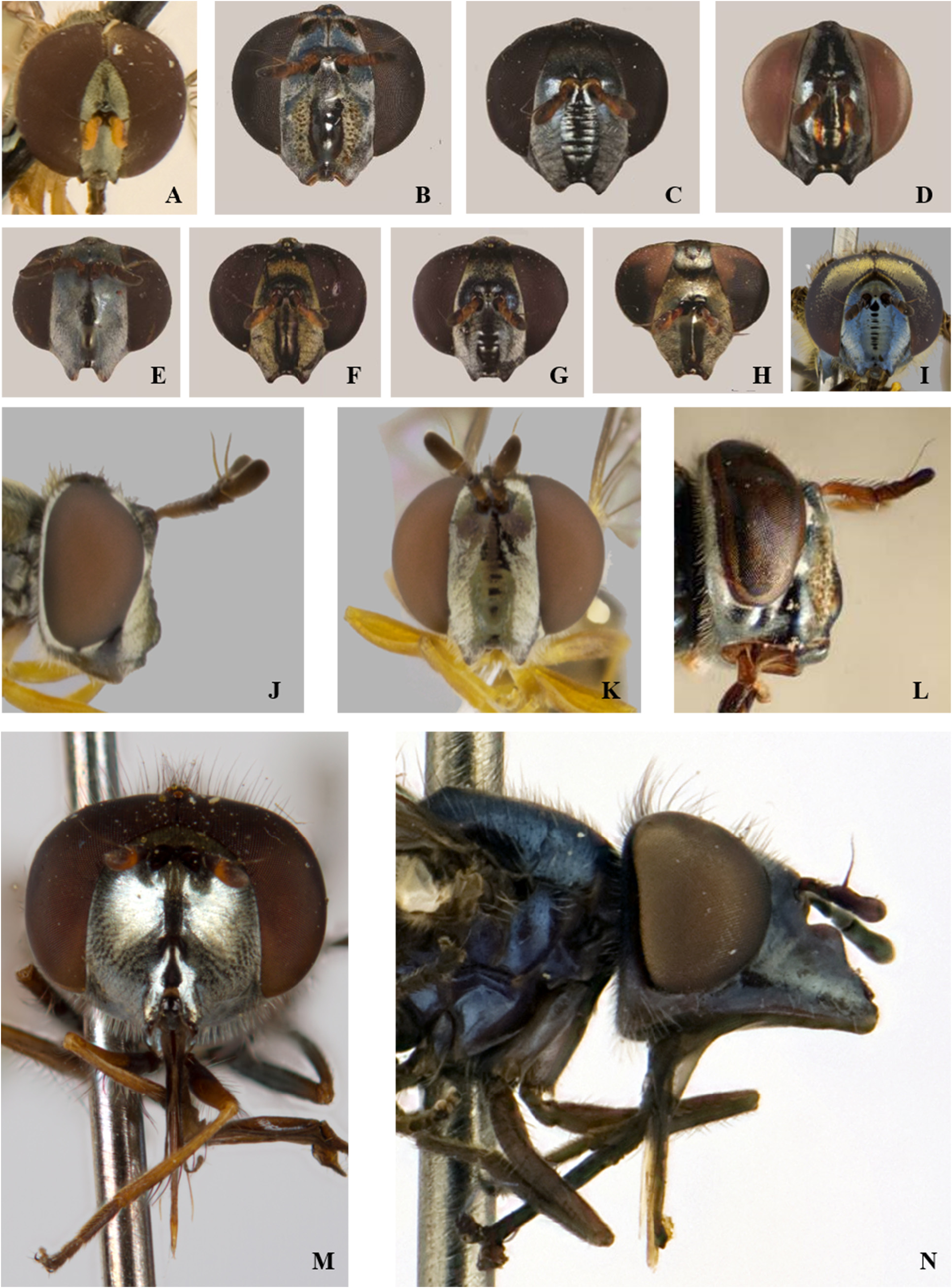
FIGURE 4. Differences in head morphology, colouration and pollinosity patterns, frontal view: A. Leucopodella sp. (♁); B. Argentinomyia longicornis (♀);C. Argentinomyia rugosonasa (♀);D. Argentinomyia jamaicensissp. nov. (♀);E. Argentinomyia testaceipes (♀); F. Argentinomyia tropica (♀); G. Argentinomyia nigrans (♀); H. Argentinomyia browni (♀); I. Argentinomyia crenulata (♀); J–K. Argentinomyia norrbomi sp. nov. (♁): J. lateral view, K. frontal view; L. Argentinomyia longicornis (♁), lateral view; M. Platycheirus (Carposcalis) (♁), frontal view; N. Platycheirus (Tuberculanostoma) (♁), lateral view. Scale bar: 5 mm.

FIGURE 5. Female postabdomen. A. Lateral view of Argentinomyia norrbomi sp. nov.; B. Posterior view of the abdominal apex of Argentinomyia occidentalis sp. nov.; C. Dorsal view of distended abdomen of Argentinomyia luculenta. Abbreviations: Ap: apodeme of epiproct; Cer = Cercus; Ep: Epiproct; Hp: Hypoproct; Scl: sclerotized area; Sper: Spermatheca; Tg: tergite. Scale bars: 0.05 mm.
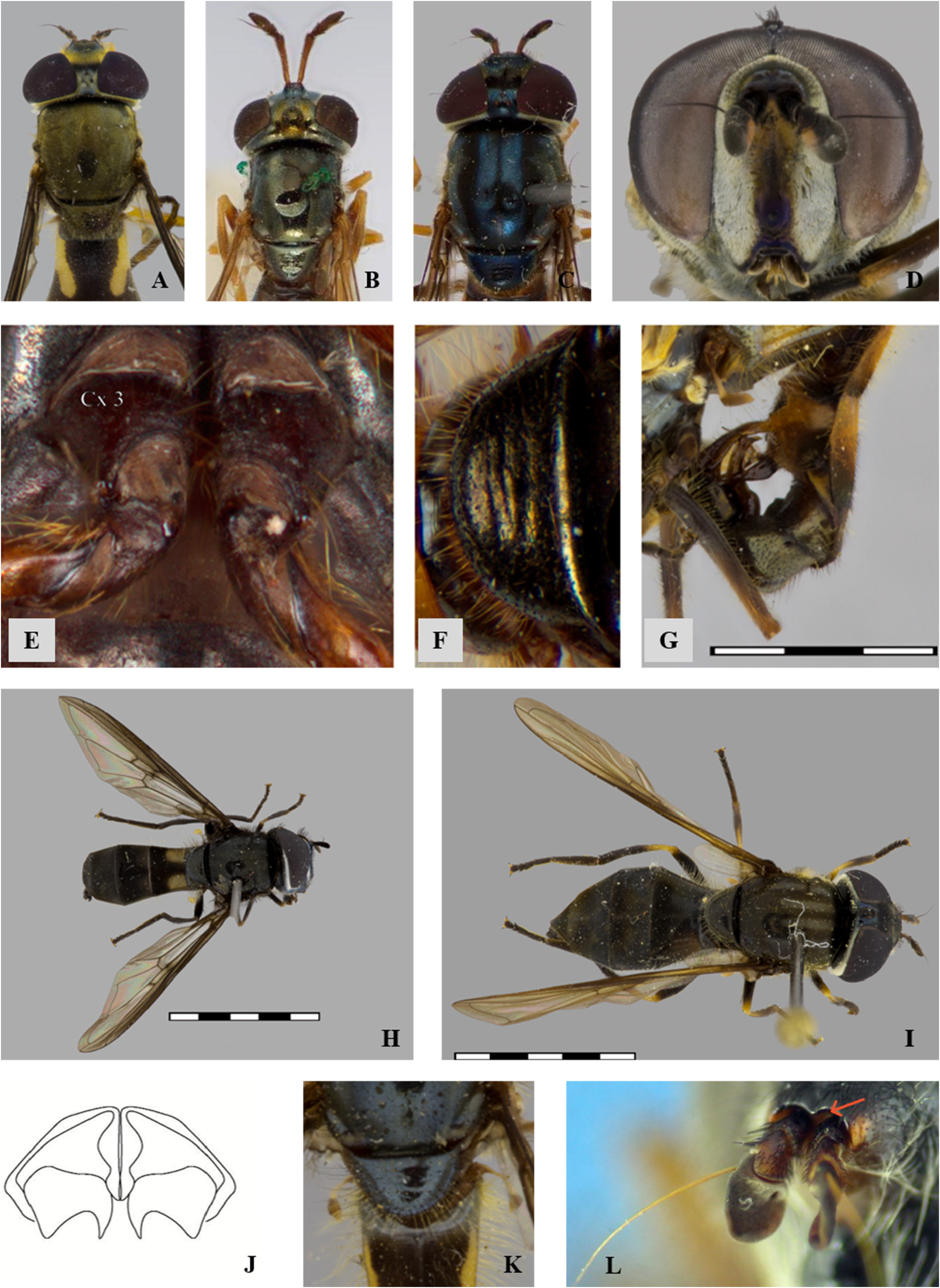
FIGURE 6. Differences in antennae morphology, dorsal view: A. Argentinomyia luculenta (♀); B. Argentinomyia testaceipes (♀); C. Argentinomyia catabomba (♀); D–G. Talahua fervida (♁): D. Head, frontal view, E. Metacoxa pile tuft, ventral view, F. Scutellum emarginated, dorsal view, G. Genitalia, lateral view; H. Argentinomyia CR-18 (♁); I. Argentinomyia sagoti sp. nov. (♀); J. Metasternum of Melanostoma sp.; K. Scutellar pile of Argentinomyia rex; L. Antennal cavities broadly confluent of Xanthandrus sp. Scale bar: 1 mm.
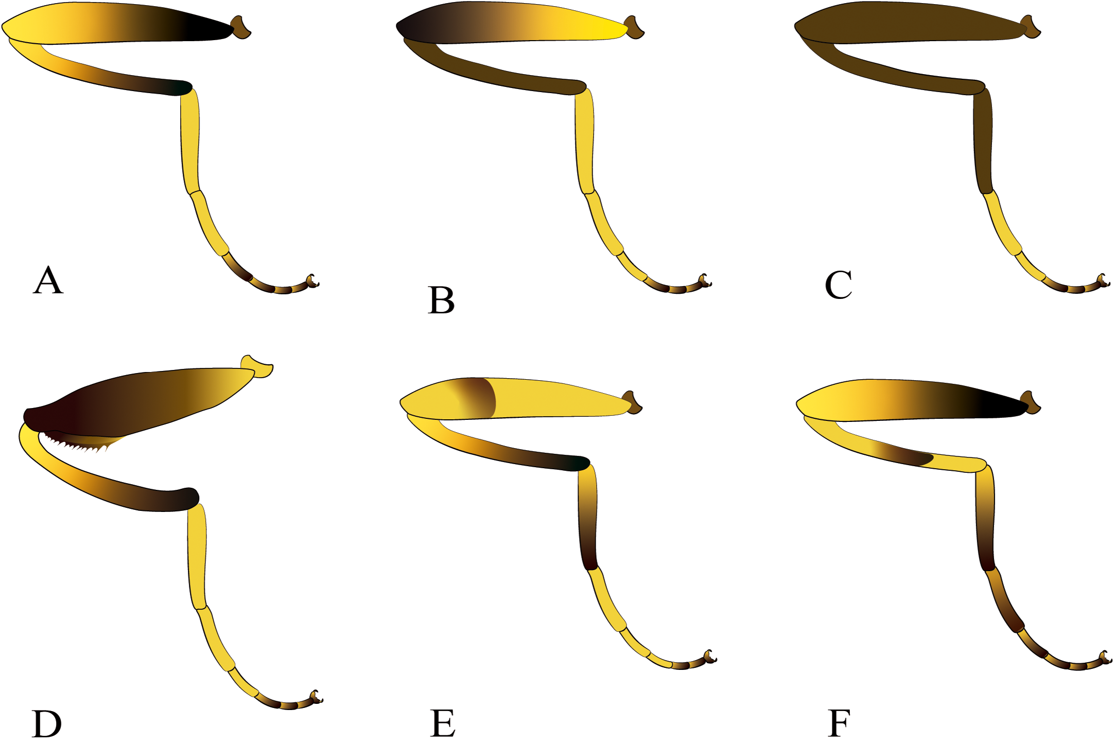
FIGURE 7. Schematic drawings showing colouration patterns of the posterior leg, lateral view.A. Argentinomyia crenulata; B. Argentinomyia longicornis; C. Argentinomyia ivani sp. nov.; D. Argentinomyia spinifemorata sp. nov.; E. Detail of black ring or smudge on the apical margin of metafemur of Argentinomyia praeusta; F. Argentinomyia occidentalis sp. nov.
FIGURE 8. Differences in wing macula pattern, lateral view. A. Argentinomyia crenulata (♀); B. Argentinomyia sagoti sp. nov. (♁); C. Argentinomyia CR-18 (♁).

FIGURE 9. Pattern of abdominal terga, dorsal view: A. Talahua fervida (♀); B. Argentinomyia norrbomi sp. nov. (♀); C. Triangular maculae in Argentinomyia nigrans (♀); D. Argentinomyia rugosonasa (♁); E. Argentinomyia belmira sp. nov. (♀); F. Lineal maculae in Argentinomyia lineata (♁); G. Oblique maculae in Argentinomyia longicornis (♀); H. Argentinomyia andina (♁), head frontal view; I. Argentinomyia currani (♁), abdomen lateral view. Scale bar: 1 mm.
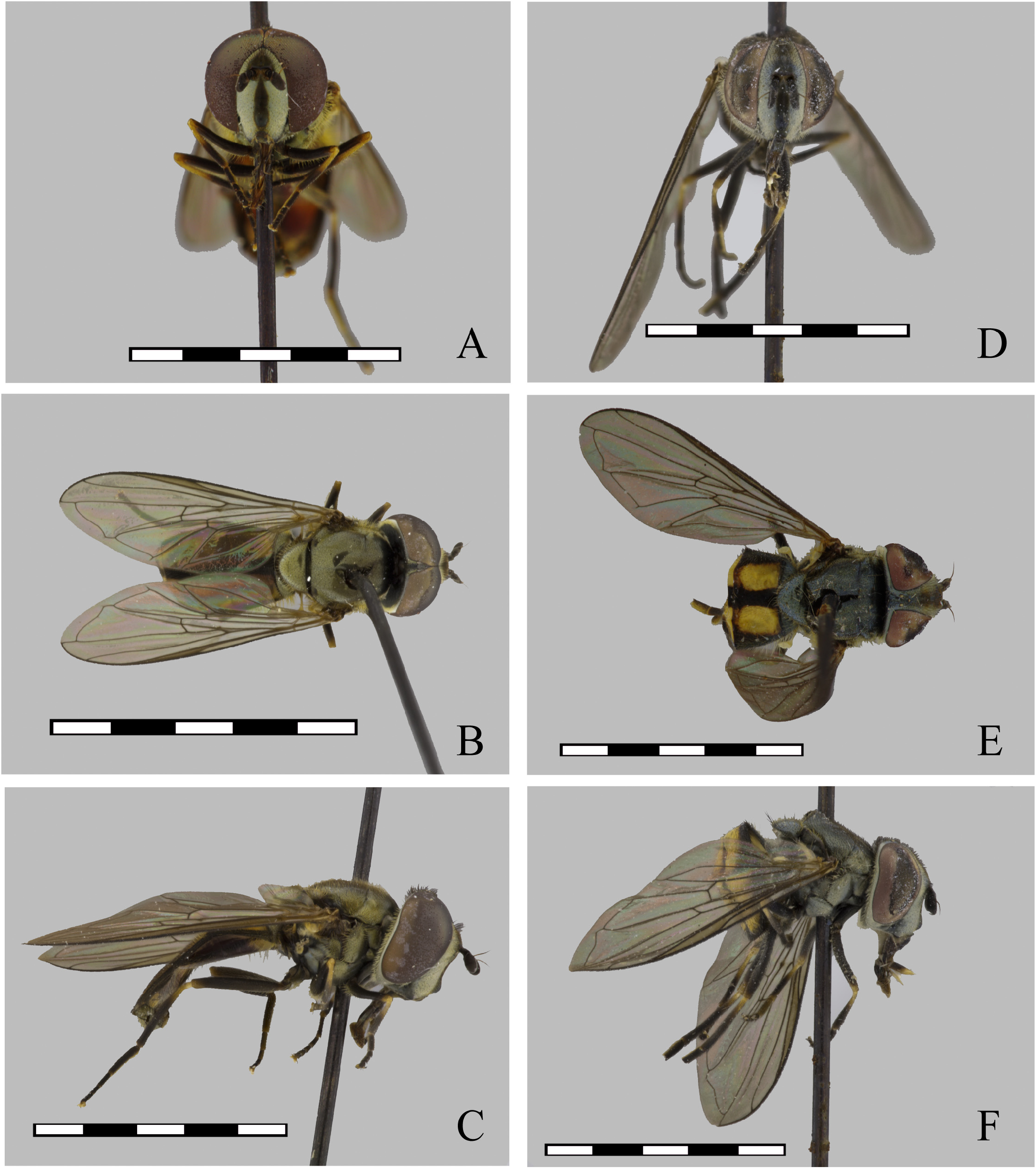
FIGURE 10. Argentinomyia agonis (Walker, 1849), male (CNC DIPTERA 24684): A. Head, frontal view; B. Dorsal view; C. Lateral view. Female (CNC DIPTERA 246840): D. Head, frontal view; E. Dorsal view; F. Lateral view. Scale bars: 5 mm.

FIGURE 11. Argentinomyia agonis (Walker, 1849), male genitalia. A. Whole genitalia including epandrium, cercus and surstylus, lateral view; B. Epandrium, dorsal view; C. Hypandrium, ventral view.Abbreviations used in male genitalia structures are as follows: Ahp = apex of hypandrium (superior lobes); Cer = Cercus; Epd = Epandrium; Hyp = Hypandrium; Led = Aedeagal lobe; Sur = Surstyle. Scale bar: 0.05 mm. D–E. Natural habitus of male and female specimens in Puerto Villamil, Galapagos, Ecuador (iNaturalist catalogue number37254474; https://www.inaturalist.org/observations/37254474). Photo taken by ©Robert Siegel.
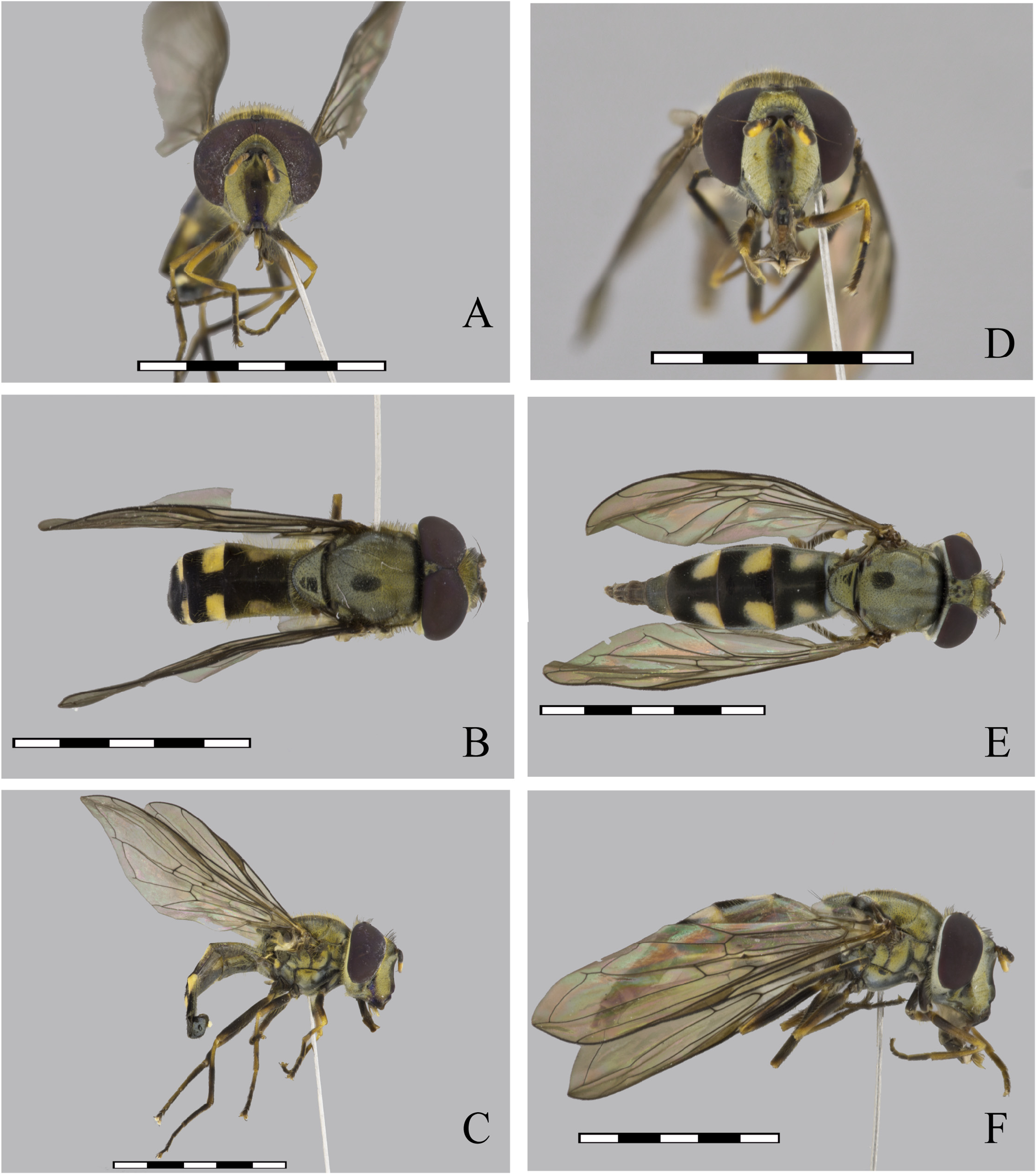
FIGURE 12. Argentinomyia altissima (Fluke, 1945), male (CEUA 93080): A. Head, frontal view; B. Dorsal view; C. Lateral view. Female (CEUA 43354): D. Head, frontal view; E. Dorsal view; F. Lateral view. Scale bars: 5 mm.
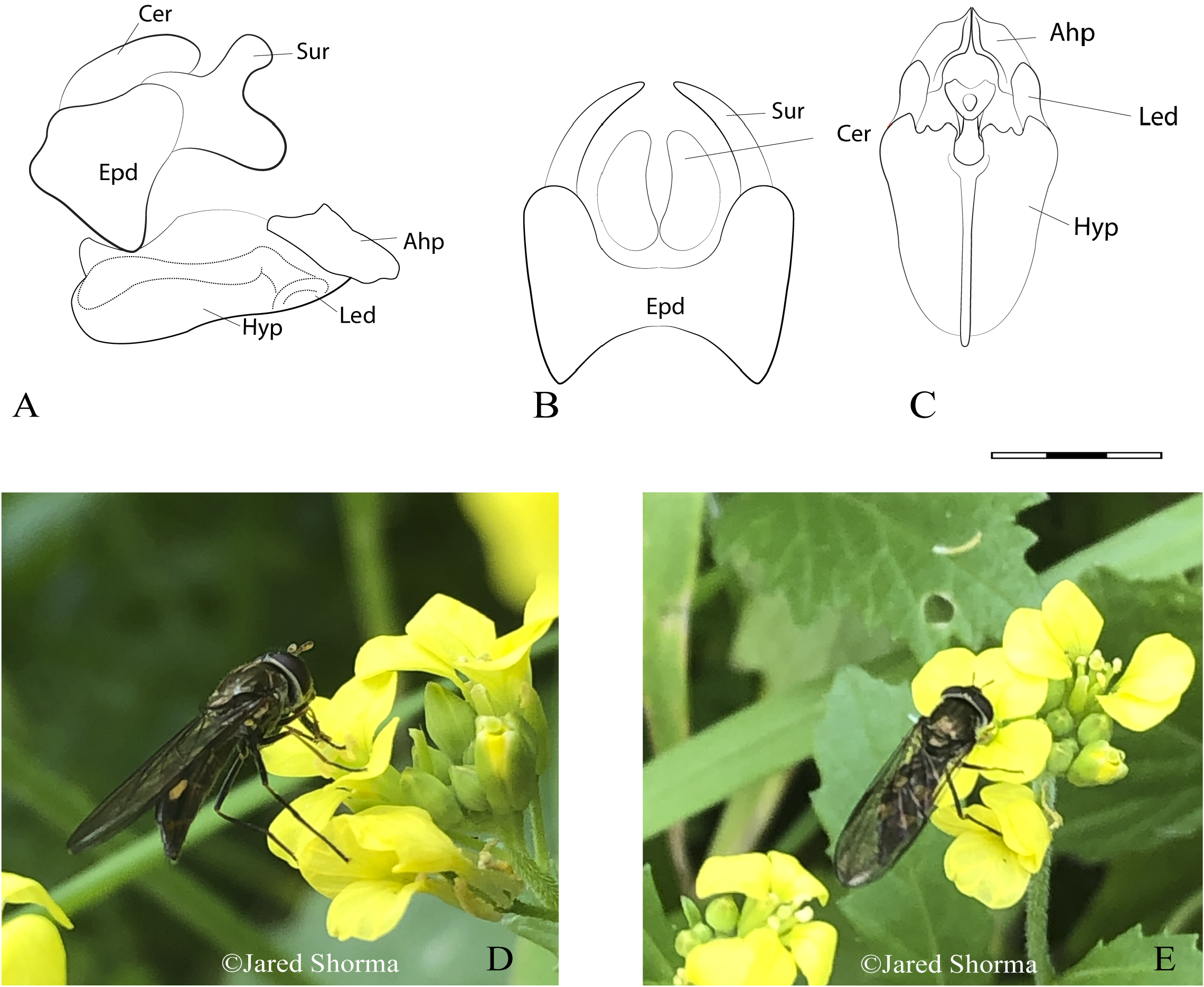
FIGURE 13. Argentinomyia altissima (Fluke, 1945), male genitalia. A. Whole genitalia, lateral view; B. Epandrium, dorsal view; C. Hypandrium, ventral view. Scale bars: 1 mm. D–E. Natural habitus of a female specimen in Pumapaccha, Cuzco, Perú (iNaturalist catalogue number# 37168068; https://www.inaturalist.org/observations/37168068). Photo taken by ©Jared Shorma.

FIGURE 14. Argentinomyia aurifacies sp. nov., male Holotype (USNM ENT 01443646): A. Head, frontal view; B. Dorsal view; C. Lateral view. Female (USNM ENT 00038431): D. Head, frontal view; E. Dorsal view; F. Lateral view. Scale bars: 5 mm.

FIGURE 15. Argentinomyia aurifacies sp. nov., male Holotype, genitalia. A. Whole genitalia, lateral view; B. Epandrium, dorsal view; C. Hypandrium, ventral view. Scale bars: 1 mm.
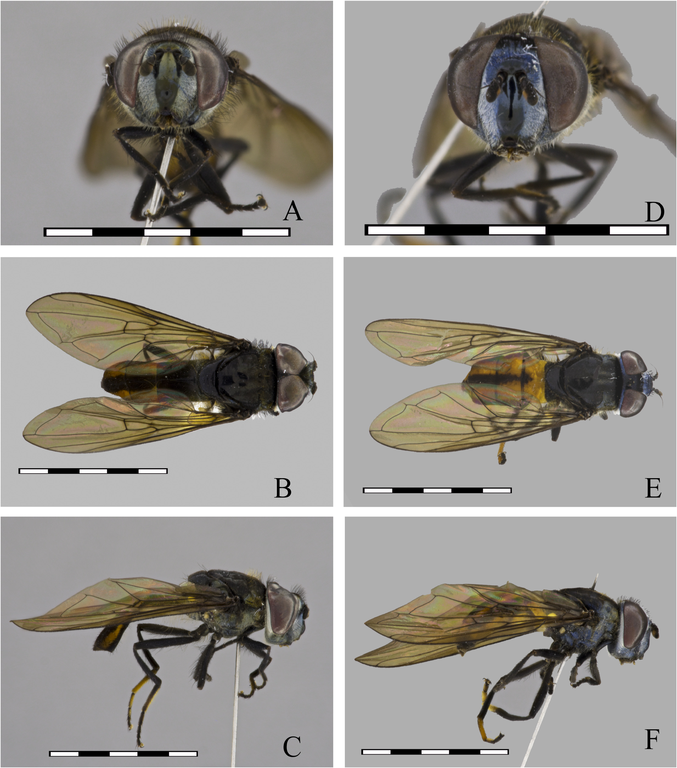
FIGURE 16. Argentinomyia belmira sp. nov., male Holotype (CEUA 95284): A. Head, frontal view; B. Dorsal view; C. Lateral view. Female (CEUA 95481): D. Head, frontal view; E. Dorsal view; F. Lateral view. Scale bars: 5 mm.
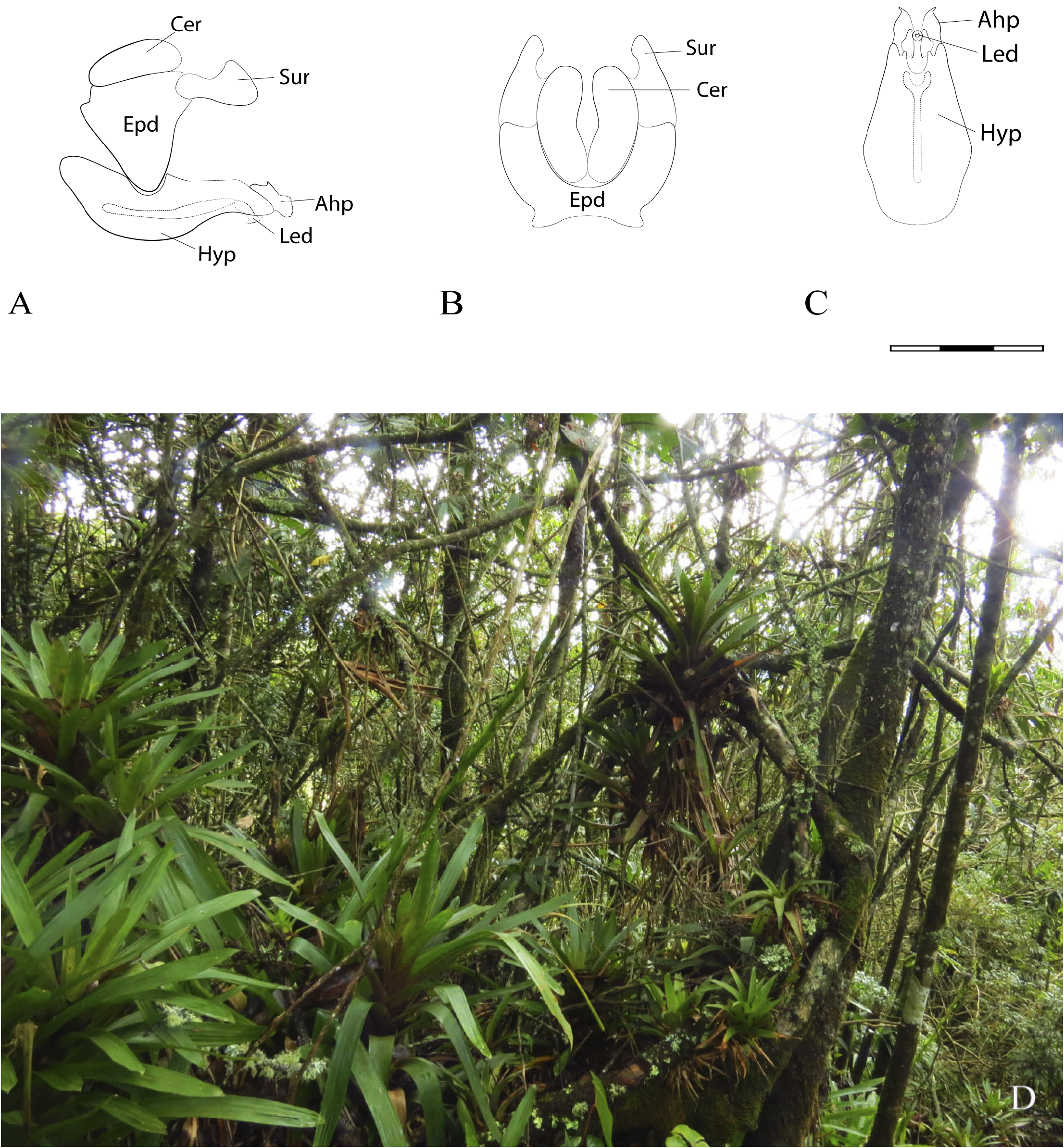
FIGURE 17. Argentinomyia belmira sp. nov., male Holotype, genitalia A. Whole genitalia, lateral view; B. Epandrium, dorsal view; C. Hypandrium, ventral view. Scale bars: 1 mm. D. Type locality in a pristine Andean forest at the Páramo of Santa Inés Belmira in Antioquia, Colombia. The specimens were collected flying in leks over the Native Bromeliad species, Guzmania coriostachya (Bromeliaceae).
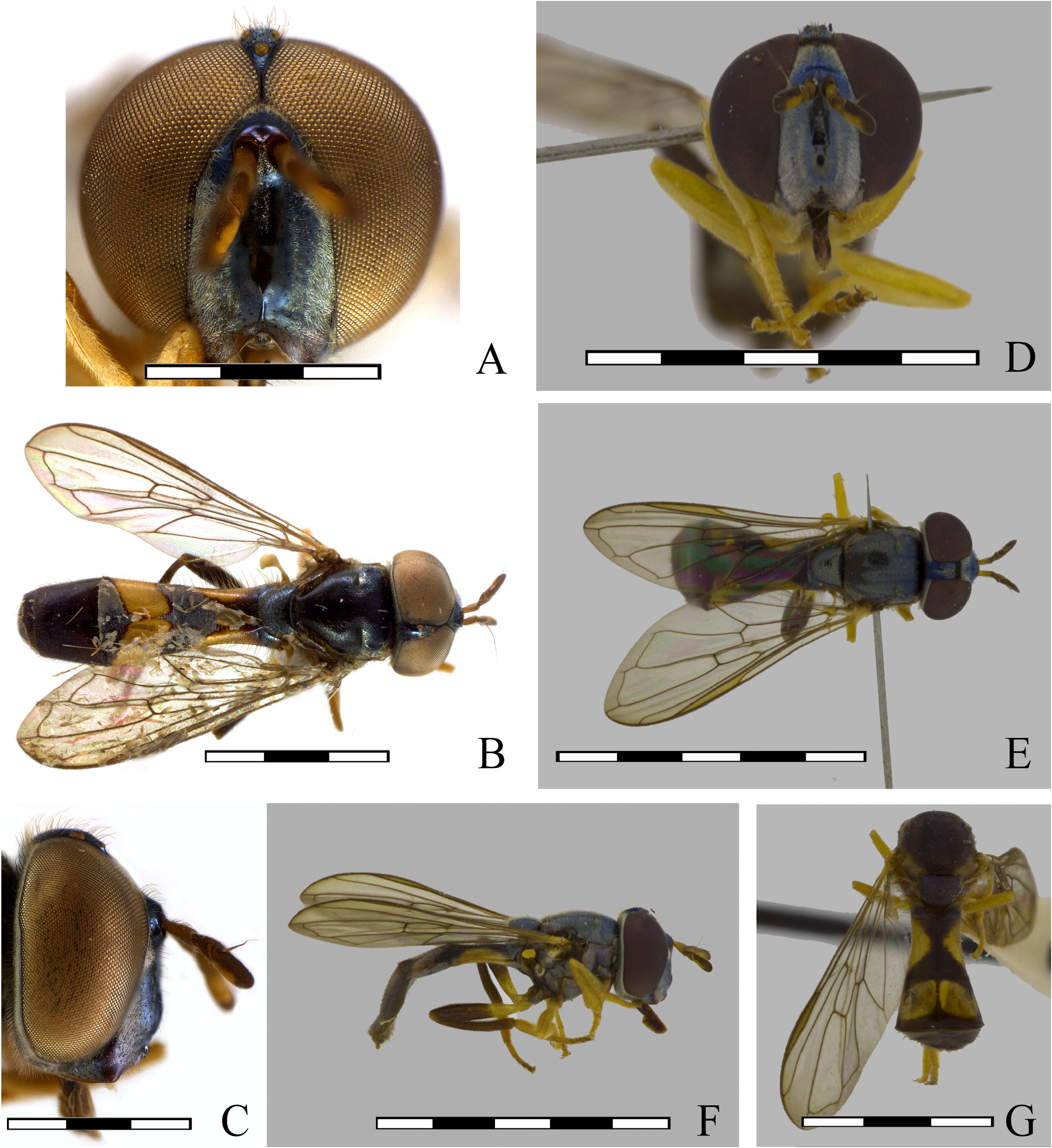
FIGURE 18. Argentinomyia berthae (Lima, 1946), male Neotype (INPA–DIP 000095): A. Head, frontal view B. Dorsal view; C. Head, lateral view. Photo taken by ©Gil Felipe Miranda in Miranda (2017). Female (USNM ENT 01443843): D. Head, frontal view; E. Dorsal view; F. Lateral view; G. Posterodorsal view (USNM ENT01443843). Scale bars: each bar is equal to 1 mm.

FIGURE 19. Argentinomyia berthae (Lima, 1946), male Neotype, genitalia. A. Whole genitalia, lateral view; B. Epandrium, dorsal view; C. Hypandrium, ventral view. Scale bars: 1 mm.
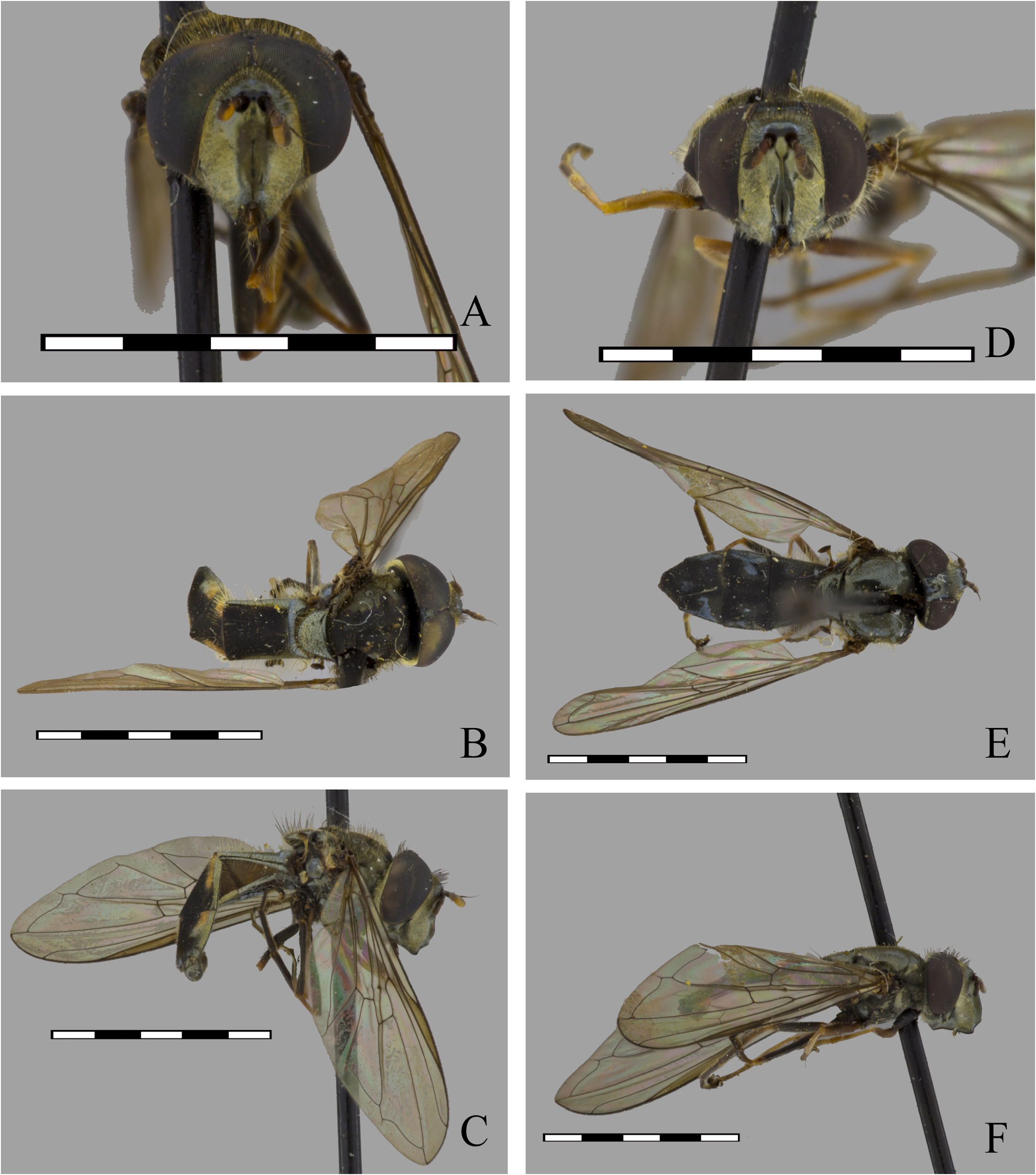
FIGURE 20. Argentinomyia bolivariensis (Fluke, 1945), male (USNM ENT 0144380): A. Head, frontal view; B. Dorsal view; C. Lateral view. Female (USNM ENT 00058848): D. Head, frontal view; E. Dorsal view; F. Lateral view. Scale bars: 5 mm.
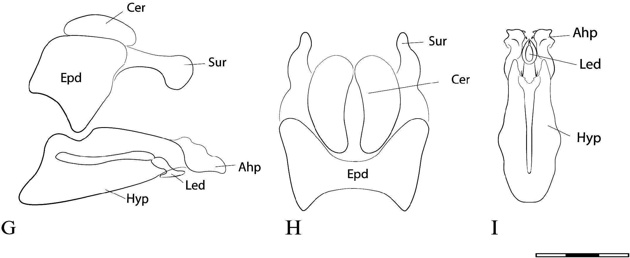
FIGURE 21. Argentinomyia bolivariensis (Fluke, 1945), male genitalia. A. Whole genitalia, lateral view; B. Epandrium, dorsal view; C. Hypandrium, ventral view. Scale bars: 1 mm.
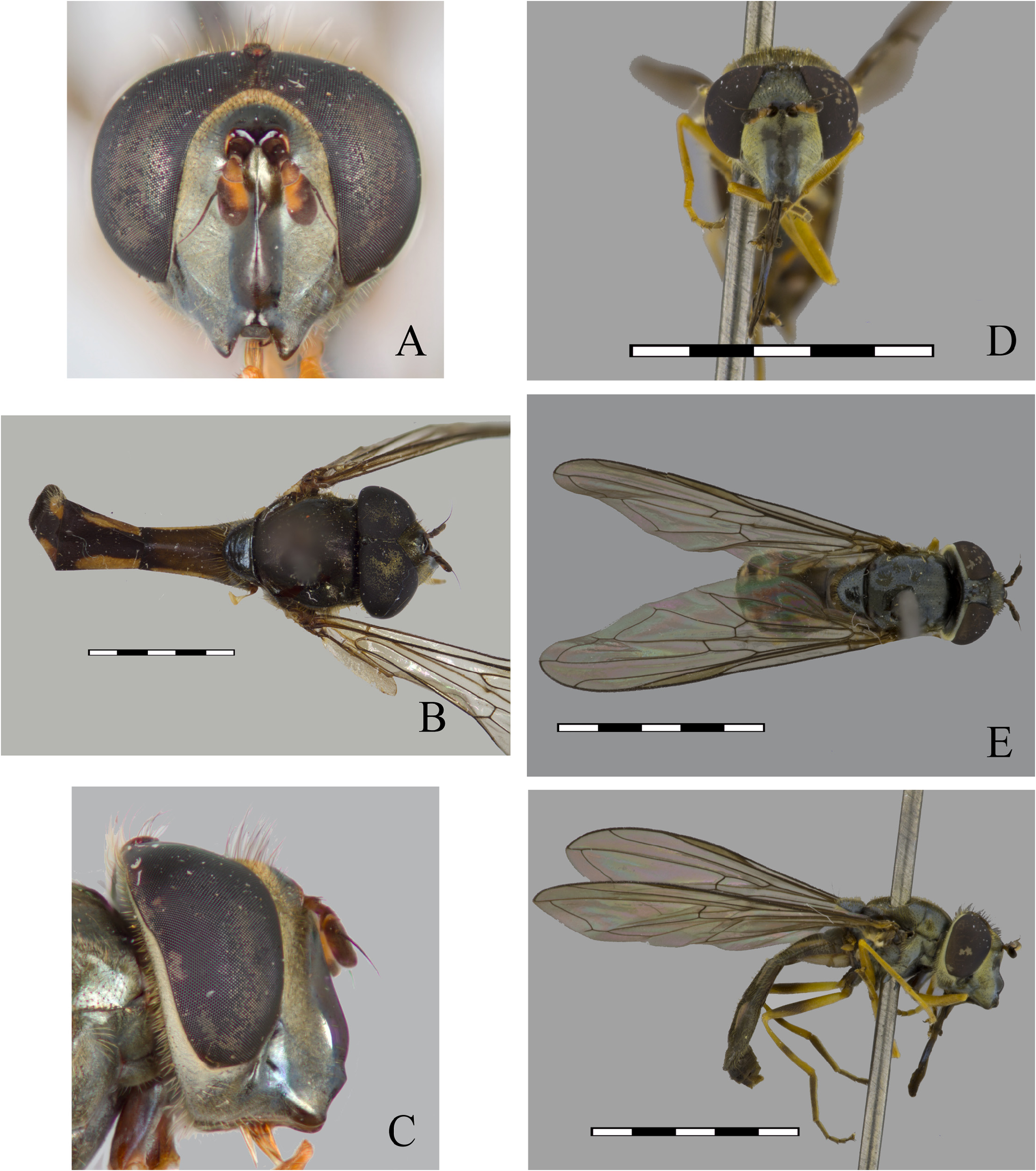
FIGURE 22. Argentinomyia browni (Fluke, 1945), male (USNM ENT 00058849): A. Head, frontal view; B. Dorsal view; C. Lateral view. Female (USNM ENT 01443719): D. Head, frontal view; E. Dorsal view; F. Lateral view. Scale bars: 5 mm.
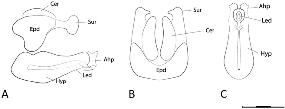
FIGURE 23. Argentinomyia browni (Fluke, 1945), male genitalia. A. Whole genitalia, lateral view; B. Epandrium, dorsal view; C. Hypandrium, ventral view. Scale bars: 1 mm.
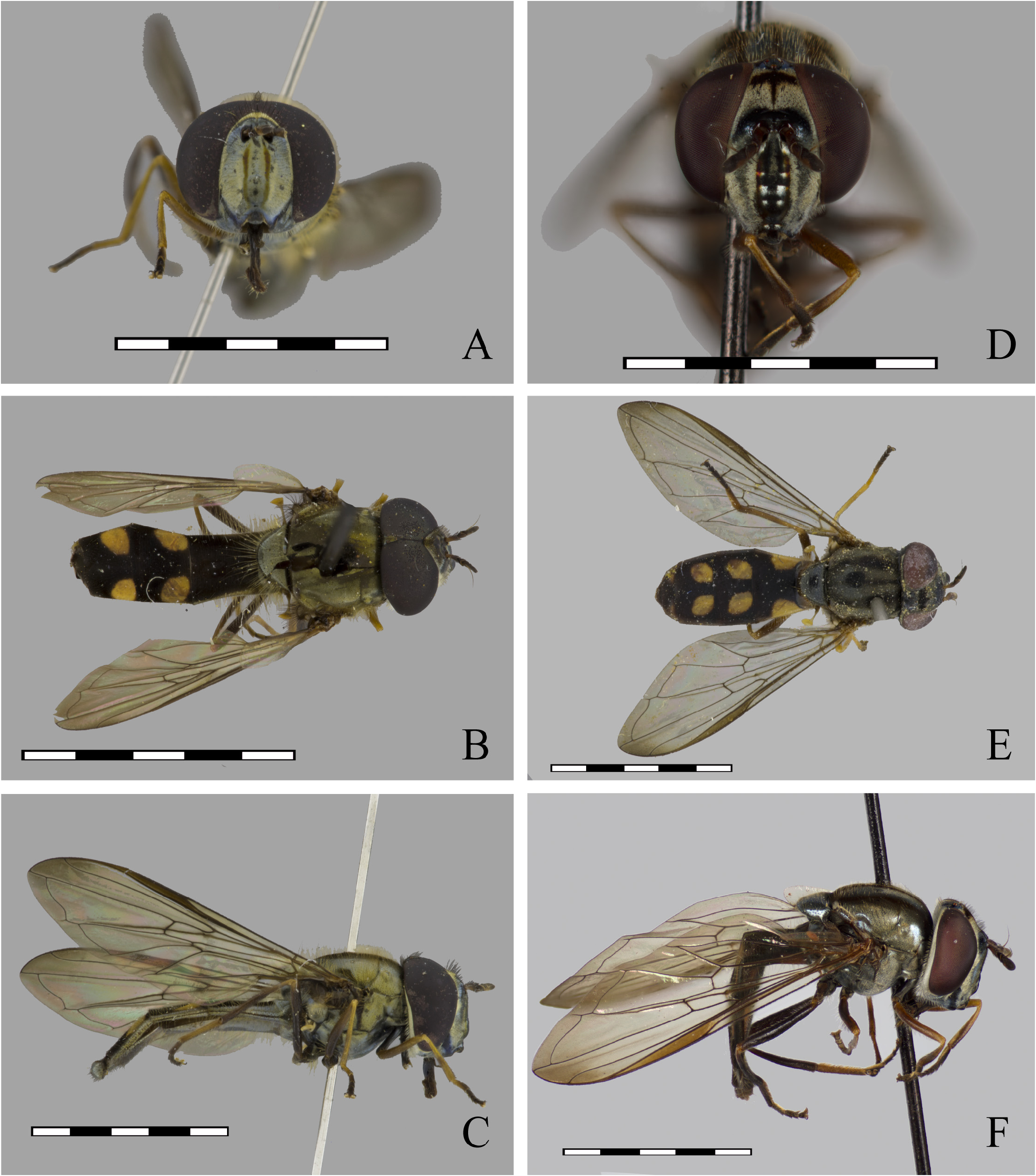
FIGURE 24. Argentinomyia catabomba (Williston, 1891), male (ECO–TAP–E 483): A. Head, frontal view; B. Dorsal view; C. Lateral view. Female (ECO–TAP–E 1715): D. Head, frontal view; E. Dorsal view; F. Lateral view (CNC DIPTERA 112220). Scale bars: 5 mm.
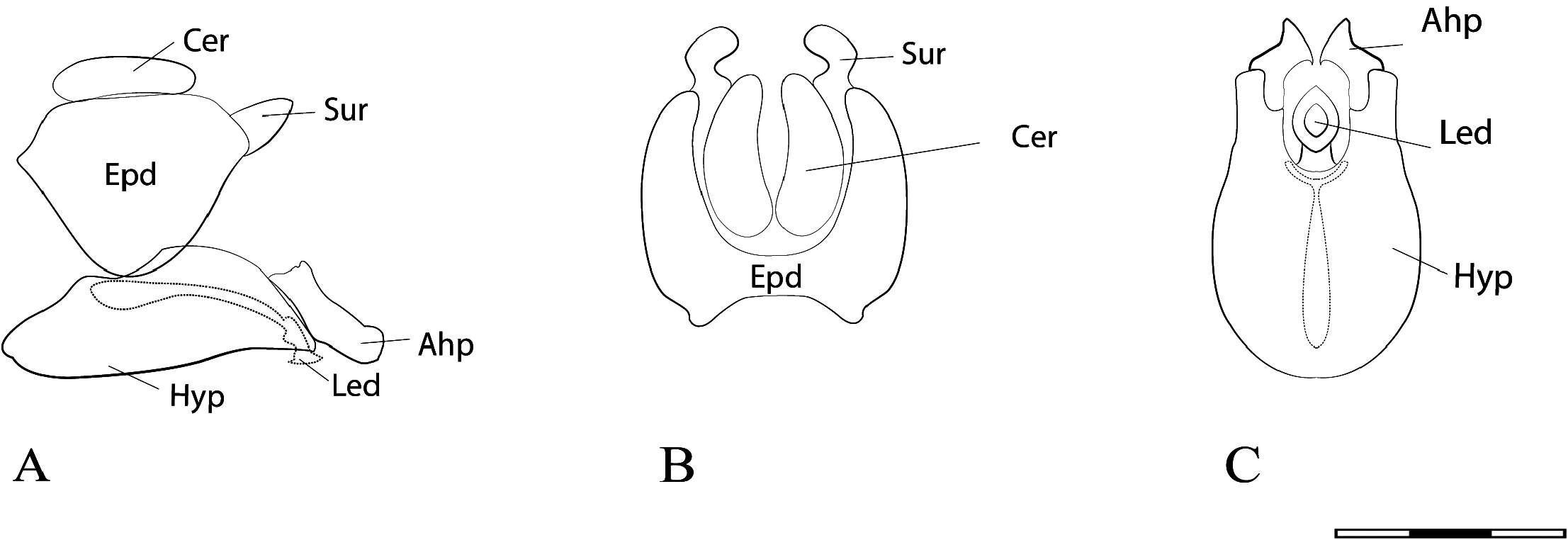
FIGURE 25. Argentinomyia catabomba (Williston, 1891), male genitalia. A. Whole genitalia, lateral view; B. Epandrium, dorsal view; C. Hypandrium, ventral view.
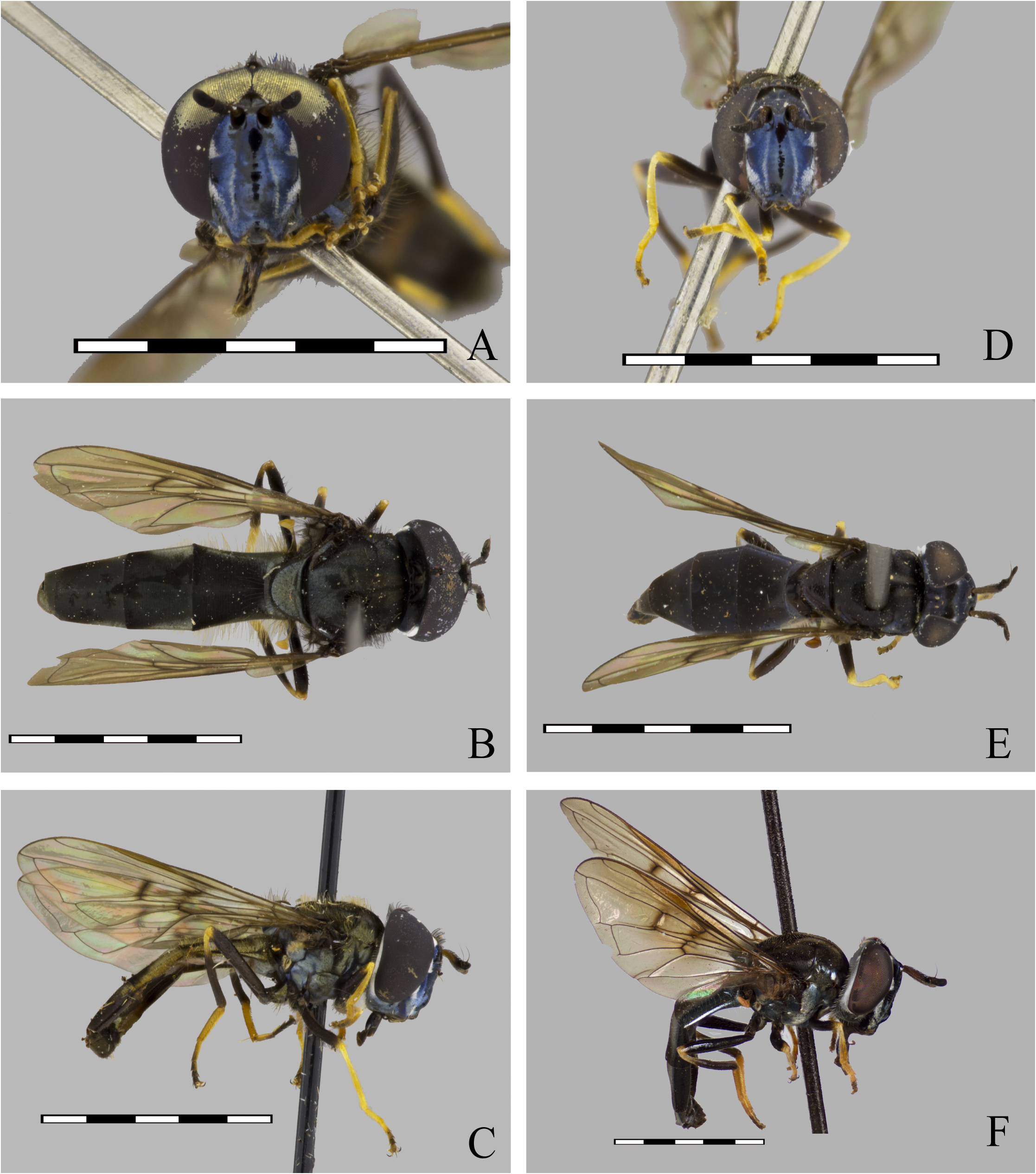
FIGURE 26. Argentinomyia crenulata (Williston, 1891), male (INBio CRI 002568658): A. Head, frontal view; B. Dorsal view; C. Lateral view. Female (INBio CRI 000409982): D. Head, frontal view; E. Dorsal view; F. Lateral view. Scale bars: 5 mm.
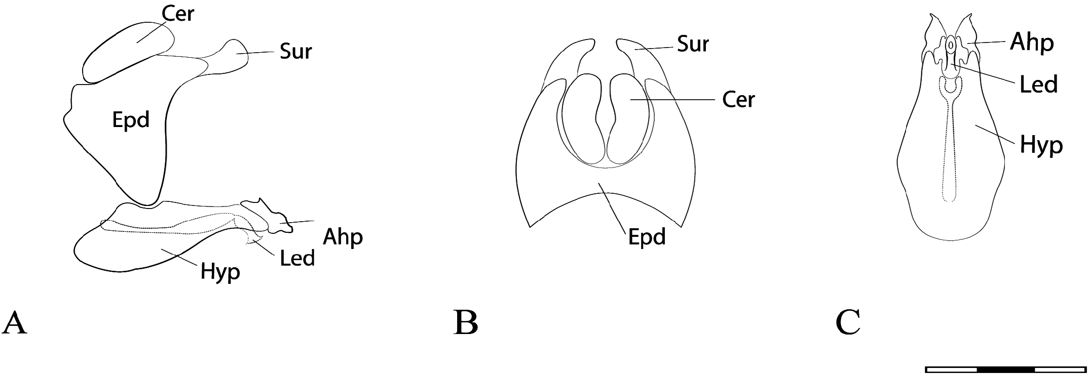
FIGURE 27. Argentinomyia crenulata (Williston, 1891), male genitalia. A. Whole genitalia, lateral view; B. Epandrium, dorsal view; C. Hypandrium, ventral view. Scale bars: 1 mm.
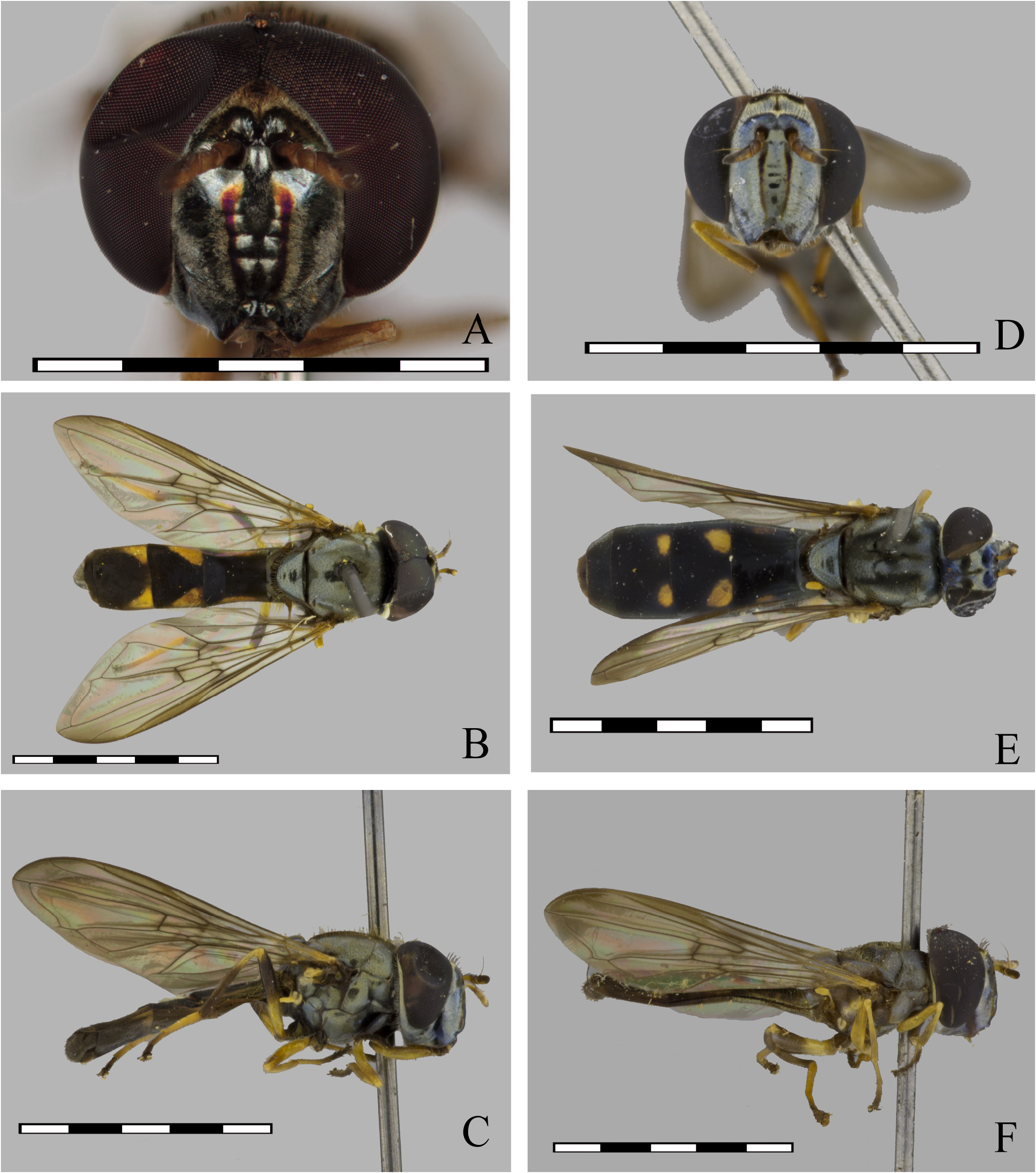
FIGURE 28. Argentinomyia currani (Fluke, 1937), male (USNM ENT 01406227): A. Head, frontal view; B. Detail of triangular maculae, dorsal view; C. Lateral view (Detail of black ring or smudge on the apical margin of metafemur). Female (USNM ENT 01406289): D. Head, frontal view; E. Detail of triangular maculae, dorsal view; F. Lateral view. Scale bars: 5 mm.
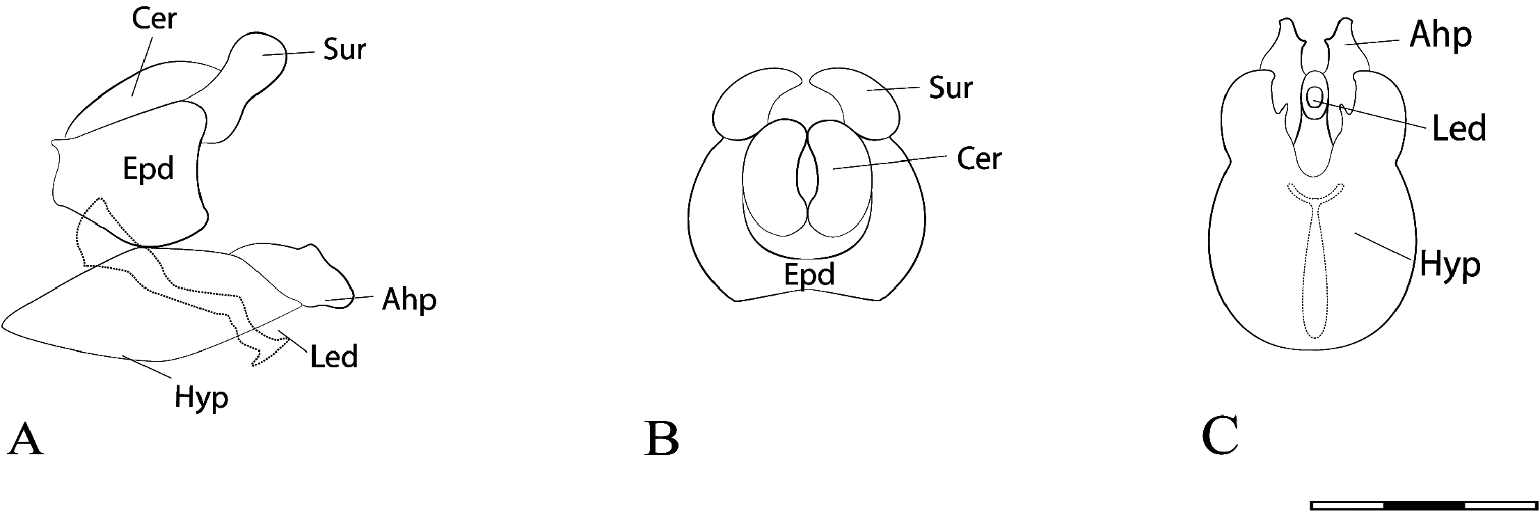
FIGURE 29. Argentinomyia currani (Fluke, 1937), male genitalia. A. Whole genitalia, lateral view; B. Epandrium, dorsal view; C. Hypandrium, ventral view. Scale bars: 1 mm.
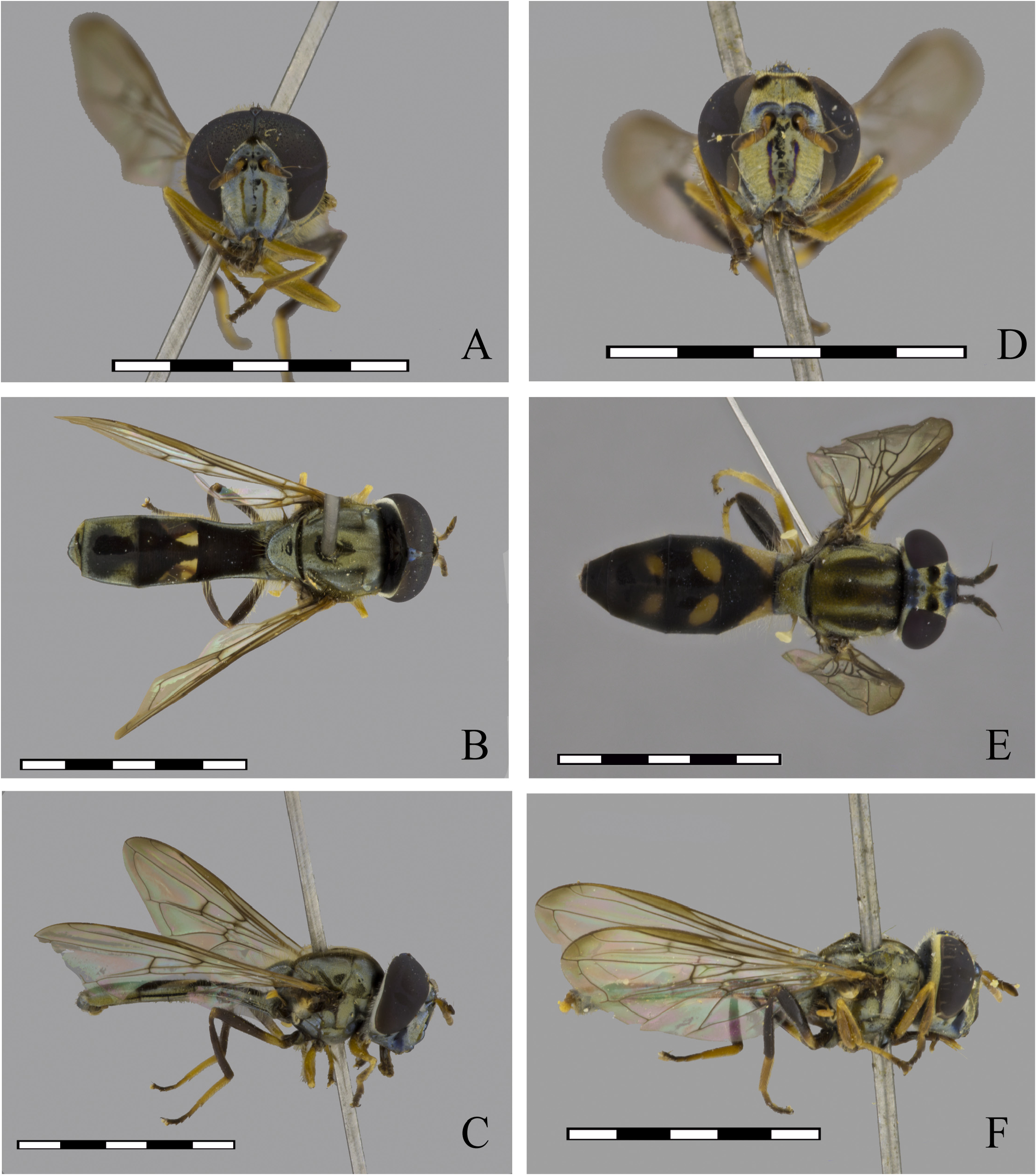
FIGURE 30. Argentinomyia fastigata (Fluke, 1945), male (USNM ENT 01406314): A. Head, frontal view; B. Dorsal view; C. Lateral view. Female (USNM ENT 01406314): D. Head, frontal view; E. Dorsal view (CEUA 103448); F. Lateral view (USNM ENT 01406314). Scale bars: 5 mm.

FIGURE 31. Argentinomyia fastigata (Fluke, 1945), male genitalia. A. Whole genitalia, lateral view; B. Epandrium, dorsal view; C. Hypandrium, ventral view. Scale bars: 1 mm. Male (USNM ENT 01443847): D. Head, frontal view; E. Dorsal view; F. Lateral view. Scale bars: 5 mm.

FIGURE 32. Argentinomyia festiva (Fluke, 1945), male (USNM ENT 000055190): A. Head, frontal view; B. Dorsal view; C. Lateral view. Female (USNM ENT 00023642): D. Head, frontal view; E. Dorsal view; F. Lateral view. Scale bars: 5 mm.

FIGURE 33. Argentinomyia festiva (Fluke, 1945), male genitalia. A. Whole genitalia, lateral view; B. Epandrium, dorsal view; C. Hypandrium, ventral view. Scale bars: 1 mm.
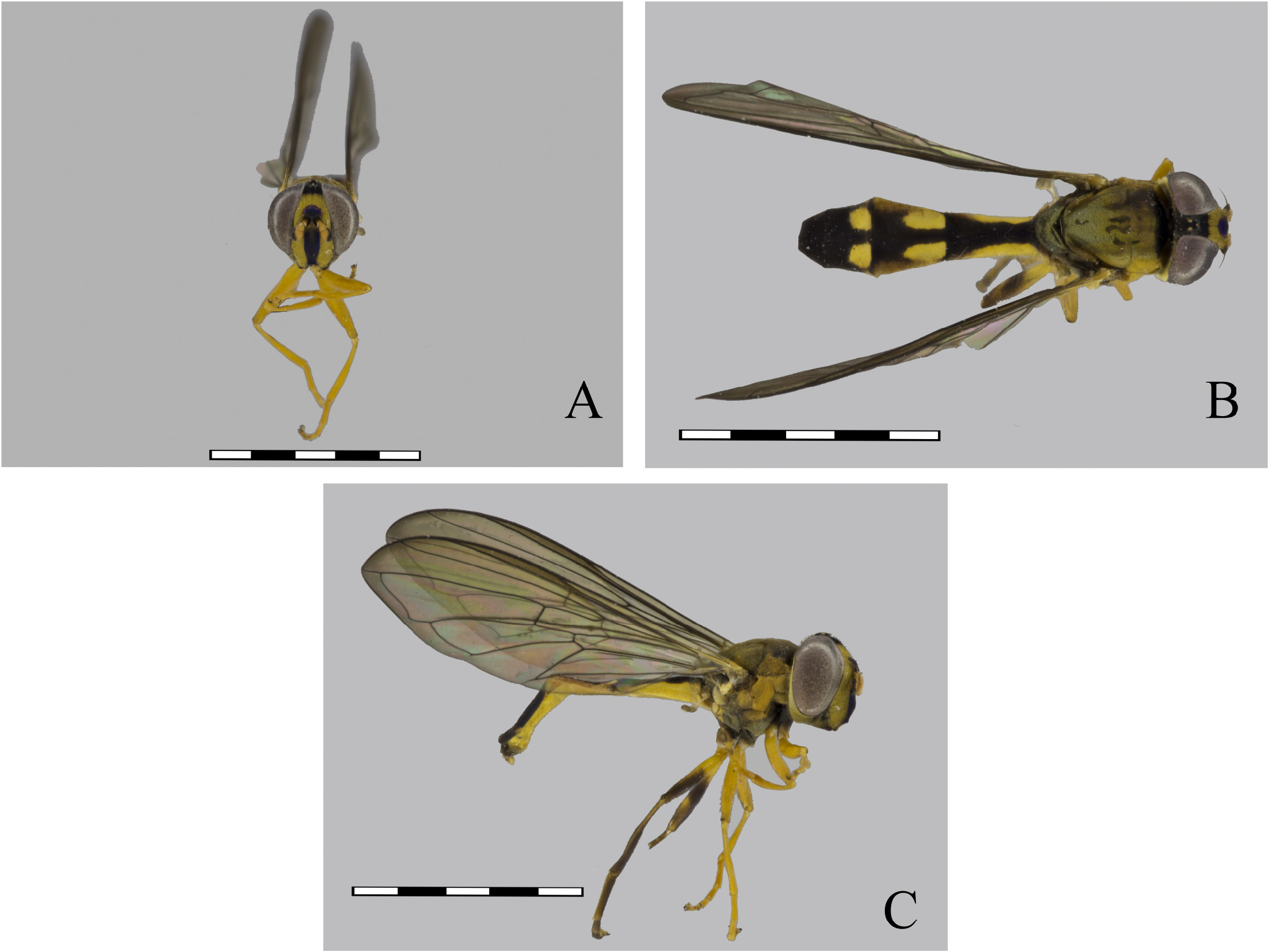
FIGURE 34. Argentinomyia humboldti sp. nov., female Holotype (CEUA 98063): A. Head, frontal view; B. Dorsal view; C. Lateral view. Scale bars: 5 mm.
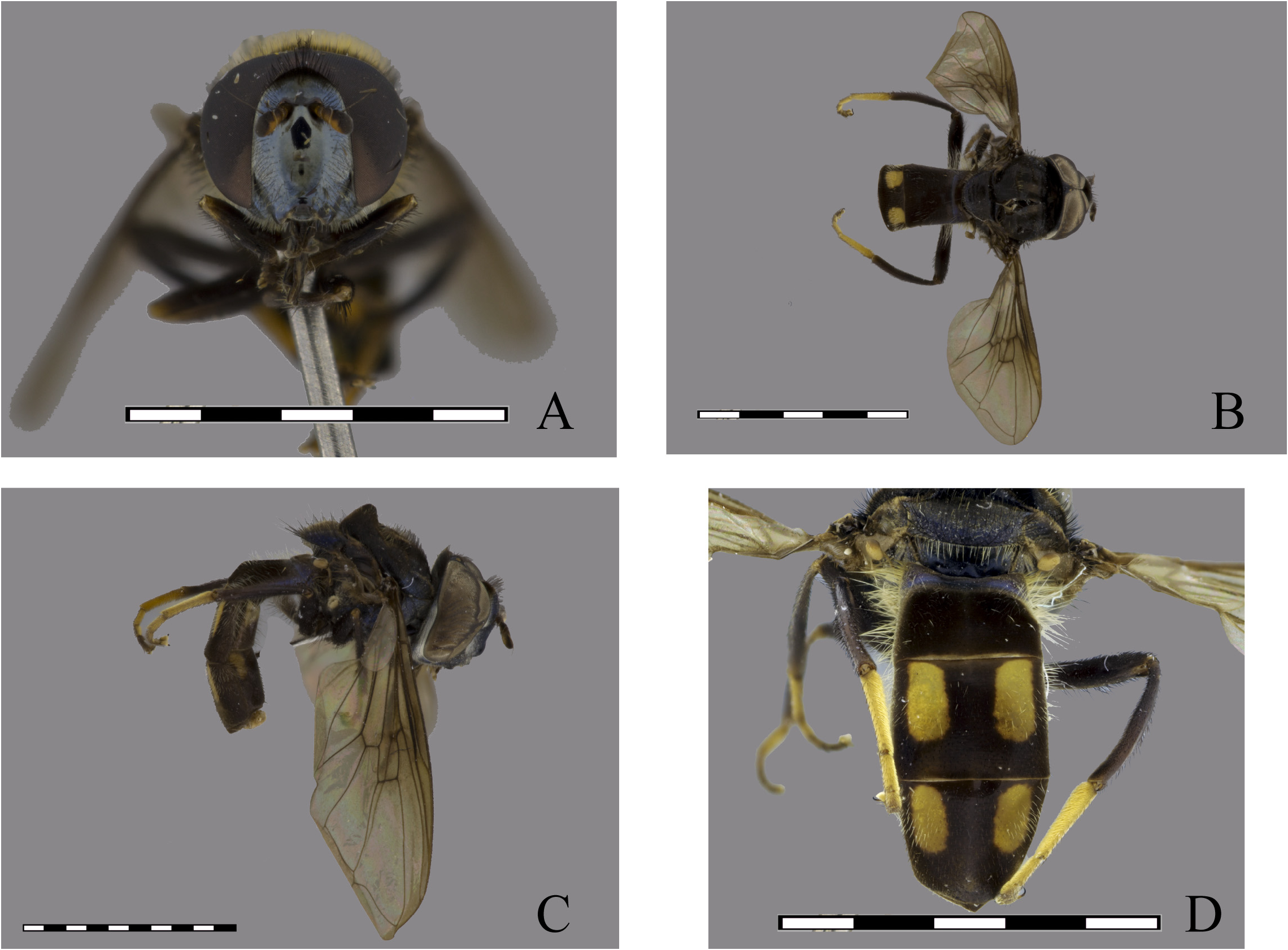
FIGURE 35. Argentinomyia ivani sp. nov., male Holotype (CEUA 95457): A. Head, frontal view; B. Dorsal view; C. Lateral view; D. Posterior view, detail of maculae on 3rd and 4th terga. Scale bars: 5 mm.
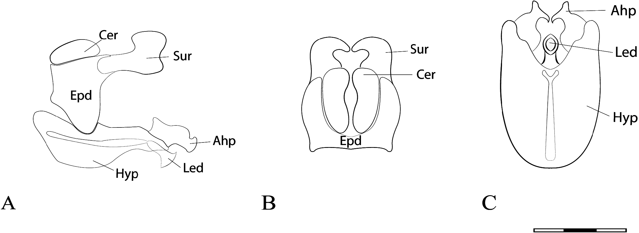
FIGURE 36. Argentinomyia ivani sp. nov., male genitalia. A. Whole genitalia, lateral view; B. Epandrium, dorsal view; C. Hypandrium, ventral view. Scale bars: 1 mm.

FIGURE 37. Argentinomyia jalcaensis sp. nov., male Holotype (Natural History Museum of Denmark, Copenhagen): A. Head, frontal view; B. Dorsal view; C. Lateral view.
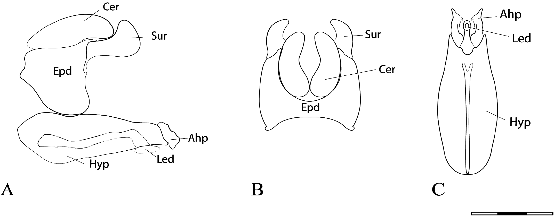
FIGURE 38. Argentinomyia jalcaensis sp. nov., male genitalia. A. Whole genitalia, lateral view; B. Epandrium, dorsal view; C. Hypandrium, ventral view. Scale bars: 1 mm.

FIGURE 39. Argentinomyia jamaicensis sp. nov., female Holotype (CNC DIPTERA 112271): A. Head, frontal view; B. Dorsal view; C. Lateral view. Scale bars: 5 mm.

FIGURE 40. Argentinomyia lanei (Fluke, 1936), male (USNM ENT 01364776): A. Head, frontal view; B. Dorsal view; C. Lateral view. Female (USNM ENT 0146360): D. Head, frontal view; E. Dorsal view; F. Lateral view. Scale bars: 5 mm.

FIGURE 41. Argentinomyia lanei (Fluke, 1936), male genitalia. A. Whole genitalia, lateral view; B. Epandrium, dorsal view; C. Hypandrium, ventral view. Scale bars: 1 mm.

FIGURE 42. Argentinomyia lineata (Fluke, 1937), male (USNM ENT 01406413): A. Head, frontal view; B. Dorsal view; C. Lateral view. Female (USNM ENT 01406353): D. Head, frontal view; E. Dorsal view; F. Lateral view. Scale bars: 5 mm.

FIGURE 43. Argentinomyia lineata (Fluke, 1937), male genitalia. A. Whole genitalia, lateral view; B. Epandrium, dorsal view; C. Hypandrium, ventral view. Scale bars: 1 mm.
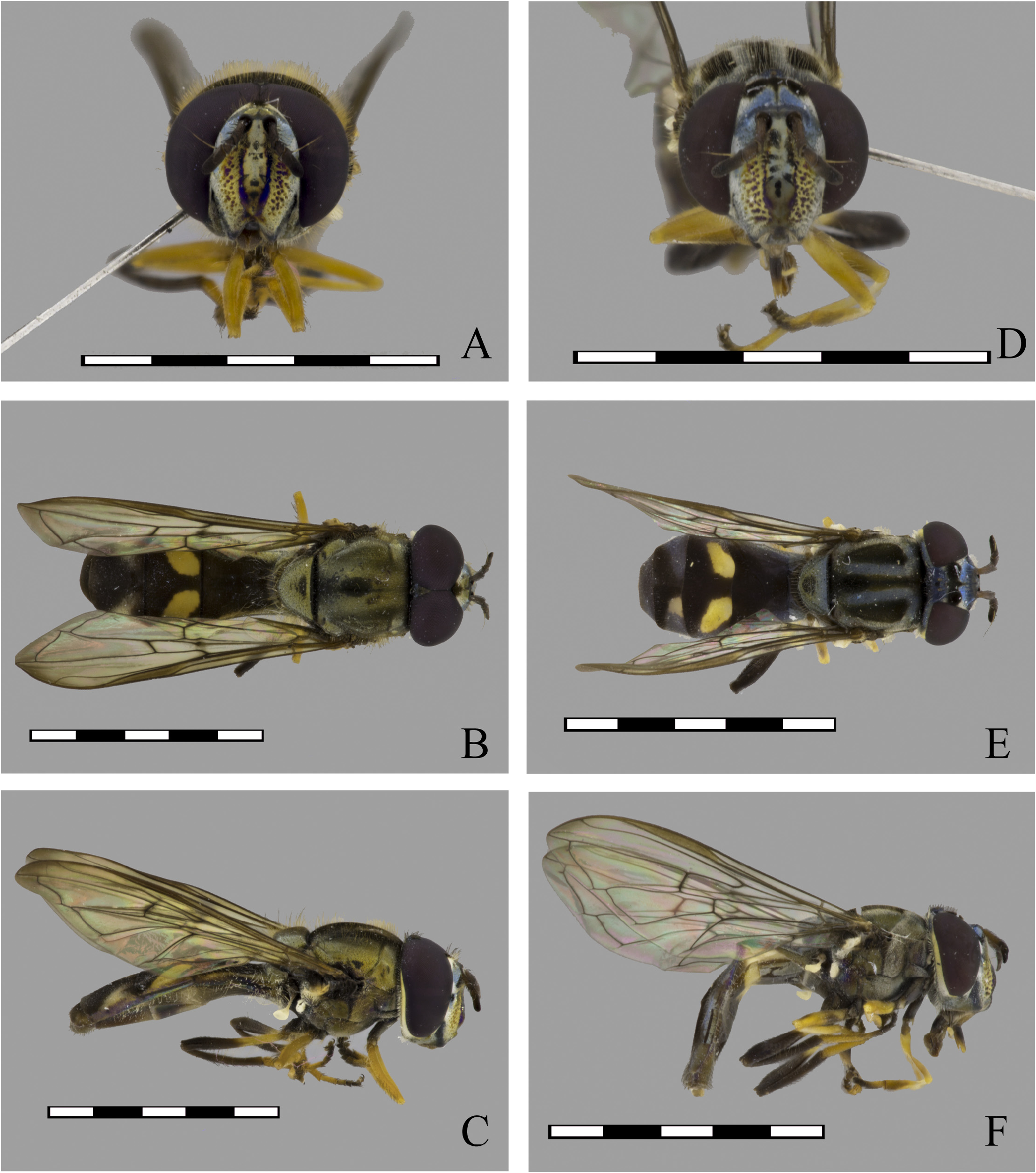
FIGURE 44. Argentinomyia longicornis (Walker, 1836), male (CEUA 47451): A. Head, frontal view; B. Dorsal view; C. Lateral view. Female (USNM ENT 00007888): D. Head, frontal view; E. Dorsal view; F. Lateral view. Scale bars: 5 mm.
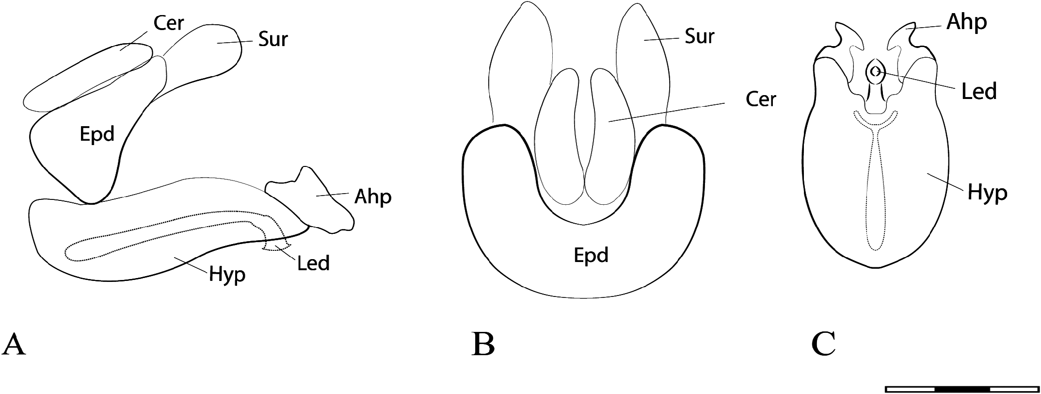
FIGURE 45. Argentinomyia longicornis (Walker, 1836), male genitalia. A. Whole genitalia, lateral view; B. Epandrium, dorsal view; C. Hypandrium, ventral view. Scale bars: 1 mm.
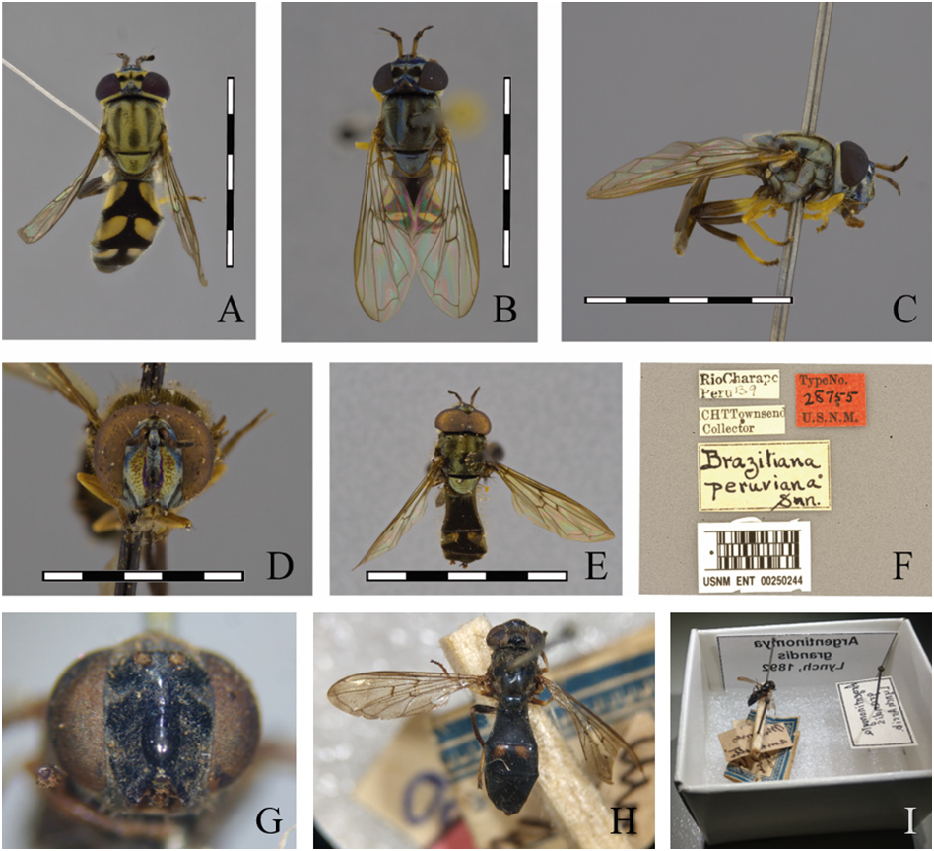
FIGURE 46. Argentinomyia longicornis (Walker, 1836), Female: A–B. Dorsal view of variation pattern in female: A. (CEUA 103438); B. (USNM ENT 00007888); C. Lateral view (USNM ENT 00007888). D–F. Braziliana peruviana Shannon, 1927, male Holotype (USNM ENT 00250244). D. Frontal view; E. Dorsal view; F. Label; G–I. Argentinomyia grandis, female Holotype (MACN). G. Frontal view; H. Dorsal view; I. Holotype box. Scale bars: 5 mm.
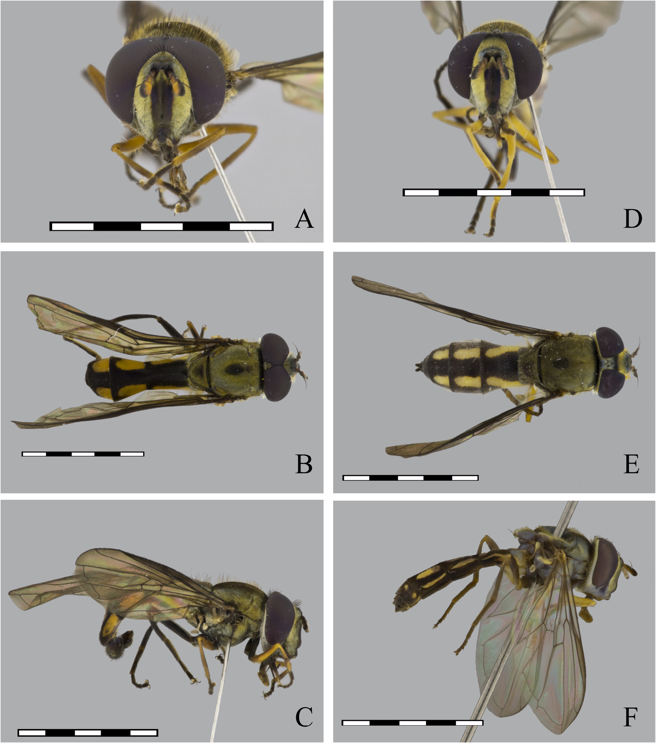
FIGURE 47. Argentinomyia luculenta (Fluke, 1945), male (CEUA 47454): A. Head, frontal view; B. Dorsal view; C. Lateral view. Female: D. Head, frontal view; E. Dorsal view (CEUA 87095); F. Lateral view (USNM ENT 0143772). Scale bars: 5 mm.
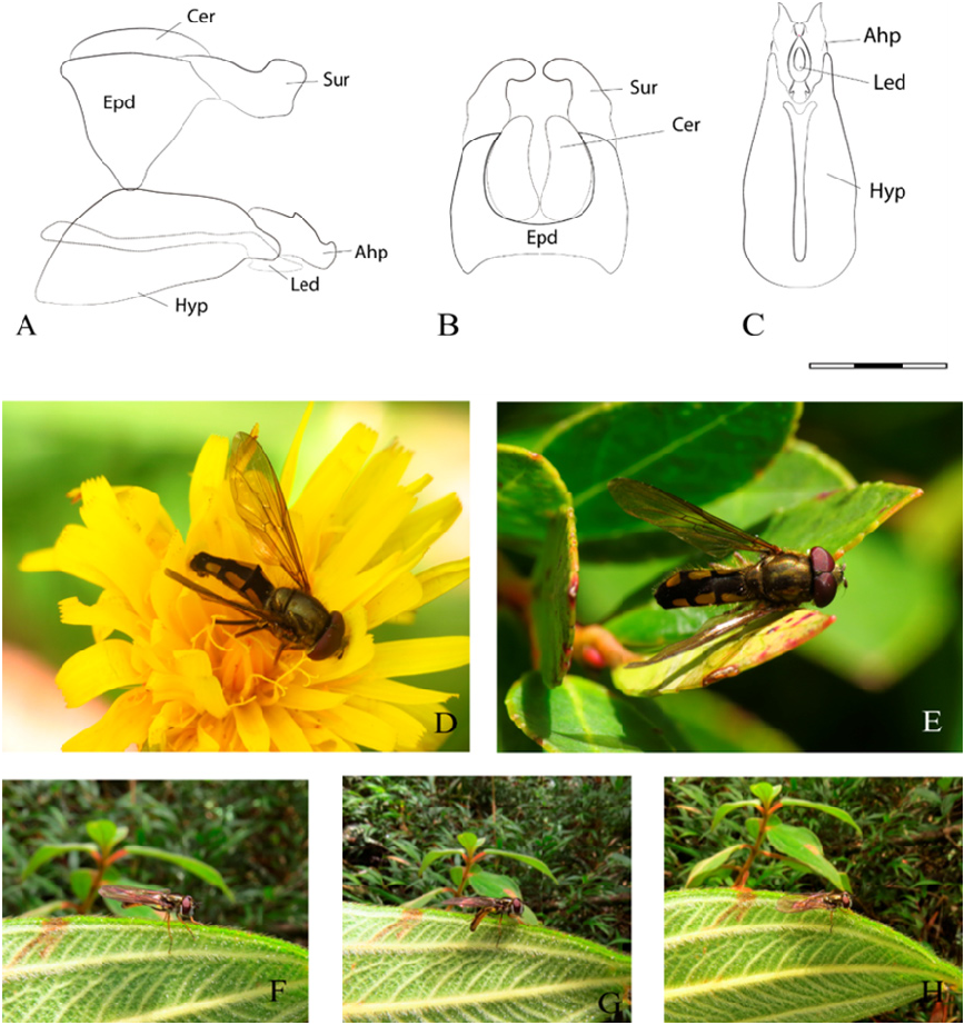
FIGURE 48. Argentinomyia luculenta (Fluke, 1945), male genitalia. A. Whole genitalia, lateral view; B. Epandrium, dorsal view; C. Hypandrium, ventral view. Scale bars: 1 mm. D–E. Natural habitus of a male specimen in the Páramo de Santa Ines Belmira in Antioquia, Colombia: D. Dorsolateral view of a male specimen visiting a flower of “Dandelion” Taraxacum officinale (Asteraceae). E. Dorsal view of a male resting on an Ericaceae leaf. F–H. Natural habitus of a female specimen in a clearing within the forest in the Páramo complex of San José de la Montaña in Antioquia Colombia.
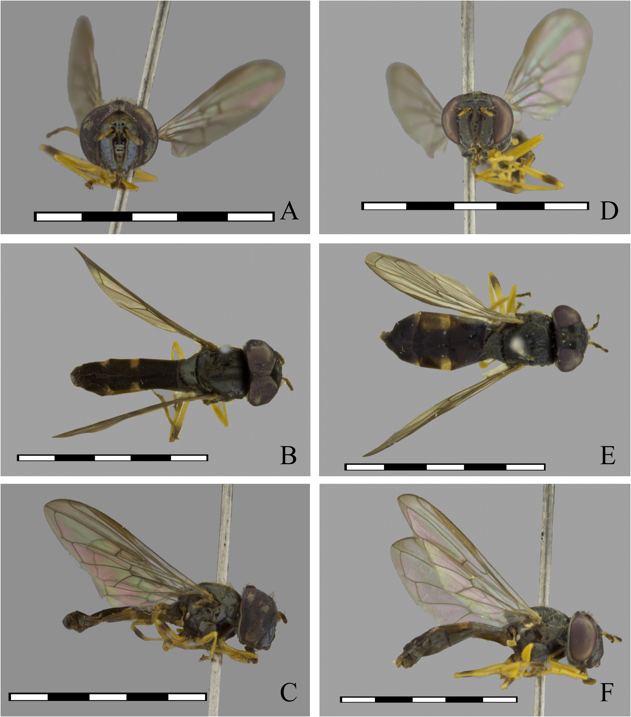
FIGURE 49. Argentinomyia maculata (Walker, 1852), male (USNM ENT 01406339): A. Head, frontal view (Detail of black ring or smudge on apical margin of metafemur); B. Detail of triangular maculae, dorsal view; C. Lateral view. Female (USNM ENT 01406336): D. Head, frontal view; E. Detail of triangular maculae, dorsal view; F. Lateral view. Scale bars: 5 mm.
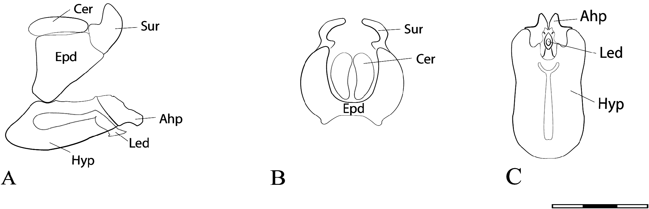
FIGURE 50. Argentinomyia maculata (Walker, 1852), male genitalia. A. Whole genitalia, lateral view; B. Epandrium, dorsal view; C. Hypandrium, ventral view. Scale bars: 1 mm.

FIGURE 51. Argentinomyia neotropica (Curran, 1937), male (USNM ENT 01409270): A. Head, frontal view; B. Dorsal view; C. Lateral view. Female (USNM ENT 01384792): D. Head, frontal view; E. Dorsal view; F. Lateral view. Scale bars: 5 mm.

FIGURE 52. Argentinomyia neotropica (Curran, 1937), male genitalia. A. Whole genitalia, lateral view; B. Epandrium, dorsal view; C. Hypandrium, ventral view. Scale bars: 1 mm. D. Natural habitus of a female specimen in Magdalena Partido, Buenos Aires Province, Argentina (iNaturalist catalogue number 6307133; https://www.inaturalist.org/observations/6307133). Photo taken by ©Gerónimo Martín Alonso.

FIGURE 53. Argentinomyia nigrans (Fluke, 1945), male (USNM ENT 01384670): A. Head, frontal view; B. Dorsal view; C. Lateral view. Female (USNM ENT 01406441): D. Head, frontal view; E. Dorsal view; F. Lateral view. Scale bars: 5 mm.
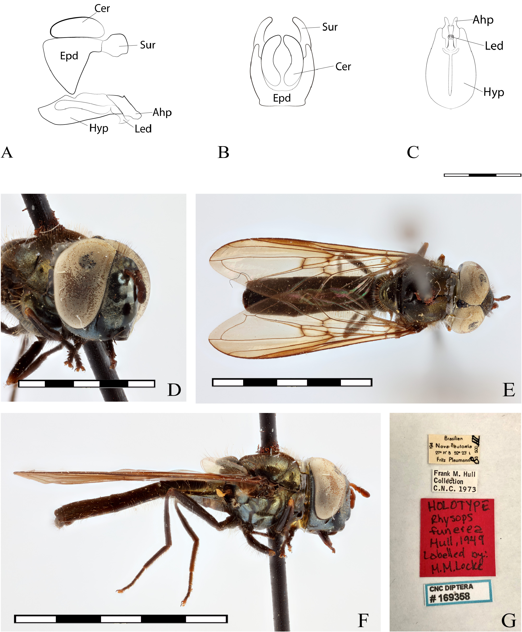
FIGURE 54. Argentinomyia nigrans (Fluke, 1945), male genitalia. A. Whole genitalia, lateral view; B. Epandrium, dorsal view; C. Hypandrium, ventral view. Scale bars: 1 mm. D–G. Rhysops funerea (Hull, 1949), male Holotype (CNC DIPTERA 169358). D. Latero–frontal view; E. Dorsal view; F. Lateral view; G. Label.
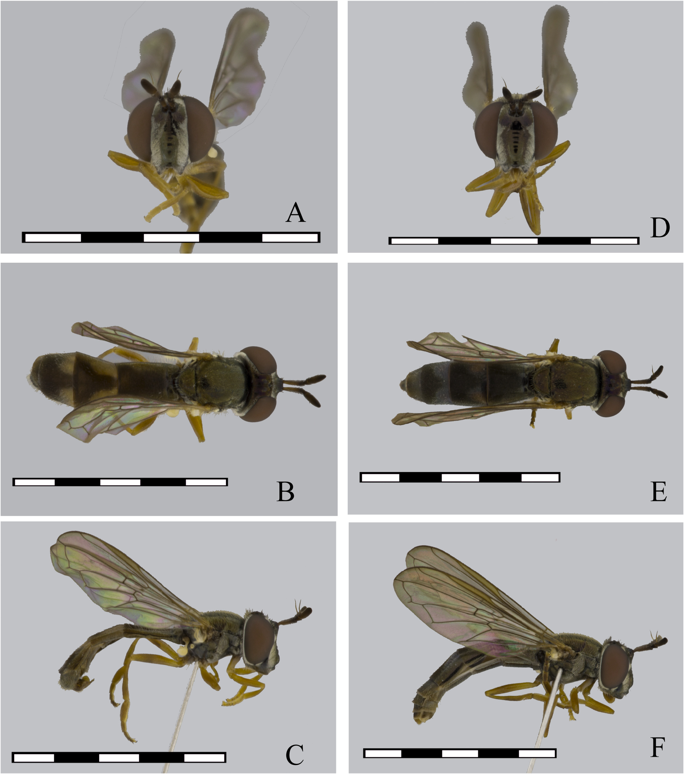
FIGURE 55. Argentinomyia norrbomi sp. nov., male Holotype (USNM ENT 01443832): A. Head, frontal view; B. Dorsal view; C. Lateral view. Female (USNM ENT 01443831): D. Head, frontal view; E. Dorsal view; F. Lateral view. Scale bars: 5 mm.

FIGURE 56. Argentinomyia norrbomi sp. nov., male Holotype, genitalia A. Whole genitalia, lateral view; B. Epandrium, dorsal view; C. Hypandrium, ventral view. Scale bars: 1 mm.

FIGURE 57. Argentinomyia occidentalis sp. nov., male Holotype (CEUA 47448): A. Head, frontal view; B. Dorsal view; C. Lateral view. Female (CEUA 47447): D. Head, frontal view; E. Dorsal view; F. Lateral view. Scale bars: 5 mm.
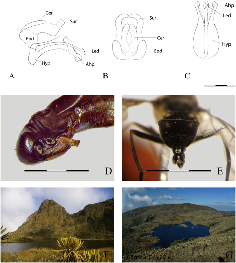
FIGURE 58. Argentinomyia occidentalis sp. nov., male genitalia. A. Whole genitalia, lateral view; B. Epandrium, dorsal view; C. Hypandrium, ventral view; D. Male genitalia, lateral view; E. Female abdomen, posterior view, detail of maculae on 4th and 5th tergum. Scale bars: 1,5 mm. F–G. Type localities. F. Páramo Santa Rica in Andes Antioquia, Colombia. Photo taken by ©Augusto L. Montoya; G. Páramo del Sol in Urrao Antioquia, Colombia. Photo taken by ©Esteban Domínguez.

FIGURE 59. Argentinomyia opaca (Fluke, 1945), male (USNM ENT 00035708): A. Head, frontal view; B. Dorsal view; C. Lateral view. Female (USNM ENT 00035708): D. Head, frontal view; E. Dorsal view; F. Lateral view. Scale bars: 5 mm.
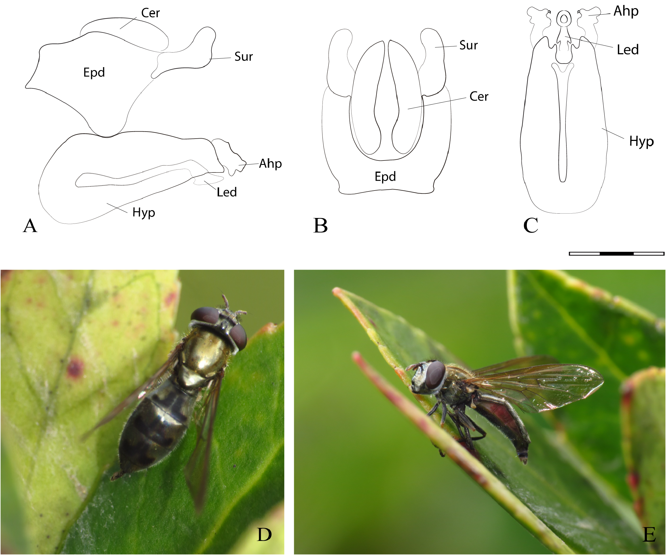
FIGURE 60. Argentinomyia opaca (Fluke, 1945), male genitalia. A. Whole genitalia, lateral view; B. Epandrium, dorsal view; C. Hypandrium, ventral view. Scale bars: 1 mm. D–E. Natural habitus of a female specimen at the Páramo ecosystem Santa Inés Belmira in Antioquia, Colombia: D. Dorsal view; E. Lateral view.
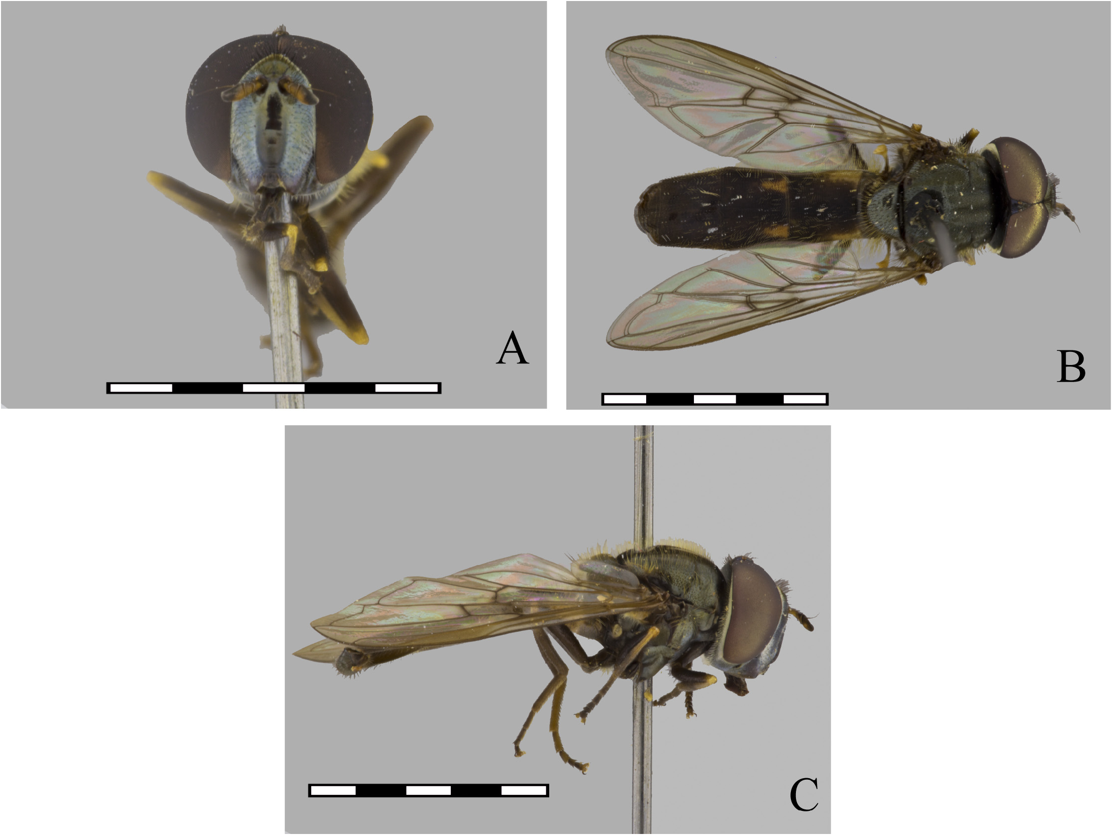
FIGURE 61. Argentinomyia plaumanni sp. nov., male Holotype (USNM ENT 00036936): A. Head, frontal view; B. Dorsal view; C. Lateral view. Scale bars: 5 mm.
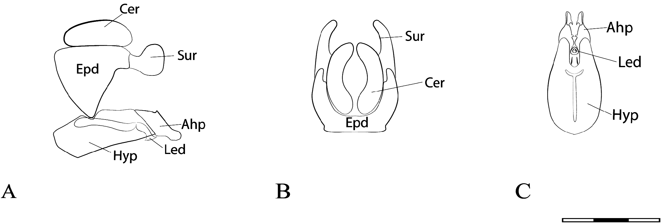
FIGURE 62. Argentinomyia plaumanni sp. nov. male Holotype, genitalia. A., Whole genitalia, lateral view; B. Epandrium, dorsal view; C. Hypandrium, ventral view. Scale bars: 1 mm.
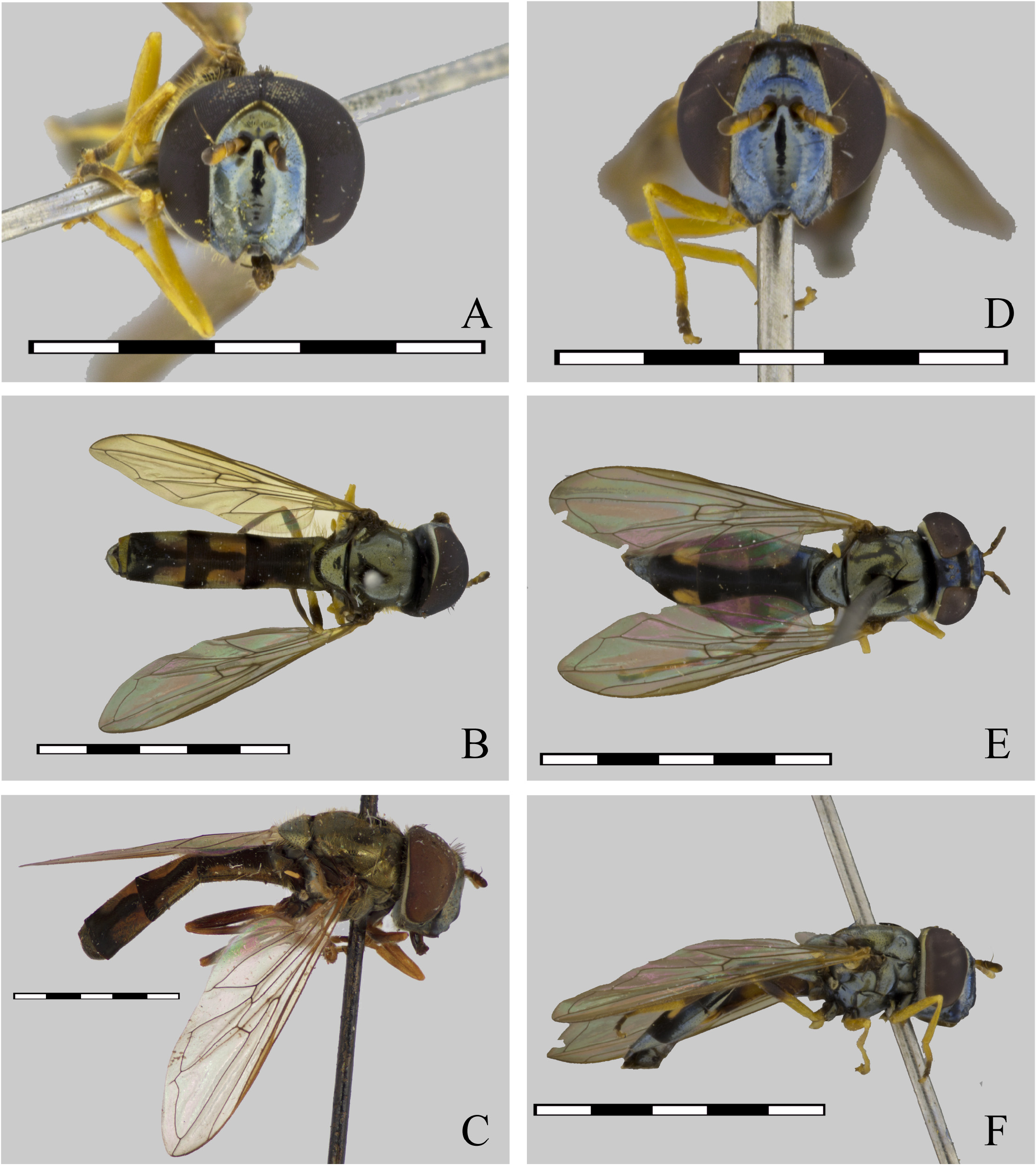
FIGURE 63. Argentinomyia pollinosa (Hull, 1942), male (USNM ENT 01406570): A. Head, frontal view; B. Dorsal view; C. Lateral view. Female (USNM ENT 01406570): D. Head, frontal view; E. Dorsal view; F. Lateral view. Scale bars: 5 mm.
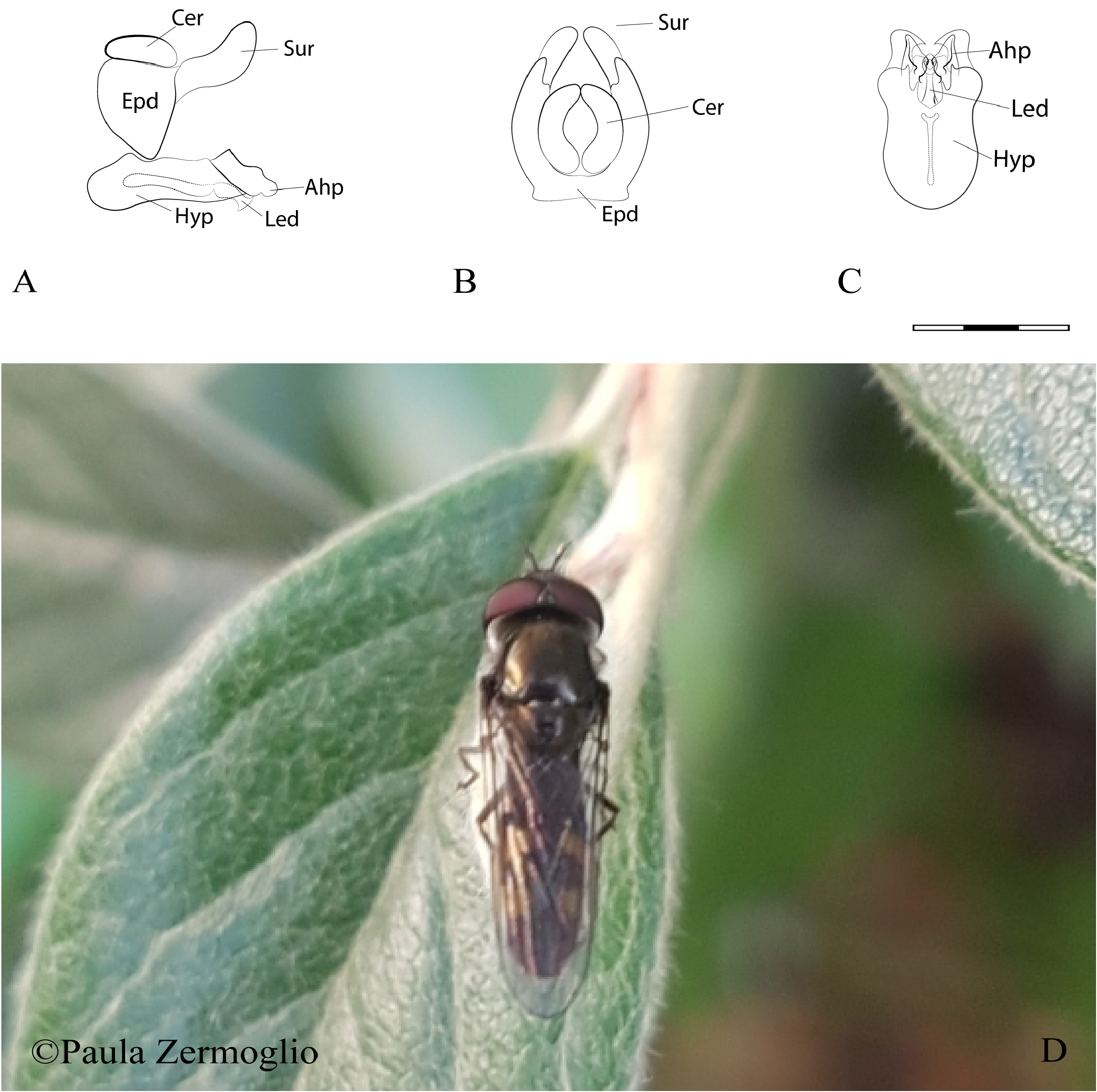
FIGURE 64. Argentinomyia pollinosa (Hull, 1942), male genitalia. A. Whole genitalia, lateral view; B. Epandrium, dorsal view; C. Hypandrium, ventral view. Scale bars: 1 mm. D. Natural habitus of a male specimen in Mozart 424, San Carlos de Bariloche, Río Negro, Argentina (iNaturalist catalogue number 37828673; https://www.inaturalist.org/observations/37828673). Photo taken by ©Paula Zermoglio.
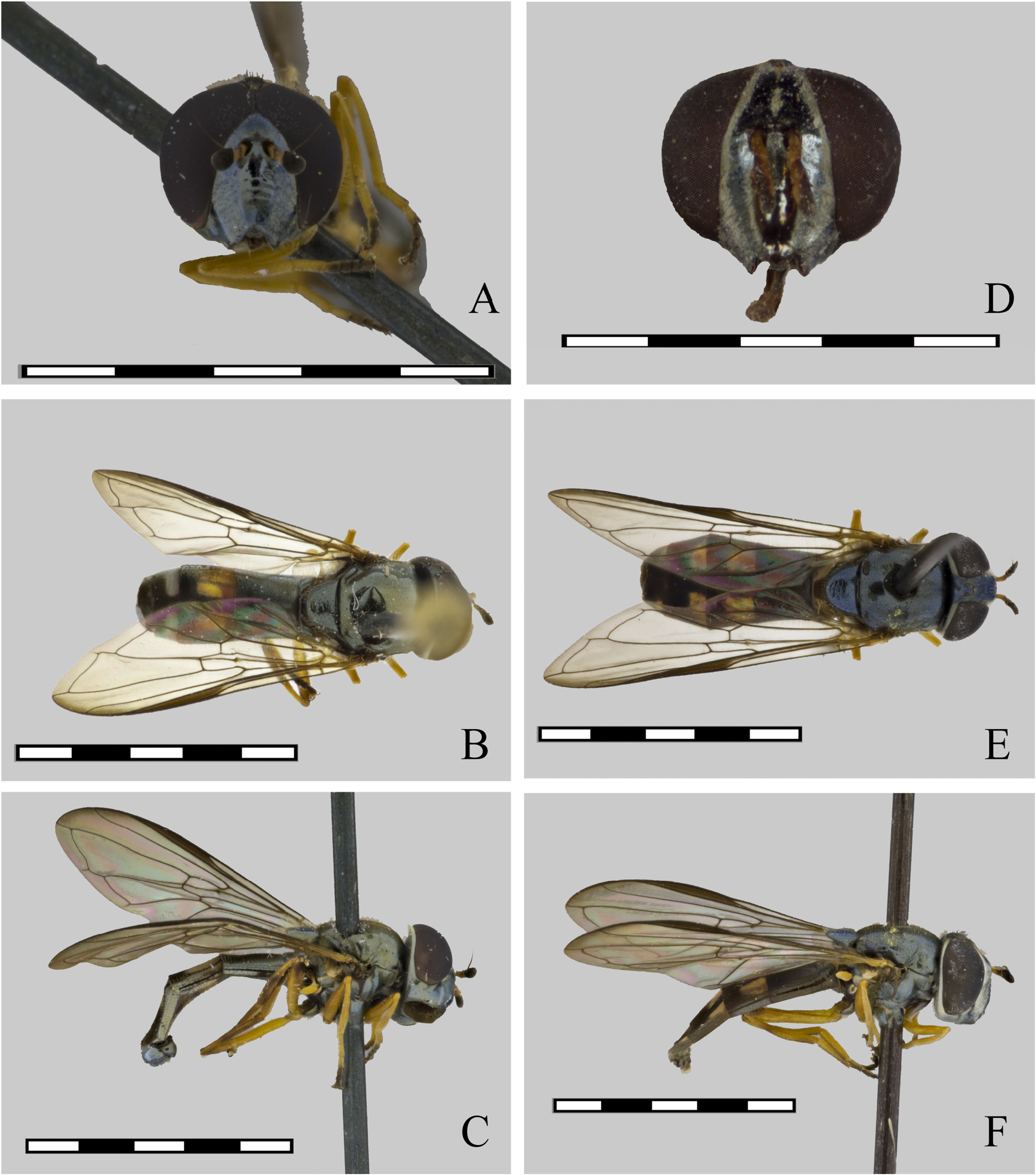
FIGURE 65. Argentinomyia praeusta (Loew, 1866), male (USNM ENT 01443776): A. Head, frontal view; B. Dorsal view; C. Lateral view. Female (USNM ENT 01443777): D. Head, frontal view; E. Dorsal view; F. Lateral view. Scale bars: 5 mm.
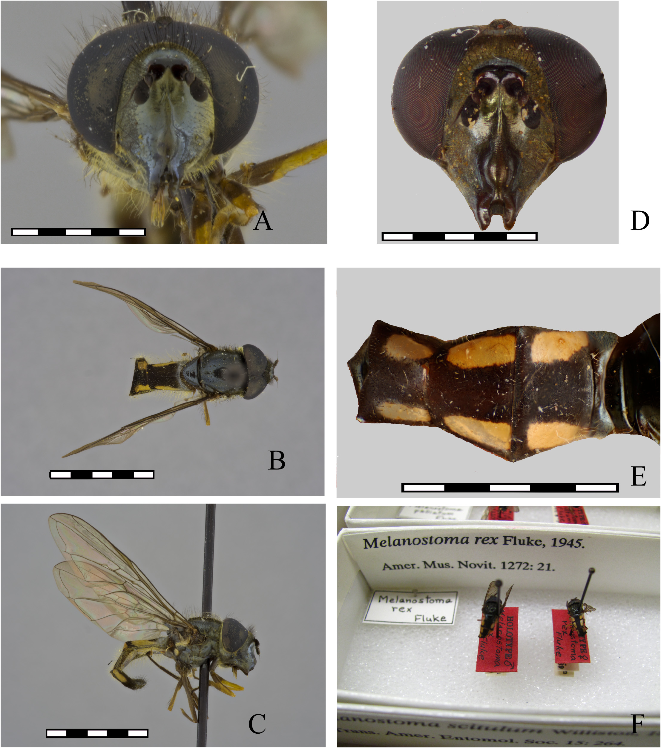
FIGURE 67. Argentinomyia rex (Fluke, 1945), male (CEUA 87097): A. Head, frontal view; B. Dorsal view; C. Lateral view. Female (AMNH): D. Head, frontal view; E. Dorsal view; F. Holotype and Allotype. Scale bars: 5 mm.
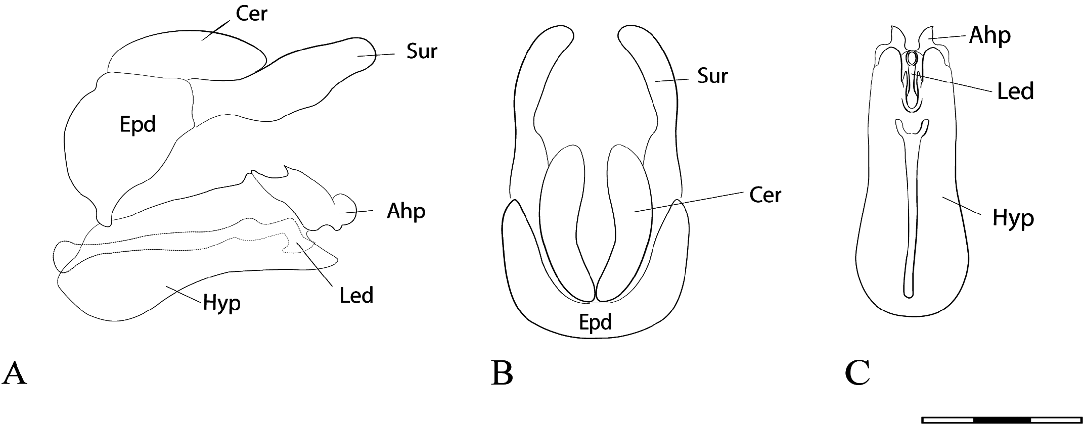
FIGURE 68. Argentinomyia rex (Fluke, 1945), male genitalia. A. Whole genitalia, lateral view; B. Epandrium, dorsal view; C. Hypandrium, ventral view. Scale bars: 1 mm.

FIGURE 69. Argentinomyia rugosonasa (Williston, 1891), male: A. Head, frontal view (INBio CRI 002452405); B. Dorsal view (ECO–TAP–E 24469); C. Lateral view (INBio CRI 002452405). Female (INBio CRI 0003753634): D. Head, frontal view; E. Dorsal view; F. Lateral view. Scale bars: 5 mm. [Note: The coloration of female legs in figure 69D is mainly due to the conservation of the specimen]
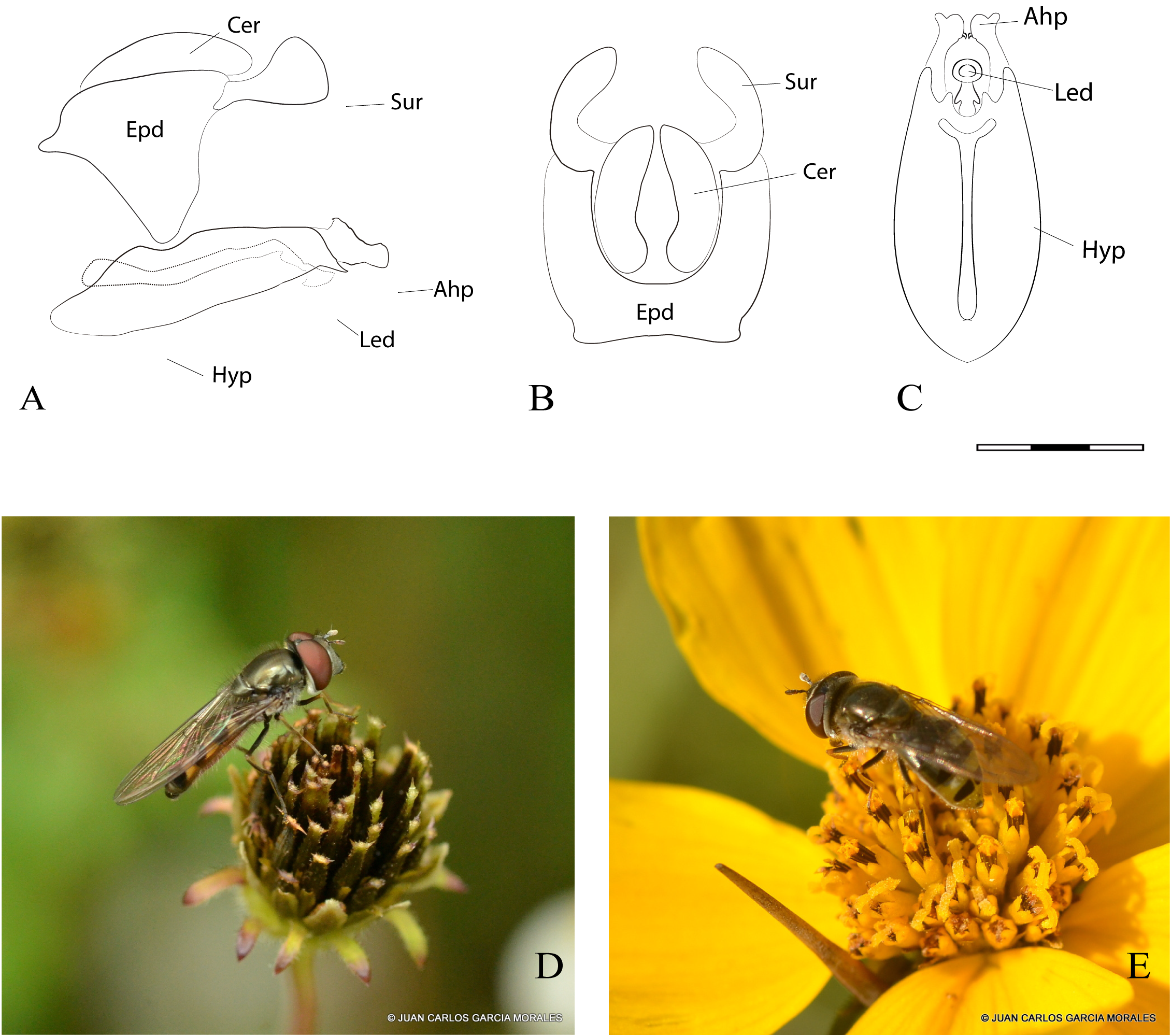
FIGURE 70. Argentinomyia rugosonasa (Williston, 1891), male genitalia. A. Whole genitalia, lateral view; B. Epandrium, dorsal view; C. Hypandrium, ventral view. Scale bars: 1 mm. D. Natural habitus of a male specimen in El Calvario, Toluca, México (iNaturalist catalogue number 17119892; https://www.inaturalist.org/observations/17119892); E. Natural habitus of a female specimen in Toluca de Lerdo, México (iNaturalist catalogue number 8506135; https://www.inaturalist.org/ observations/8506135). Photos taken by ©Juan Carlos García Morales.
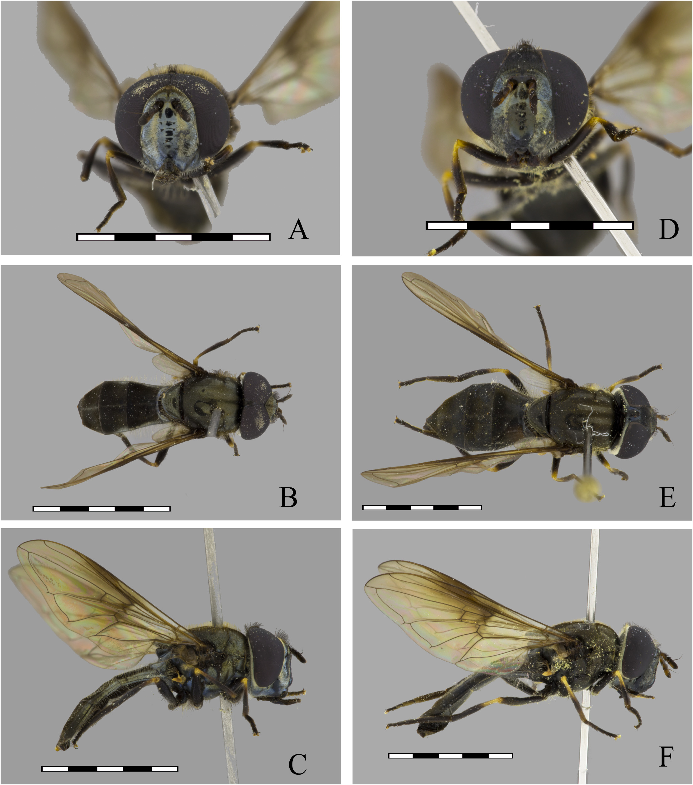
FIGURE 71. Argentinomyia sagoti sp. nov., male Holotype (ECOSCE): A. Head, frontal view; B. Dorsal view; C. Lateral view. Female (ECOSCE 24470): D. Head, frontal view; E. Dorsal view; F. Lateral view. Scale bars: 5 mm.
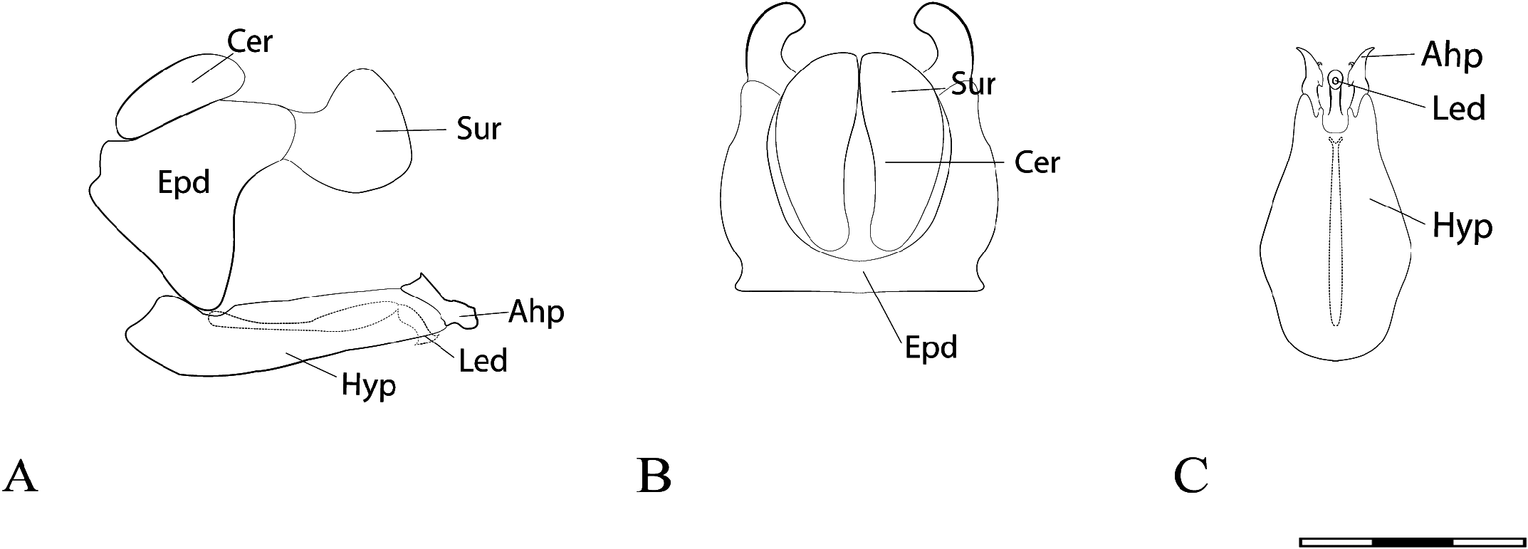
FIGURE 72. Argentinomyia sagoti sp. nov., male Holotype, genitalia. A. Whole genitalia, lateral view; B. Epandrium, dorsal view; C. Hypandrium, ventral view. Scale bars: 1 mm.
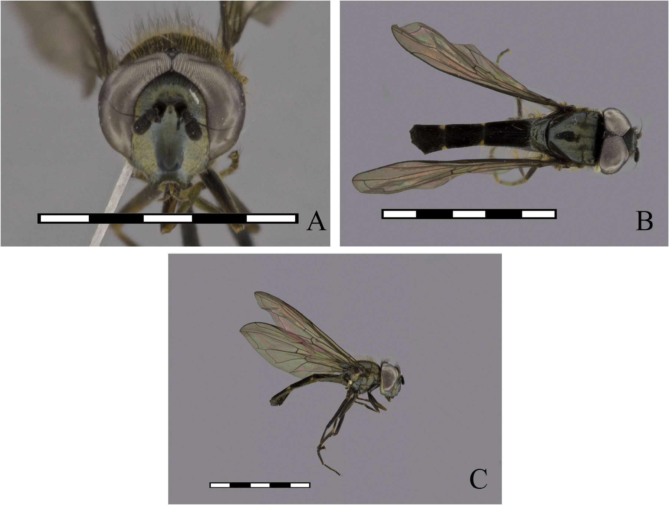
FIGURE 74. Argentinomyia serendipia sp. nov., male Holotype (CEUA 98284): A. Head, frontal view; B. Dorsal view; C. Lateral view.
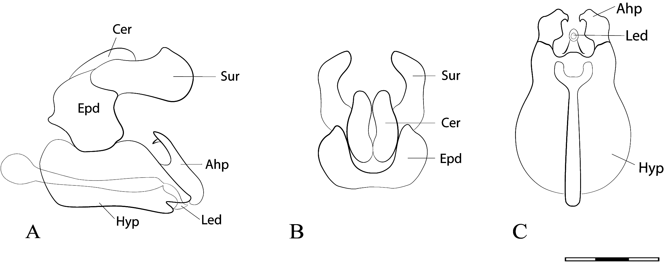
FIGURE 75. Argentinomyia serendipia sp. nov., male Holotype, genitalia. A. Whole genitalia, lateral view; B. Epandrium, dorsal view; C. Hypandrium, ventral view. Scale bars: 1 mm.
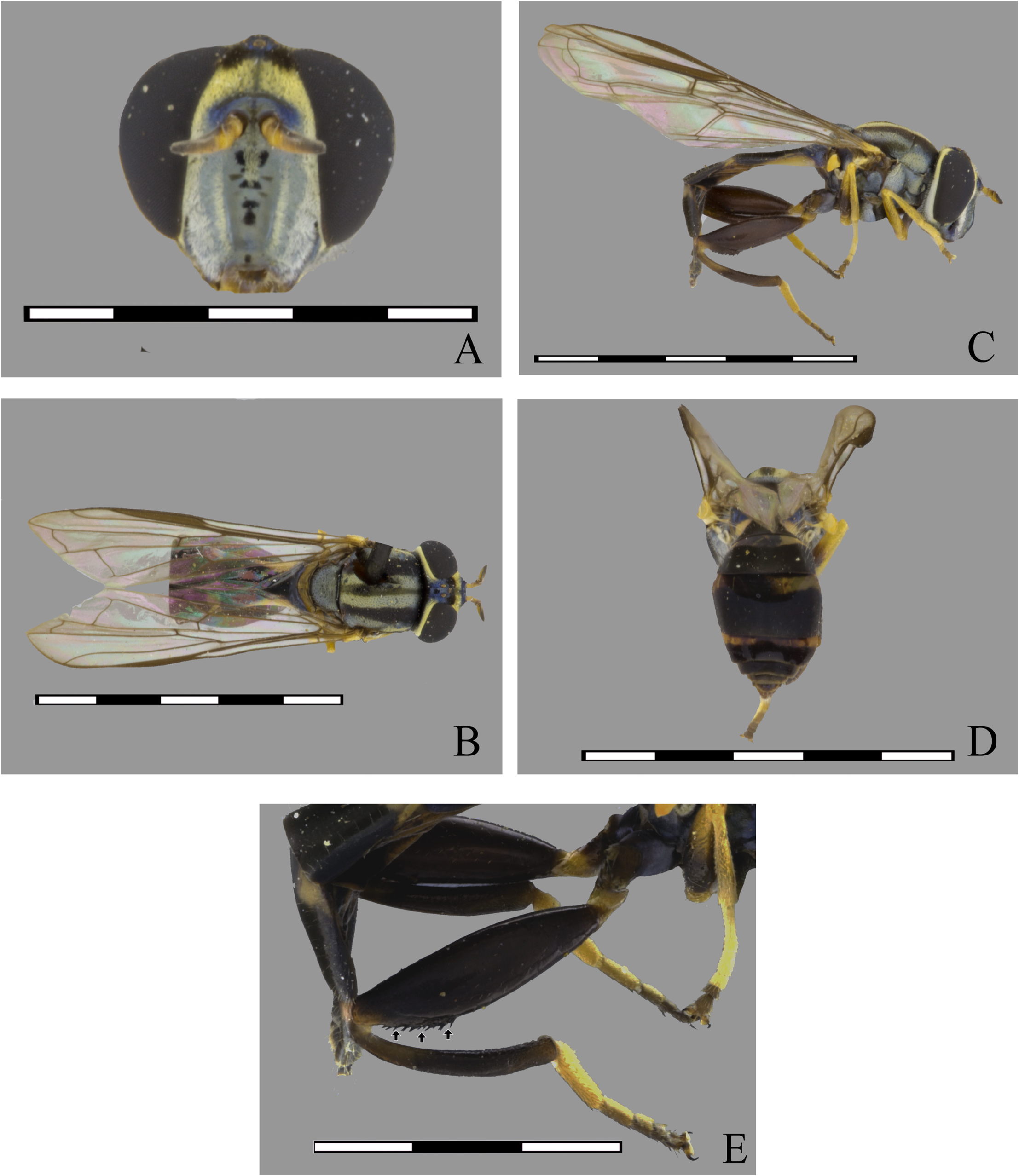
FIGURE 76. Argentinomyia spinifemorata sp. nov., female Holotype (MEFLG 27270): A. Head, frontal view; B. Dorsal view; C. Lateral view; D. Vista posterodorsal. Scale bars: 5 mm. E. Posterior leg, detail of ventral spines (Black arrows indicating ventral spines). Scale bars: 2 mm.
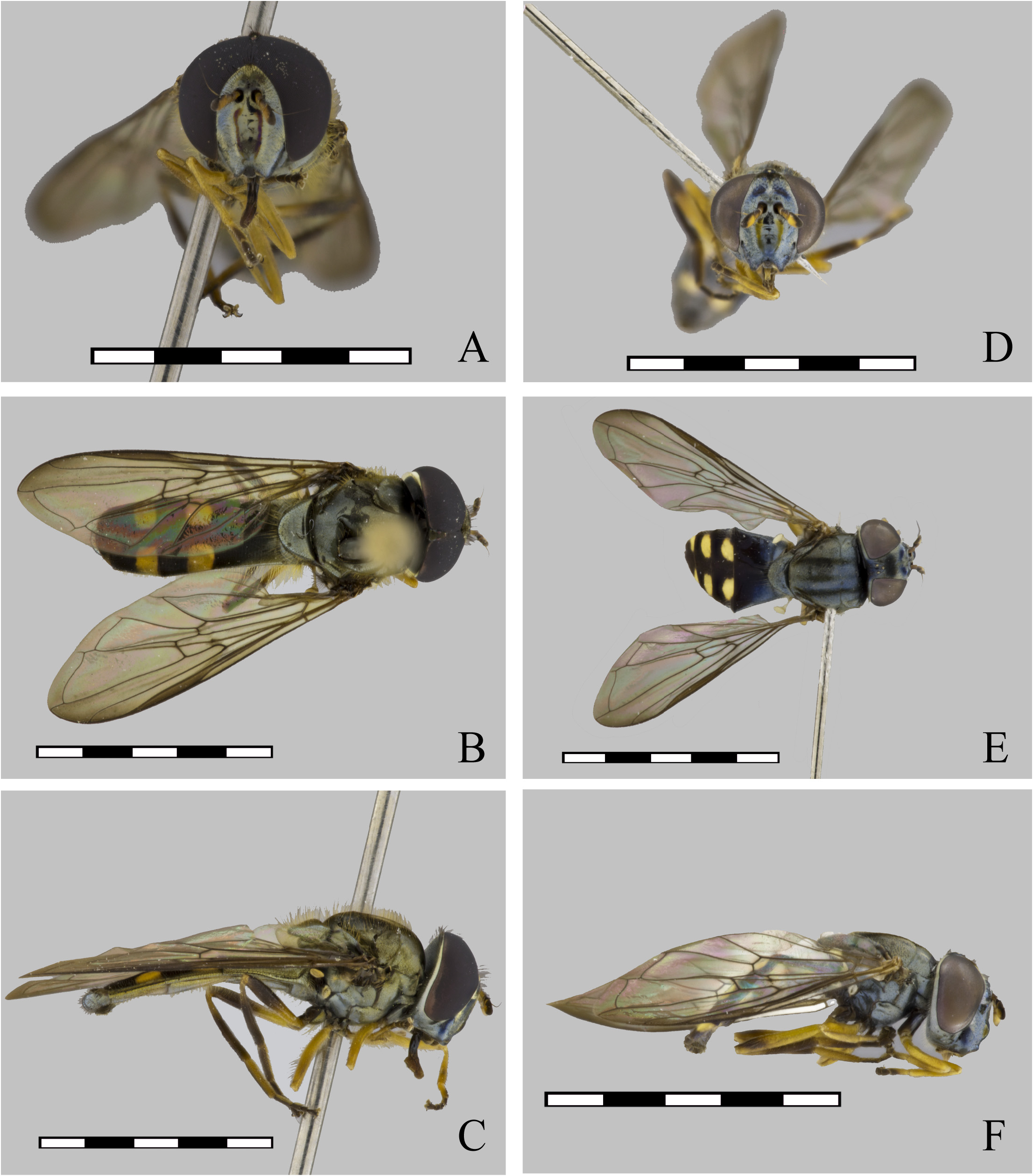
FIGURE 77. Argentinomyia taina sp. nov., male Holotype (USNM ENT 01443645): A. Head, frontal view; B. Dorsal view; C. Lateral view. Female (USNM ENT 01443644): D. Head, frontal view; E. Dorsal view; F. Lateral view. Scale bars: 5 mm.
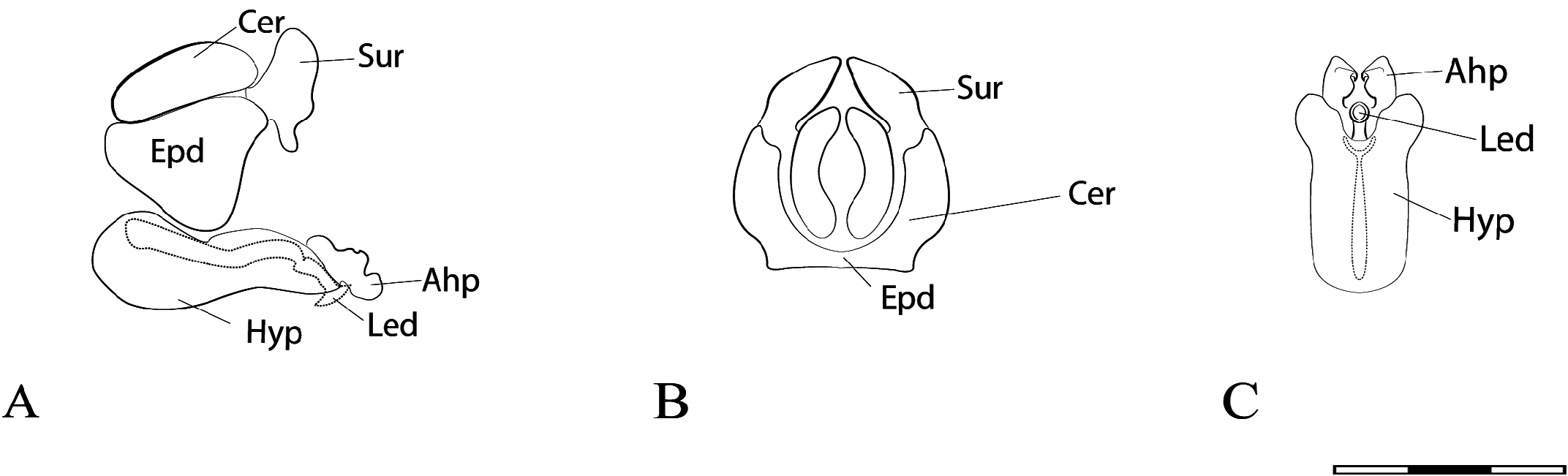
FIGURE 78. Argentinomyia taina sp. nov., male Holotype, genitalia. A. Whole genitalia, lateral view; B. Epandrium, dorsal view; C. Hypandrium, ventral view. Scale bars: 1 mm.
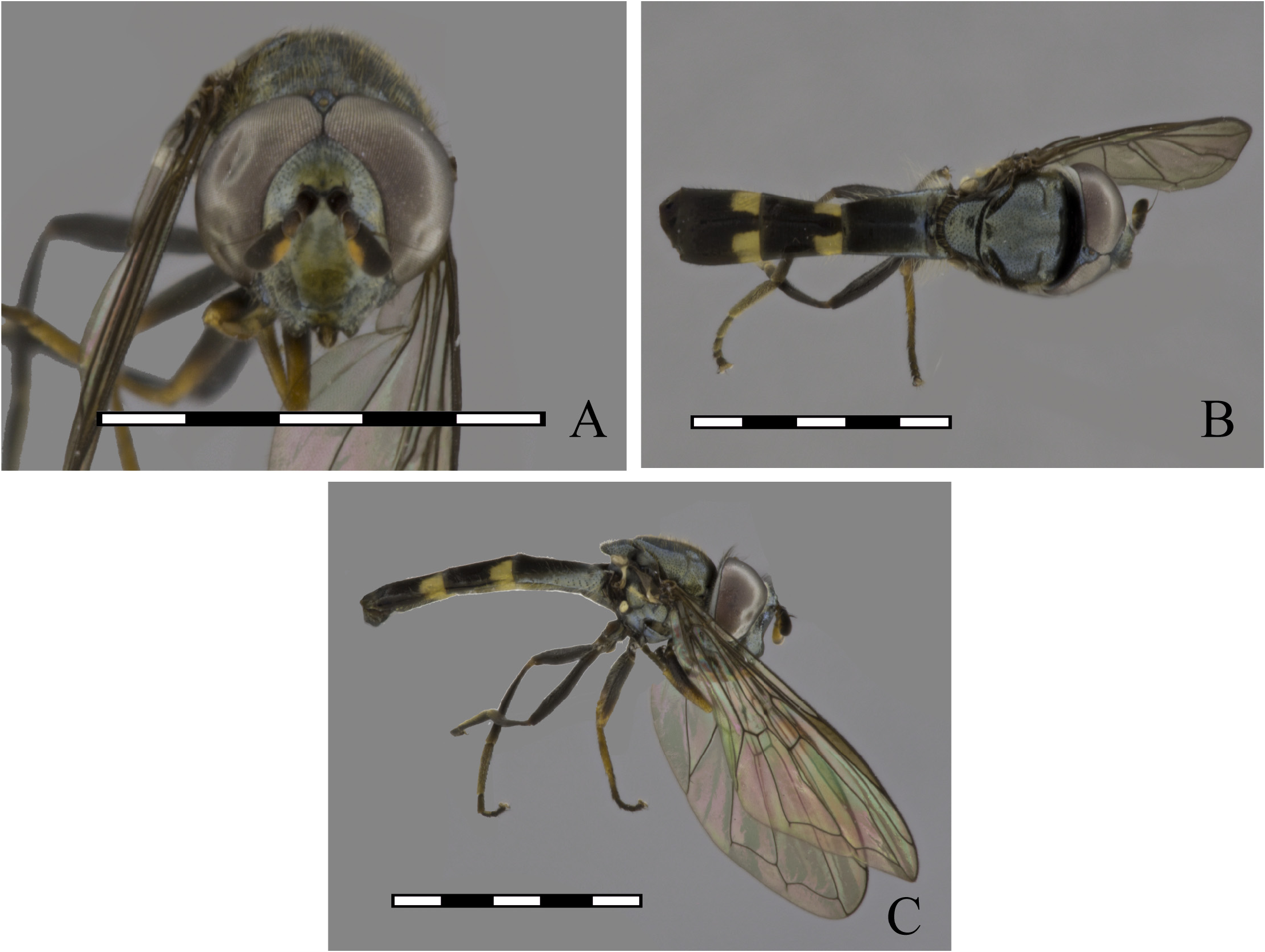
FIGURE 79. Argentinomyia teresae sp. nov., male Holotype (CEUA 98384): A. Head, frontal view; B. Dorsal view; C. Lateral view.
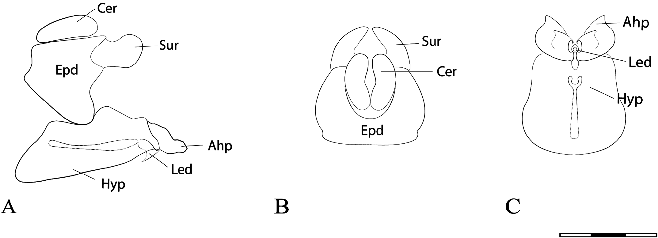
FIGURE 80. Argentinomyia teresae sp. nov., male Holotype, genitalia. A. Whole genitalia, lateral view; B. Epandrium, dorsal view; C. Hypandrium, ventral view. Scale bars: 1 mm.
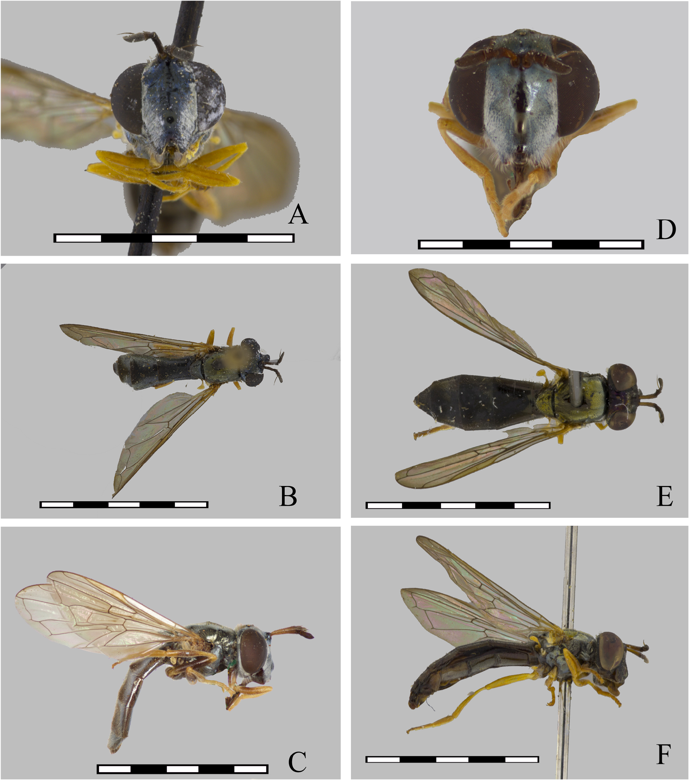
FIGURE 81. Argentinomyia testaceipes Lynch Arribálzaga, 1891, male (USNM ENT 01443767): A. Head, frontal view; B. Dorsal view. Female (CEUFLA): C. Cabeza, Lateral view D. Head, frontal view; E. Dorsal view; F. Lateral view. Scale bars: 5 mm.
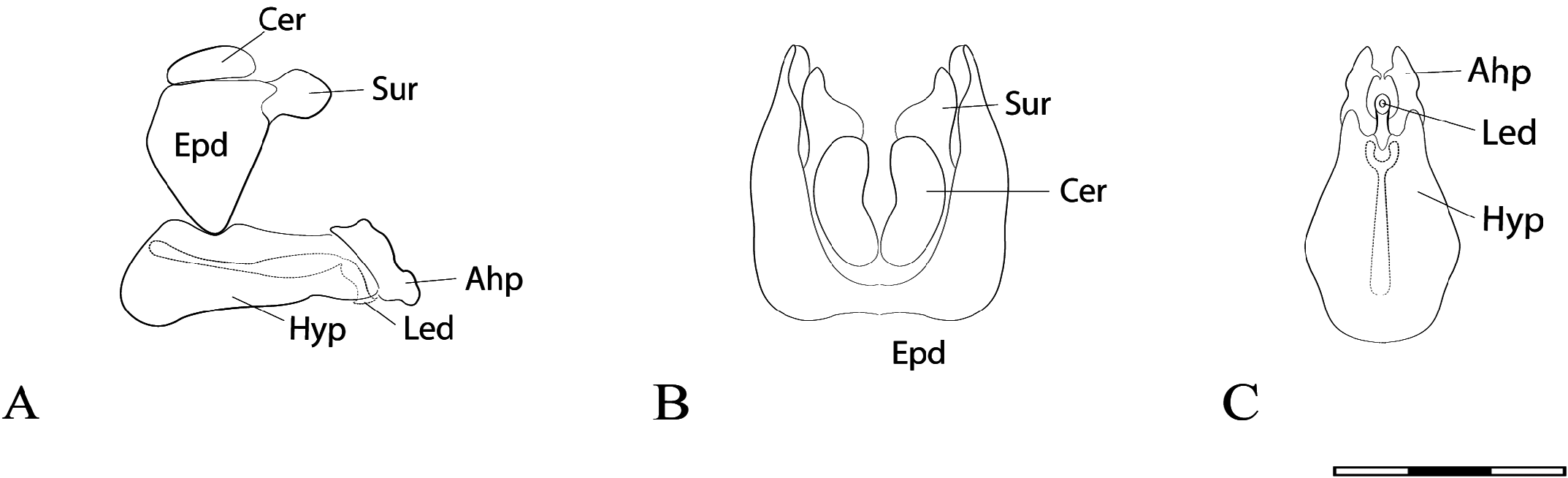
FIGURE 82.Argentinomyia testaceipes LynchArribálzaga, 1891, male genitalia.A. Wholegenitalia, lateral view; B. Epandrium, dorsal view; C. Hypandrium, ventral view. Scale bars: 1 mm.
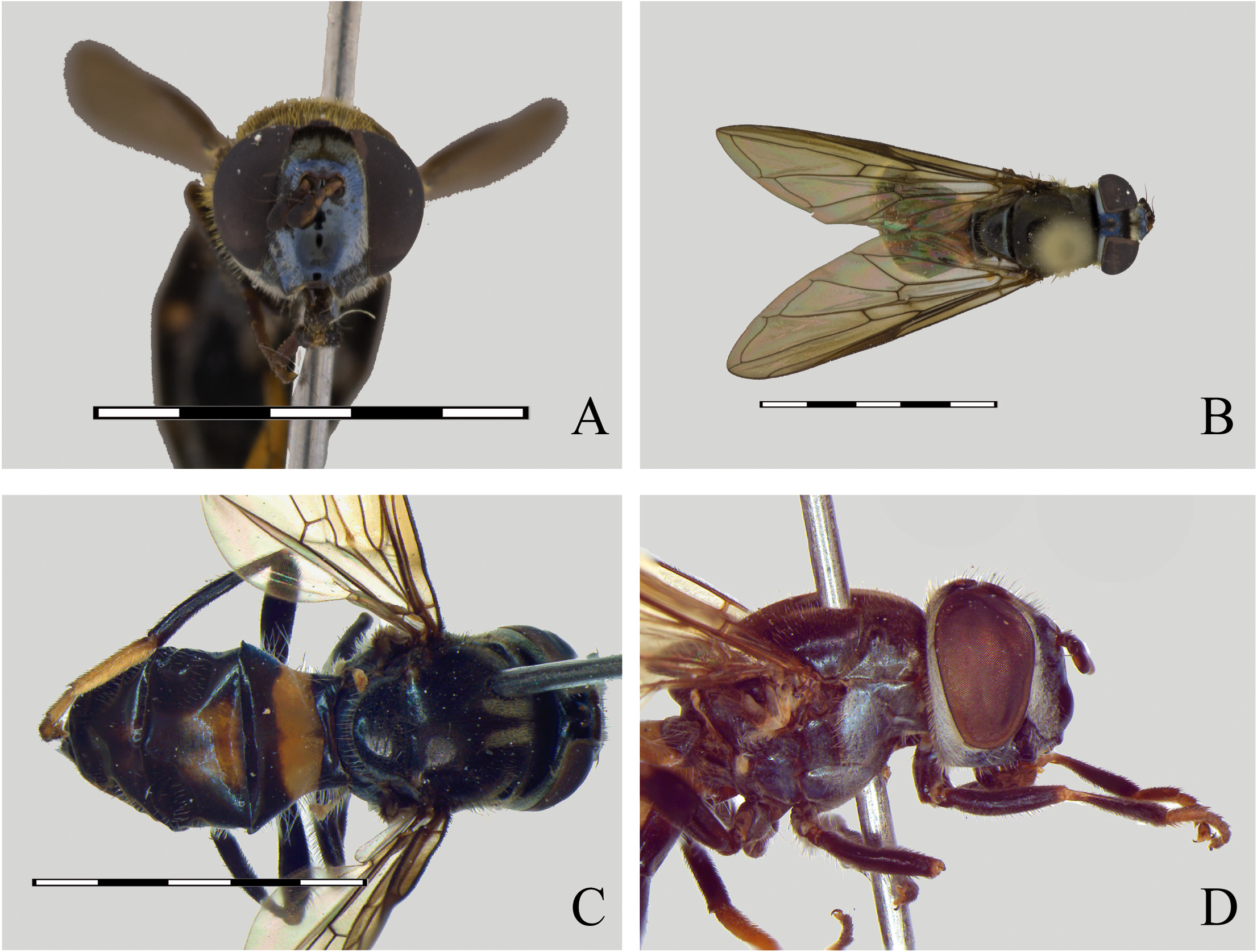
FIGURE 83. Argentinomyia transversalis sp. nov., female. A. Head, frontal view; B. Dorsal view (USNMENT 00055996). C. Posterior view, detail of maculae pattern on abdomen; D. Lateral view (CEUA 87110). Scale bars: 5 mm.
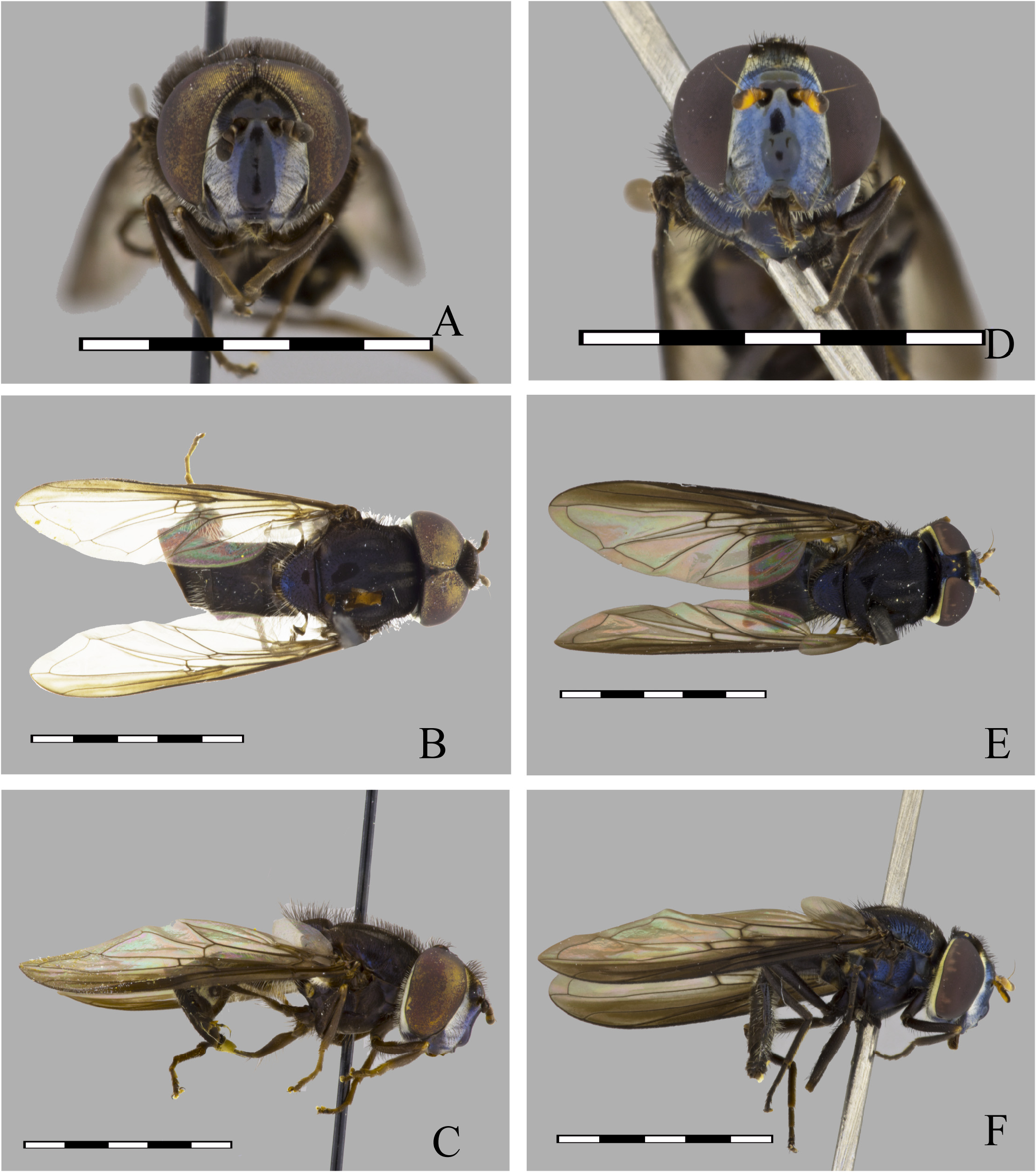
FIGURE 84. Argentinomyia tropandeana sp. nov., male Holotype (CEUA 13210): A. Cabeza, Vista frontal; B. Dorsal view; C. Lateral view. Female (QCAZ 103714): D. Head, frontal view; E. Dorsal view; F. Lateral view. Scale bars: 5 mm.
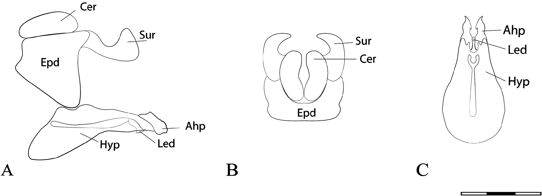
FIGURE 85. Argentinomyia tropandeana sp. nov., male Holotype, genitalia. A. Whole genitalia, lateral view; B. Epandrium, dorsal view; C. Hypandrium, ventral view. Scale bars: 1 mm.
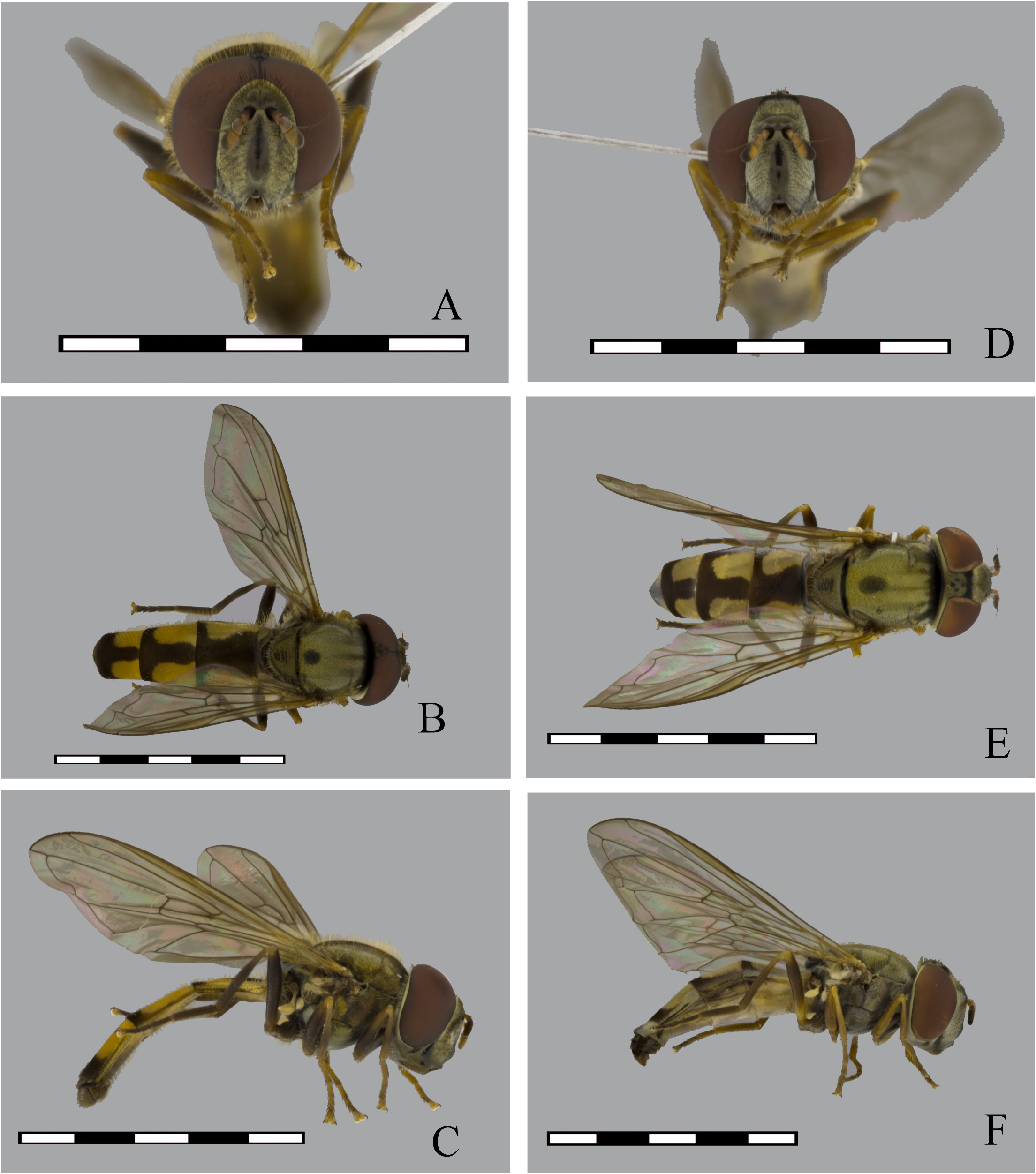
FIGURE 86. Argentinomyia tropica (Curran, 1937), male (CEUA 93084): A. Head, frontal view; B. Dorsal view; C. Lateral view. Female (CEUA 69474): D. Head, frontal view; E. Dorsal view; F. Lateral view. Scale bars: 5 mm.
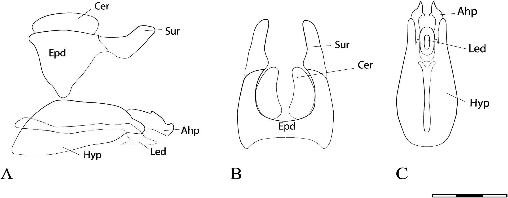
FIGURE 87. Argentinomyia tropica (Curran, 1937), male genitalia. A. Whole genitalia, lateral view; B. Epandrium, dorsal view; C. Hypandrium, ventral view. Scale bars: 1 mm.
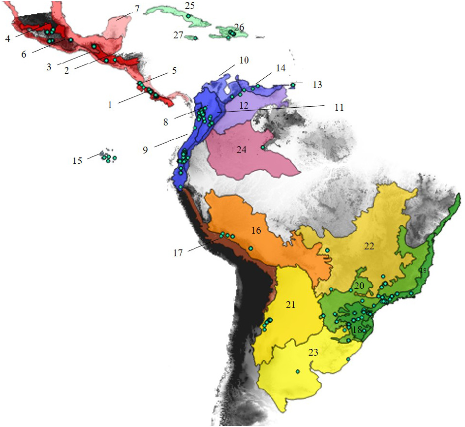
FIGURE 88. Biogeographic distribution of Argentinomyia by domains and provinces in the Neotropics: A= Mesoamerican or Central America [Red] (1= Puntarenas-Chiriquí, 2= Chiapas Highlands, 3= Pacific Lowlands, 4= Trans-Mexican Volcanic Belt Pacific, 5= Guatuso-Talamanca, 6= Sierra Madre del Sur, 7= Veracruzan); B= North Andes [Blue] (8= Magdalena, 9= Cauca, 10= Guajira, 11= North Andean Páramo, 12= Sabana, 13= Trinidad, 14= Venezuelan, 15= Galápagos Islands); C= Central Andes [Orange] (16= Rondônia, 17= Yungas); D= Paraná [Green] (18= Araucaria Forest, 19= Atlántico Forest, 20= Paraná Forest); E= Chaco [Yellow] (21= Chaco 22= Cerrado, 23= Pampa); F= Amazonía [Purple] (24= Imerí); G= West Indies [Light blue] (25= Cuba, 26= Hispaniola, 27= Jamaica). The biogeographical provinces follow the proposal of Morrone (2014). The light blue dots represent the distribution of Argentinomyia. The color intensity represents the coincidence of occurrence between Argentinomyia species associated with a specific province.


No known copyright restrictions apply. See Agosti, D., Egloff, W., 2009. Taxonomic information exchange and copyright: the Plazi approach. BMC Research Notes 2009, 2:53 for further explanation.