
Afrogethes Audisio & Cline, 2009
|
publication ID |
https://doi.org/10.5281/zenodo.5319334 |
|
persistent identifier |
https://treatment.plazi.org/id/03BE87CC-F648-FFAC-BA9C-FBE3FD5CF9A6 |
|
treatment provided by |
Felipe (2021-08-28 07:26:47, last updated by Plazi 2023-11-05 05:53:56) |
|
scientific name |
Afrogethes Audisio & Cline |
| status |
gen. nov. |
10. Afrogethes Audisio & Cline View in CoL View at ENA , gen. nov.
( Figs. 10 a–h View Fig )
Type species. Meligethes reticulatus Reitter, 1872: 243 , 253 (by present designation) [= Afrogethes reticulatus (Reitter, 1872) comb. nov.].
Generic description and diagnosis. Inclusive species vary greatly in size (1.4–4.4 mm length), and share the following combination of characters.
Body color and pubescence: pubescence silvery-whitish, highly variable, short and fine, faintly distinct to long and dense, recumbent, in a few species partly obscuring the blackish (rarely reddish-brown) dorsal body surface; pronotal and elytral sides narrowly flattened, typically the same color as disc. Lateral margin of pronotum and elytra with a series of more or less distinct, small and short setae, each seta usually 0.3–0.5× as long as those on elytral disc; posterior margin of pronotum comprising moderately long, usually distally trifid to multifid and stellate microsetae, microsetae uniformly distributed on middle region anterior to scutellum ( Fig. 10e View Fig ).
Dorsal habitus: body more or less convex, highly variable in shape ( Figs. 10a, k View Fig ; Figs. 1–13 View Fig View Fig View Fig View Fig View Fig View Fig View Fig View Fig View Fig View Fig View Fig View Fig View Fig , 15– 16 View Fig View Fig in AUDISIO 1997b); dorsal punctures on discal portion of pronotum as large as or larger than eye facets, usually deeply impressed and densely distributed, rarely fine, sparse, and shallow; anterior margin of clypeus usually moderately arcuately emarginate, distinctly and narrowly bordered ( Fig. 10b View Fig ), usually with a small, faintly distinct medial bulge, slightly protruding anteriorly; circum-ocular furrows (occipital sulci) on dorsal side of head not developed, absent or indistinct ( Fig. 10b View Fig ); eyes large and usually moderately projecting laterally ( Figs. 10a, b, d, k View Fig ); pronotum with distinct obtuse posterior angles, never posteriorly directed ( Figs. 10a, k View Fig ); scutellum more or less regularly and sparsely punctured at least in posterior half of exposed portion ( Fig. 10k View Fig ); elytra with highly variable punctation, completely transversely strigose or with simple punctures; elytral humeral angle moderately distinct, not protruding laterally ( Figs. 10a, k View Fig ); elytral humeral striae usually distinct; elytral pre-sutural striae visible, originating slightly posterior to scutellar vertex, terminating close to elytral apex, and delimiting on each elytron a more or less distinct, flat, unraised sutural border, widest at posterior third and nearly as wide as proximal portion of 3 rd antennomere; elytral apices truncately rounded in both sexes ( Figs. 10a, k View Fig ); pygidium partially exposed, moderately convex, apically rounded in both sexes ( Figs. 10a, k View Fig ).
Ventral habitus: antennal furrows markedly delimited, nearly parallel-sided, slightly divergent posteriorly; mentum subpentagonal ( Fig. 10d View Fig ); prosternal antennal furrows of anterior margin of prosternum more or less strongly raised but relatively short ( Fig. 10d View Fig ); prosternal process variable, usually relatively narrow, subapical dilated portion 2.0–2.5× as wide as maximum width of 1 st antennomere, usually distally blunt ( Fig. 10c View Fig ); lateral borders of prosternal process delimiting moderately deeply impressed and distinct furrows, distally terminating over predistal lateral expansions, frequently nearly reaching posterior margin ( Fig. 10c View Fig ), which is usually microscopically denticulate; posterior margin of mesoventrite simple, never incised medially; male impressions on metaventrite moderately developed; first two visible abdominal ventrites simple in both sexes, without tufts of setae; caudal marginal lines of metacoxal cavities simple, moderately narrowly paralleling metacoxal cavities, comprising moderately deep arched impression of outer ‘axillary’ line ( Fig. 10g View Fig ); ‘axillary’ space on first abdominal ventrite moderately developed, ‘axillary’ angle usually broadly obtuse ( Fig. 10g View Fig ); relatively large, long, and deeply impressed arched impressions on basal portion of last visible abdominal ventrite, typically partially covered by distal portion of penultimate visible abdominal ventrite ( Fig. 10f View Fig ).
Appendages: male 1 st antennomere 0.8–0.9× as long as width of protibiae excluding distal teeth ( Figs. 10a, k View Fig ); 3 rd antennomere in both sexes usually only 2.0–2.1× as long as wide, 0.9–1.0× as long but distinctly thinner than 2 nd antennomere ( Fig. 10d View Fig ); 4 th and 5 th antennomeres in both sexes subequal, short, nearly as long as wide; antennal club compact, small, simple, comprising last 3 antennomeres in both sexes (8 th antennomere scarcely widened, 0.5–0.6× as wide as 9 th antennomere) ( Figs. 10a, d View Fig ), slightly or distinctly narrower than width of protibiae, sexual dimorphism absent; labial palpi relatively short in both sexes ( Fig. 10d View Fig ), terminal segment nearly1.8× as long as wide; maxillary palpi moderately long and slender in both sexes ( Fig. 10d View Fig ), terminal segment 2.1–2.2× as long as wide; mandible mid-sized ( Fig. 10d View Fig ), apex moderately acuminate, no sexual dimorphism present; tarsal claws highly variable, simple, bluntly toothed at base, or strongly and acutely toothed; tarsi of normal size and shape, 0.6–0.7× as long as corresponding tibiae ( Figs. 10a, k View Fig ); protibiae with a series of usually large, uneven, long and variably shaped (blunt to sharply acuminate) teeth on lateral margin ( Figs. 10a, k View Fig ; Figs. 18–19 View Fig View Fig and 23–32 View Fig View Fig View Fig View Fig View Fig View Fig View Fig View Fig View Fig in EASTON 1960; Figs. 129 f–l, m–n in AUDISIO 1993b; Figs. 17–29 View Fig View Fig View Fig View Fig View Fig View Fig View Fig View Fig View Fig View Fig View Fig View Fig View Fig in AUDISIO 1997b); meso- and metatibiae on lateral margin bearing a single and usually moderately even row of large and robust pegs ( Fig. 10h View Fig ), without U-shaped sinuosity at distal third; meso- and metatibiae of variable width, usually moderately slender and narrow ( Figs. 10a, h, k View Fig ), never subtrapezoidal or axe-shaped; male metatibiae more sinuate than in females; tarsal plates of prolegs usually distinctly wider in males; posterior margin of metafemora simple in both sexes, without tubercles or projections.
Male genitalia: processes along inner side of parameres absent (Figs. 70–73 and 76–93 in EASTON 1960; Figs. 140 and 141 m –p in AUDISIO 1993b; Figs. 30–80 in AUDISIO 1997b), with more or less deeply incised or truncate distal margin, without deep median longitudinal desclerotization from proximal portion of tegmen extending to medial distal V-shaped excision; median lobe of aedeagus variable, without lateral emargination, narrowed and variably shaped distally.
Female genitalia (ovipositor): highly variable in shape, usually large; styli long and pigmented, or short and unpigmented, simple, cylindrical. Afrogethes howdeni ( Kirejtshuk, 1990) from South Africa with entire ovipositor absent and replaced by an unsclerotized tubular structure ( AUDISIO 1997b). Afrogethes africanus ( Kraatz, 1895) from Western Africa, with abruptly and widely truncate apex (AUDISIO unpublished data). Styli situated close to apex of usually contiguous gonostyloids, each gonostyloid lightly sclerotized and not darkly pigmented distally. Outer portion of basicoxites simple, never indentate (Figs. 112–116 and 118–127 in EASTON 1960; Figs. 157 a–f in AUDISIO 1993b; Figs. 81–93 in AUDISIO 1997b), with a single, narrow, scarcely pigmented and sclerotized arcuate area along outer subdistal portion of gonostyloids. ‘Central point’ of ovipositor usually more distad than middle, with or without proximad directed spicule.
Etymology. The generic name is derived from the Latin ‘ africanus ’ (= African), to emphasize the probable African origin of the ancestor for all inclusive species and species-groups, and from ‘- gethes ’, to emphasize its phylogenetic relationship with Meligethes . Gender masculine.
Biology. The biology of most inclusive species is only partially known, and likely being heterogeneously expressed within this large taxon. Members of the large Afrogethes reticulatus / forcipatus / coronatus species-group are likely all associated with flowers of Verbenaceae , while members of the A. planiusculus species-group are all associated with the related Boraginaceae , in particular with Echium L., Trichodesma R. Brown and allied genera ( AUDISIO 1993b; WILLIAMS 2002; AUDISIO et al. 2009b). Members of the large African A. amplicollis and A. aethiopicus species-groups are, on the contrary, all associated with Fabaceae , except the single southern African species, A. breithenbachae Audisio, 1997 , which is likely associated ( AUDISIO 1997b, AUDISIO & DE BIASE 2004a) with phylogenetically related Polygalaceae ( JUDD et al. 1994, 2002).
Phylogenetic position. Available molecular and morphological datasets provide moderately concordant evidence for a possible monophyletic clade including Afrogethes gen. nov., as well as Fabogethes gen. nov., Genistogethes gen. nov., Sagittogethes gen. nov., and Thymogethes gen. nov. ( AUDISIO et al. 2009b, TRIZZINO et al. 2009). However, phylogenetic relationships between these taxa and between this clade and Aristogethes gen. nov. are still unclear.
Taxonomy and geographic distribution. Afrogethes gen. nov. is the largest genus of the ‘ Meligethes s. l. ’ generic complex, including ~120 described and some additional 30 undescribed species. The genus is mainly distributed in Tropical Africa and Madagascar, with a few species-groups marginally penetrating into Europe, eastern Asia, and North America ( AUDISIO et al. 2009b). The tentatively included species below are attributed to at least five formerly recognized species-groups, i.e. the ‘ Meligethes reticulatus / forcipatus / coronatus ’,
‘ M. planiusculus ’, ‘ M. aethiopicus ’, ‘ M. voeltzkowi ’, and ‘ M. amplicollis ’ species-groups (EAS- TON 1954b, 1959b, 1960; AUDISIO 1994, 1997b; KIREJTSHUK 2001; AUDISIO et al. 2009b).
Afrogethes abstractus (Grouvelle, 1908) comb. nov. Ethiopia, Eritrea, Somaliland
Afrogethes adenensis (Easton, 1954) comb. nov. S Arabian Peninsula
Afrogethes adversus (Easton, 1959) comb. nov. Ethiopia
Afrogethes aethiopicus (Grouvelle, 1908) comb. nov. Central and S Africa
Afrogethes africanus ( Kraatz, 1895) comb. nov. Togo
Afrogethes alani (Kirejtshuk, 1988) comb. nov. South Africa: KwaZulu-Natal, Mpumalanga Afrogethes alluaudi ( Grouvelle, 1896) comb. nov. Gambia, Senegal
Afrogethes altercatio (Easton, 1959) comb. nov. Ethiopia
Afrogethes amplicollis ( Boheman, 1851) comb. nov. South Africa: KwaZulu-Natal, E Cape
Afrogethes amplimanus ( Easton, 1960) comb. nov. Tanzania
Afrogethes annae (Kirejtshuk, 1996) comb. nov. South Africa: KwaZulu-Natal
Afrogethes arabicus ( Jelínek, 1988) comb. nov. Arabian Peninsula
Afrogethes arcopenis (Kirejtshuk, 1996) comb. nov. Namibia
Afrogethes aspalathi (Audisio & De Biase, 2004) comb. nov. South Africa: W and E Cape
Afrogethes assutus ( Easton, 1960) comb. nov. Kenya
Afrogethes astylus ( Easton, 1960) comb. nov. Kenya
Afrogethes attactus ( Kirejtshuk & Viklund, 2002) Kenya comb. nov.
Afrogethes basicollis (Easton, 1964) comb. nov. South Africa: NW Province
Afrogethes bocaki ( Audisio, Jelínek & Cooter, 2005) S China comb. nov.
Afrogethes breitenbachae (Audisio, 1997) comb. nov. South Africa: W Cape
Afrogethes breviusculus ( Kraatz, 1895) comb. nov. Tropical Africa
Afrogethes brittoni (Easton, 1954) comb. nov. S Arabian Peninsula
Afrogethes buduensis (Ganglbauer, 1899) comb. nov. E Mediterranean areas, SE Europe, Middle East, W Middle Asia
Afrogethes canadensis (Easton, 1955) comb. nov. W North America
Afrogethes canariensis (Kirejtshuk, 1997) comb. nov. Canary Islands
Afrogethes capensis (Reitter, 1872) comb. nov. South Africa: E Cape, KwaZulu-Natal
Afrogethes chevrolati (Reitter, 1872) comb. nov. South Africa: W Cape
Afrogethes clavatus (Reitter, 1872) comb. nov. South Africa: W Cape
Afrogethes clypeonitens ( Easton, 1960) comb. nov. Kenya
Afrogethes colophonoides (Audisio, 1997) comb. nov. South Africa: W Cape
Afrogethes conformis ( Spornraft & Kirejtshuk, 1993) South Africa: E Cape comb. nov.
Afrogethes cornutus ( Easton, 1960) comb. nov. Kenya, Tanzania, Uganda
Afrogethes coronatus (Easton, 1959) comb. nov. Ethiopia
Afrogethes curtulus ( Grouvelle, 1916) comb. nov. Southern Central Africa
(= Meligethes tutimanus Easton, 1964 )
Afrogethes dahlgreni (Audisio, 1997) comb. nov. South Africa: W Cape
Afrogethes debiasei (Audisio, 1997) comb. nov. South Africa: E Cape, KwaZulu-Natal
Afrogethes dentellus ( Spornraft & Kirejtshuk, 1993) Swaziland comb. nov.
Afrogethes desperatoides ( Audisio, 1994) comb. nov. Sierra Leone
Afrogethes desperatus (Easton, 1964) comb. nov. Congo
Afrogethes edwardsi ( Easton, 1960) comb. nov. Kenya, Uganda
Afrogethes elgonensis ( Easton, 1960) comb. nov. Uganda
Afrogethes exiguus ( Kirejtshuk, 1990) comb. nov. Ethiopia
Afrogethes fistuca ( Kirejtshuk & Viklund, 2002) comb. nov. Kenya
Afrogethes floralimimus (Audisio, 1997) comb. nov. South Africa: E Cape, KwaZulu-Natal
Afrogethes floralis (Reitter, 1872) comb. nov. South Africa: E Cape, KwaZulu-Natal
Afrogethes forcipatus ( Kirejtshuk & Easton, 1988) South Africa: KwaZulu-Natal, Free State comb. nov.
Afrogethes fossilis (Easton, 1959) comb. nov. Ethiopia
Afrogethes fritschii (Reitter, 1872) comb. nov. South Africa: W Cape
Afrogethes fruticola ( Spornraft & Kirejtshuk, 1993) South Africa: W Cape comb. nov.
Afrogethes grandicollis (Reitter, 1872) comb. nov. South Africa: E Cape?
Afrogethes gurjevae ( Kirejtshuk, 1984) comb. nov. Mongolia, E Siberia: Yakutia
Afrogethes howdeni ( Kirejtshuk, 1990) comb. nov. South Africa: KwaZulu-Natal
Afrogethes imperator (Easton, 1959) comb. nov. Ethiopia
Afrogethes inconspicuus ( Spornraft & Kirejtshuk, 1993) South Africa: Free State; Swaziland comb. nov.
Afrogethes isoplexidis ( Wollaston, 1854) comb. nov. Madeira
Afrogethes janczyki (Kirejtshuk, 1988) comb. nov. Madagascar
Afrogethes johnstoni ( Easton, 1960) comb. nov. Uganda
Afrogethes kirkspriggsi ( Audisio, 1994) comb. nov. Sierra Leone
Afrogethes largus ( Spornraft & Kirejtshuk, 1993) comb. nov. South Africa: KwaZulu-Natal
Afrogethes latimanus (Easton, 1959) comb. nov. Ethiopia
Afrogethes latissimus (Reitter, 1872) comb. nov. S Africa: W and E Cape
Afrogethes lepelleyi ( Easton, 1960) comb. nov. Kenya
Afrogethes limifer (Easton, 1959) comb. nov. Ethiopia
Afrogethes livens (Grouvelle, 1908) comb. nov. Ethiopia
Afrogethes lividus (Easton, 1959) comb. nov. Ethiopia, Kenya, Congo
Afrogethes lomaensis ( Audisio, 1994) comb. nov. Sierra Leone
Afrogethes maureenae (Easton, 1959) comb. nov. Eritrea
Afrogethes mauritii (Grouvelle, 1908) comb. nov. East Africa, Ethiopia
Afrogethes maynei ( Kirejtshuk, 1990) comb. nov. Congo, Sierra Leone
Afrogethes microclavatus (Easton, 1964) comb. nov. Congo
Afrogethes micropunctatus (Easton, 1959) comb. nov. Ethiopia
Afrogethes mimetes ( Grouvelle, 1910) comb. nov. Tanzania, Congo
Afrogethes mimoides ( Audisio, 1994) comb. nov. Sierra Leone
Afrogethes mimus (Easton, 1964) comb. nov. Congo
Afrogethes montisatris (Audisio, 1997) comb. nov. South Africa: W Cape
Afrogethes natalensis ( Spornraft & Kirejtshuk, 1993) South Africa: KwaZulu-Natal comb. nov.
Afrogethes obtusidentatus ( Spornraft & Kirejtshuk, 1993) South Africa: W Cape comb. nov.
Afrogethes pamirensis (Kirejtshuk, 1979) comb. nov. Tajikstan, N Pakistan
Afrogethes paraproctatus ( Easton, 1960) comb. nov. Kenya
Afrogethes patiens ( Easton, 1960) comb. nov. Uganda, Kenya
Afrogethes pectinatus (Schilsky, 1894) comb. nov. Turkey, Caucasus
Afrogethes planiusculus ( Heer, 1841) comb. nov. W Palaearctic areas
Afrogethes primigenius (Audisio, 1997) comb. nov. South Africa: W Cape
Afrogethes profugus ( Easton, 1960) comb. nov. Tanzania: Zanzibar
Afrogethes pseudorimulosus (Audisio, 1997) comb. nov. South Africa: W Cape
Afrogethes pygmaeus (Reitter, 1872) comb. nov. South Africa: W Cape
Afrogethes regalis (Easton, 1964) comb. nov. Congo, Sierra Leone
Afrogethes reticulatus (Reitter, 1872) comb. nov. South Africa: W Cape
Afrogethes rimulosus (Reitter, 1872) comb. nov. South Africa: W and N Cape
Afrogethes robertsoni (Audisio, 1997) comb. nov. South Africa: W Cape
Afrogethes roeri (Kirejtshuk, 1998) comb. nov. N Namibia
Afrogethes rossii ( Audisio, 1994) comb. nov. Sierra Leone
Afrogethes rugifer ( Spornraft & Kirejtshuk, 1993) South Africa: KwaZulu-Natal, Mpumalanga comb. nov.
Afrogethes rugipennis ( Spornraft & Kirejtshuk, 1993) South Africa: W and E Cape, KwaZulu-Natal comb. nov.
Afrogethes rugipusillus (Audisio, 1997) comb. nov. South Africa: W Cape
Afrogethes saevus (J. LeConte, 1859) comb. nov. N America
Afrogethes schilskyi (Reitter, 1896) comb. nov. Middle Asia, N Africa?
Afrogethes schoutedeni ( Kirejtshuk, 1990) comb. nov. Congo
Afrogethes scotti (Easton, 1954) comb. nov. S Arabian Peninsula, E Africa
Afrogethes serrator (Reitter, 1872) comb. nov. subtropical Southern Africa, Central Africa
(= Meligethes cinctus Easton, 1964 )
Afrogethes sokolovi ( Kirejtshuk, 1990) comb. nov. Kenya
Afrogethes strigulosus (Reitter, 1872) comb. nov. South Africa: W Cape
Afrogethes subcaerulescens (Grouvelle, 1908) comb. nov. E Africa
Afrogethes subexilis (Grouvelle, 1908) comb. nov. Ethiopia
Afrogethes subfloralis (Kirejtshuk, 1988) comb. nov. South Africa: W Cape
Afrogethes subtristis (Easton, 1957) comb. nov. Middle Asia
Afrogethes tatjanae (Kirejtshuk, 1982) comb. nov. Uzbekistan
Afrogethes tenuirugatus ( Spornraft & Kirejtshuk, 1993) South Africa: W and E Cape, KwaZulu-Natal comb. nov.
Afrogethes testudo (Audisio, 1997) comb. nov. South Africa: Mpumalanga
Afrogethes trapezicollis ( Kirejtshuk, 1990) comb. nov. Congo
Afrogethes tristis ( Sturm, 1845) comb. nov. Europe, N Caucasus
Afrogethes univestis ( Spornraft & Kirejtshuk, 1993) South Africa: E Cape comb. nov.
Afrogethes upembanus (Easton, 1964) comb. nov. Congo
Afrogethes vacca ( Easton, 1960) comb. nov. Uganda
Afrogethes voeltzkowi (Grouvelle, 1913) comb. nov. Tropical E Africa, NE South Africa
Afrogethes yemenensis (Easton, 1954) comb. nov. NE Africa, Arabian Peninsula, Jordan
Species ‘ incertae sedis ’. The placement of the following African and Indian species within Afrogethes gen. nov. or their assignment to separated genera remains uncertain, and requires further analyses. Members of the ‘ Meligethes ’ perfectus group ( EASTON 1960, 1961) are likely not too distantly related from Aristogethes gen. nov. ‘ Meligethes ’ rileyi Easton, 1960 from E Africa, is the only known representative of Meligethinae known to be probably associated with Proteaceae ( EASTON 1960) . The isolated ‘ Meligethes ’ heteropus Gerstaecker, 1871 from Tropical Africa, is the only known ‘ Meligethes s. l. ’ associated with Poaceae (KIRK- SPRIGGS 1985).
‘ Meligethes ’ braeti Grouvelle, 1894 N India
‘ Meligethes ’ heteropus Gerstaecker, 1871 Tropical Africa
‘ Meligethes ’ imperfectus Easton, 1960 Tanzania
‘ Meligethes ’ perfectus Easton, 1960 Tanzania
‘ Meligethes ’ rileyi Easton, 1960 Kenya, Tanzania
‘ Meligethes ’ suppar Easton, 1961 Tanzania
‘ Meligethes ’ waterhousei Grouvelle, 1908 N India
AUDISIO P. 1993 b: Coleoptera Nitidulidae - Kateretidae. Fauna d'Italia. Vol. 32. Calderini Edizione, Bologna, xvi + 971 pp.
AUDISIO P. 1994: The Meligethes of Sierra Leone (Coleoptera, Nitidulidae, Meligethinae). Quaderno Accademia Nazionale dei Lincei 267: 343 - 356.
AUDISIO P. 1997 b: New and little-known South African Meligethes of the M. amplicollis group from South Africa (Coleoptera, Nitidulidae, Meligethinae). Fragmenta Entomologica 29: 313 - 363.
AUDISIO P. & DE BIASE A. 2004 a: The South African Meligethes of the M. amplicollis-complex (Coleoptera: Nitidulidae: Meligethinae). African Entomology 12: 231 - 242.
AUDISIO P., JELINEK J. & COOTER J. 2005 c: New and little-known species of Meligethes Stephens, 1830 from China (Coleoptera: Nitidulidae). Acta Entomologica Musei Nationalis Pragae 45: 111 - 127.
AUDISIO P., DE BIASE A., TRIZZINO M., KIRK-SPRIGGS A. H., CLINE A. R., ANTONINI G. & MANCINI E. 2009 b: Molecular biogeography of Mediterranean and southern African disjunctions as exemplified by pollen beetles of the Meligethes planiusculus species-complex (Coleoptera: Nitidulidae; Meligethinae). Biogeographia, Lavori della Societa Italiana di Biogeografia, Nuova Serie 29: in press.
BOHEMAN C. H. 1851: Insecta Caffraria annis 1838 - 1845 a J. A. Wahlberg collecta, amici auxilio suffultus, descripsit Carolus H. Boheman. Pars I., Fasciculus 2. Coleoptera (Buprestides, Elaterides, Cebrionites, Rhipicerides, Cyphonides, Lycides, Lampyrides, Telephorides, Melyrides, Clerii, Terediles, Ptiniores, Palpatores, Silphales, Histeres, Scaphidilia, Nitidulariae, Cryptophagidae, Byrrhii, Dermestini, Parnidae, Hydrophilidae). Officina Norstedtiana, Holmiae, pp. 299 - 626, pl. 2.
EASTON A. M. 1960: The Meligethes of East Africa (Coleoptera: Nitidulidae). Transactions of the Royal Entomological Society of London 112: 263 - 318.
EASTON A. M. 1961: A new species of Meligethes Stephens from East Africa (Coleoptera: Nitidulidae). Proceedings of the Royal Entomological Society of London (B) 30: 131 - 132.
GROUVELLE A. 1896: Descriptions de Clavicornes d'Afrique et du Madagascar. Annales de la Societe Entomologique de France 65: 71 - 94.
GROUVELLE A. 1910: Coleoptera. 15. Clavicornes. Pp. 309 - 334. In: SJOSTEDT Y. (ed.): Wissenschaftliche Ergebnisse der Schwedischen zoologischen Expedition nach dem Kilimandjaro, dem Meru und den umgebenden Massaisteppen Deutsch-Ostafrikas 1905 - 1906, unter leitung von prof. dr. Yngve Sjostedt. Vol. 1. Herausgegeben mit Unterstutzung von der Koniglichen Schwedischen Akademie der Wissenschaften, Stockholm.
GROUVELLE A. 1916: Clavicornes du Musee du Congo. Deuxieme memoire. Revue Zoologique Africaine 4 (1914 - 1916): 129 - 141.
HEER O. 1841: Fauna Coleopterorum Helvetica. Pars 1., fasciculus 3. Impensis Orelii, Fuesslini et Sociorum, Turici, pp. 361 - 652 pp.
JELINEK J. 1988: Coleoptera: Nitidulidae of Saudi Arabia (Part 2). Fauna of Saudi Arabia 9: 42 - 51.
JUDD W. S., SANDERS R. W. & DONOGHUE M. J. 1994: Angiosperm family pairs: preliminary cladistic analyses. Harvard Papers of Botany 5: 1 - 51.
JUDD W. S., CAMPBELL C. S., KELLOGG E. A. & STEVENS P. F. 2002: Plant systematics: a phylogenetic approach, 2 nd ed. Sinauer Associates, Sunderland, MA, 576 pp.
KIREJTSHUK A. G. 1984: New taxa of Nitidulidae (Coleoptera) from the Indo-Malayan fauna. Annales Historico- Naturales Musei Nationalis Hungarici 76: 169 - 195.
KIREJTSHUK A. G. & EASTON A. M. 1988: Reviziya roda Anthystrix Kirejtshuk i novye vidy podsem. Meligethinae (Coleoptera, Nitidulidae) iz yuzhnoy Afriki. [Revision of the genus Anthystrix Kirejtshuk and new species of the subfamily Meligethinae (Coleoptera, Nitidulidae) from South Africa]. Trudy Vsesoyuznogo Entomologicheskogo Obshchestva 70: 41 - 55 (in Russian).
KIREJTSHUK A. G. 1990: Novye taksony zhukov-blestyanok vostochnogo polushariya. Chast' 4. [New taxa of the Nitidulidae (Coleoptera) of the Eastern Hemisphere. Part 4]. Trudy Zoologicheskogo Instituta, Akademiya Nauk SSSR 211: 84 - 103 (in Russian).
KIREJTSHUK A. G. 2001: Notes on the systematics of the African Nitidulidae (Coleoptera). Annales Historico- Naturales Musei Nationalis Hungarici 93: 17 - 89.
KIREJTSHUK A. G. & VIKLUND B. 2002: Contribution to the knowledge on the subgenus Meligethes (Clypeogethes Scholtz, 1932) from Kenya (Coleptera, Nitidulidae). Annales Historico-Naturales Musei Nationalis Hungarici 94: 9 - 21.
KRAATZ G. 1895: Nitidulidae von Togo. Deutsche Entomologische Zeitschrift 1895: 145 - 153.
SPORNRAFT K. & KIREJTSHUK A. G. 1993: Uber alte und neue sudafrikanische Meligethes-Arten (Coleoptera, Nitidulidae). Mitteilungen der Munchner Entomologischen Gesellschaft 83: 47 - 75.
STURM J. 1845: Deutschlands Fauna in Abbildungen nach der Natur mit Beschreibungen. V. Abtheilung. Die Insecten. Zweites Bandchen. Kafer. Nurnberg, 114 pp + pls. ccciv - cccix.
TRIZZINO M., AUDISIO P., ANTONINI G., DE BIASE A. & MANCINI E. 2009: Comparative analysis of sequences and secondary structures of the rRNA internal transcribed spacer 2 (ITS 2) in pollen-beetles of the subfamily Meligethinae (Coleoptera, Nitidulidae): potential use of slippage-derived sequences in molecular systematics. Molecular Phylogenetics and Evolution 51: 215 - 226.
WILLIAMS A. H. 2002: Food plant and distribution of Meligethes saevus (Coleoptera: Nitidulidae). Great Lakes Entomologist 35: 93 - 95.
WOLLASTON T. V. 1854: Insecta Maderensia; being an account of the insects of the islands of the Madeiran group. John van Voorst, London, xliii + 634 pp, 13 pls.
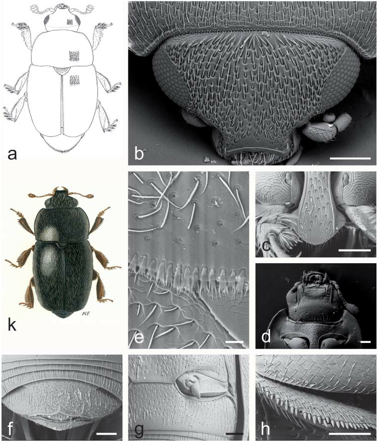
Fig. 10. Afrogethes Audisio & Cline, gen. nov.: a – A. tristis (Sturm, 1845); b–d, h – A. reticulatus (Reitter, 1872); e – A. alani (Kirejtshuk, 1988); f–g – A. planiusculus (Heer, 1841); k – A. isoplexidis (Wollaston, 1854). a, k – male habitus (a – length 2.6 mm; k – length 2.5 mm); b – dorsal view of head; c – prosternal process; d – ventral view of head and anterior portion of prosternum; e – microsetae on middle posterior margin of pronotum; f – exposed portion of last visible abdominal ventrite; g – caudal marginal lines of metacoxal cavities; h – outer margin of mesotibia. Scale bars: Figs. b, c, d, f, g, h = 100 μm; Fig. e = 20 μm.
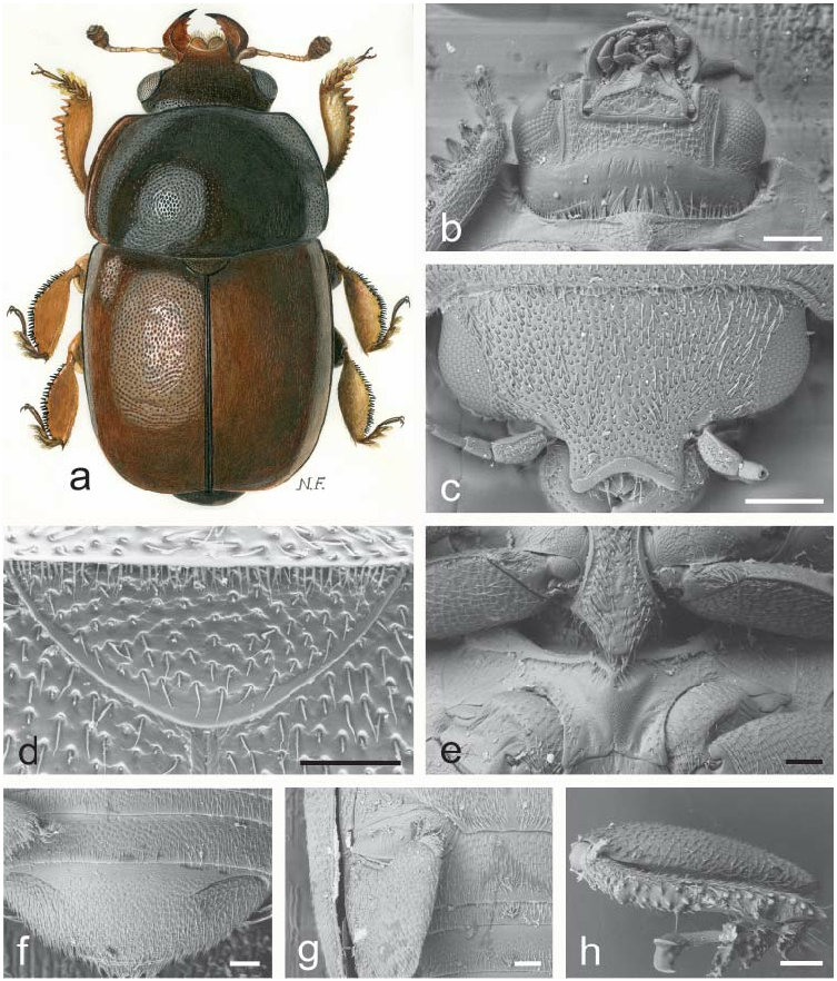
Fig. 1. Acanthogethes Reitter, 1871: a–h – A. fuscus (Olivier, 1790). a – male habitus (length 3.5 mm); b – ventral view of head and anterior portion of prosternum; c – dorsal view of head; d – scutellum and microsetae on posterior margin of pronotum; e – prosternal process and mesoventrite; f – exposed portion of last visible abdominal ventrite; g – caudal marginal lines of metacoxal cavities; h – middle leg illustrating outer margin of mesotibia. Scale bars: Figs. b, c = 200 μm; Figs. d, e, f, g, h = 100 μm.
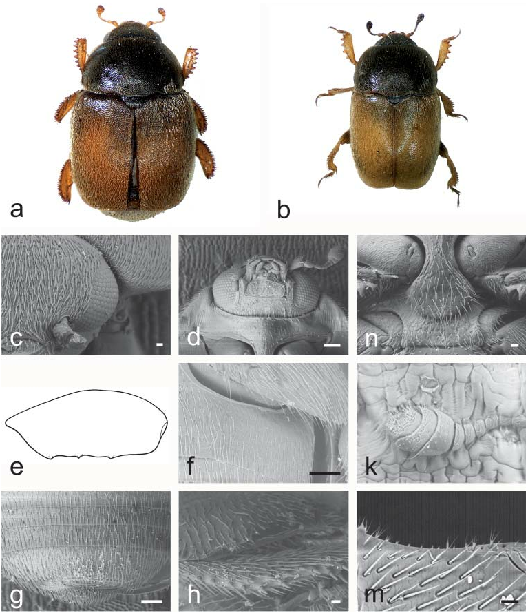
Fig. 2. Asterogethes Audisio & Cline, gen. nov.: a – A. endroedyi (Kirejtshuk & Audisio, 1995); b–d, f–n – A. arcuatus (Reitter, 1872); e – A. rufiventris (Reitter, 1872). a, b – male habitus (a – length 3.2 mm; b – length 2.4 mm); c – dorso-lateral view of head; d – ventral view of head and anterior portion of prosternum; e – outline of male metafemur (length 0.5 mm); f – caudal marginal lines of metacoxal cavities; g – exposed portion of last visible abdominal ventrite; h – middle leg with illustrating outer margin of mesotibia; k – antenna; m – pronotal setae and microsetae on posterior margin of pronotum; n – prosternal process and mesoventrite. Scale bars: Figs. c, h, m, n = 20μm; Figs. d, f, g = 100 μm.

Fig. 3. Odontholariopsis Audisio & Cline, gen. nov.: a, g – O. haagii (Reitter, 1872); b–f, h – O. nebulosus (Reitter, 1872). a – male habitus (length 2.6 mm); b – dorsal view of head; c – ventral view of head and anterior portion of prosternum; d – middle leg illustrating outer margin of mesotibia; e – scutellum and microsetae on posterior margin of pronotum; f – prosternal process and mesoventrite; g – outline of male metafemur (length 0.5 mm); h – exposed portion of last visible abdominal ventrite. Scale bars: Figs. b, c, f, h = 100 μm; Fig. d = 30 μm; Fig. e = 20 μm.

Fig. 4. Lariopsis Kirejtshuk, 1989: a – L.vultuosus (Kirejtshuk & Audisio, 1995); b–k – L. variabilis (Reitter, 1872). a – male habitus (length 3.3 mm); b, c – dorso-lateral view of head; d – ventral view of head and anterior portion of prosternum; e – prosternal process and mesoventrite; f – middle leg with outer margin of mesotibia; g – microsetae on middle posterior margin of pronotum; h – exposed portion of last visible abdominal ventrite; k – caudal marginal lines of metacoxal cavities. Scale bars: Figs. b, c, d, e, h, k = 100 μm; Fig. f = 30 μm; Fig. g = 10 μm.
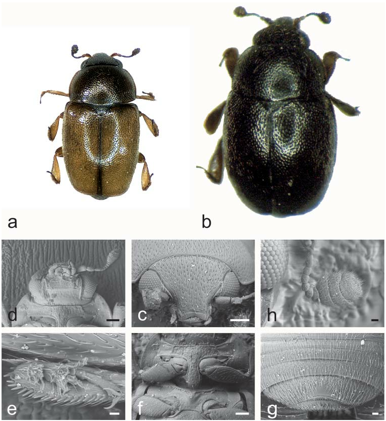
Fig. 5. Neolariopsis Audisio & Cline, gen. nov.: a, c–h – N. cercoides (Reitter, 1872); b – N. thalycroides (Kirejtshuk & Audisio, 1995). a, b – male habitus (a – length 2.1 mm, b – length 2.1 mm); c – dorsal view of head; d – ventral view of head and anterior portion of prosternum; e – middle leg with outer margin of mesotibia; f – prosternal process and mesoventrite; g – exposed portion of last visible abdominal ventrite; h – antenna. Scale bars: Figs. c, d, f = 100 μm; Figs. e, h = 20 μm; Fig. g = 30 μm.

Fig. 6. Clypeogethes Scholz, 1932 and Xerogethes Audisio & Cline, gen. nov.: a, c – C. chlorocyaneus (Jelínek & Audisio, 1977); b, d–e – C. elongatus (Rosenhauer, 1856); k–n – C. lepidii (Miller, 1851); f–g – X. osellai (Audisio & Jelínek, 2000); h – X. rotundicollis (C. N. F. Brisout de Barneville, 1863). a – male habitus (length 2.5 mm); b, c, h – ovipositors; d–e, f–g – male genitalia; k – exposed portion of last visible abdominal ventrite; m – dorsal view of head; n – ventral view of head and anterior portion of prosternum. Figs. b–h: refer to AUDISIO (1993b) and AUDISIO et al. (2000) for scale. Scale bars: Figs. k, m, n = 100 μm.

Fig. 7. Xerogethes Audisio & Cline, gen. nov.: a – X. osellai (Audisio & Jelínek, 2000); b–g – X. rotundicollis (C. N. F. Brisout de Barneville, 1863). a – male habitus (length 2.0 mm); b – dorso-lateral view of head; c – ventral view of head and anterior portion of prosternum; d – prosternal process and mesoventrite; e – exposed portion of last visible abdominal ventrite; f – caudal marginal lines of metacoxal cavities; g – microsetae on middle of posterior margin of pronotum. Scale bars: Figs. b, c, d, e, f = 100 μm; g = 30 μm.

Fig. 8. Idiogethes Kirejtshuk, 1977: a–e – I. angustitarsus Kirejtshuk, 1977. a – male habitus (length 2.2 mm); b – dorsal view of head; c – antenna; d – anterior leg; e – mesotibia. Figs. b–e: refer to KIREJTSHUK (1977a) for scale.

Fig. 9. Boragogethes Audisio & Cline, gen. nov.: a, d, k, m – B. symphyti (Heer, 1841); b–c, e–h – B. rosenhaueri (Reitter, 1871). a, b – male habitus (a – length 3.0 mm; b – length 2.5 mm); c, d – dorsal view of head; e – microsetae on posterior margin of pronotum; f – ventral view of head and anterior portion of prosternum; g – prosternal process and mesoventrite; h – exposed portion of last visible abdominal ventrite; k – caudal marginal lines of metacoxal cavities; m – outer margin of mesotibia. Scale bars: Figs. c, d, f, g, h, m = 100 μm; Fig. e = 20 μm; Fig. k = 200 μm.
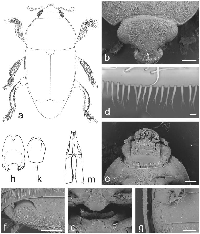
Fig. 11. Indogethes Audisio & Cline, gen. nov.: a–m – I. curvipes (Grouvelle, 1908). a – male habitus (pubescence and mandibles not illustrated; length 3.5 mm); b – dorsal view of head; c – prosternal process and mesoventrite; d – microsetae on middle of posterior margin of pronotum; e – ventral view of head and anterior portion of prosternum; f – exposed portion of last visible abdominal ventrite; g – caudal marginal line of metacoxal cavity; h–k – male genitalia (h – length 0.5 mm; k – 0.4 mm); m – ovipositor (length 0.7 mm). Scale bars: Figs. b, c, e, f, g = 200 μm; Fig. d = 10 μm.

Fig. 12. Bolbocerogethes Audisio & Cline, gen. nov.: a–e – B. pallipes (Boheman, 1851). a – male habitus (length 2.6 mm); b – ovipositor (modified from SPORNRAFT & KIREJTSHUK (1993); length 0.6 mm); c–d – male genitalia (c, d – length 0.3 mm); e – prosternal process (width 0.3 mm).

Fig. 13. Genistogethes Audisio & Cline, gen. nov.: a – G. immundus (Kraatz, 1858); b–h – G. punctatus (C. N. F. Brisout de Barneville, 1863). a – male habitus (length 2.7 mm); b – dorso-lateral view of head; c – ventral view of head and anterior portion of prosternum; d – caudal marginal line of metacoxal cavity; e – anterior portion of scutellum and microsetae on middle of posterior margin of pronotum; f – exposed portion of last visible abdominal ventrite; g – prosternal process and mesoventrite; h – middle leg with outer margin of mesotibia. Scale bars: Figs. b, c, d, f, g, h = 100 μm; Fig. e = 30 μm.

Fig. 15. Thymogethes Audisio & Cline, gen. nov.: a, h – T. egenus (Erichson, 1845); b – T. subfumatus (Ganglbauer, 1899); c–g, k – T. nigritus (Lucas, 1849). a, b – male habitus (a – length 2.5 mm; b – length 2.7 mm); c – ventral view of head and anterior portion of prosternum; d – anterior portion of scutellum and microsetae on middle of posterior margin of pronotum; e – dorsal view of head; f – prosternal process and mesoventrite; g – caudal marginal line of metacoxal cavity; h – exposed portion of last visible abdominal ventrite; k – middle leg with outer margin of mesotibia. Scale bars: Figs. c, e, f, g, h = 100 μm; Fig. d = 20 μm.
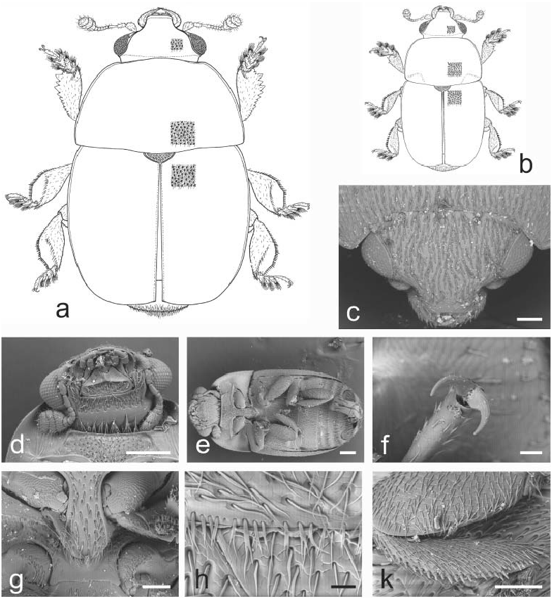
Fig. 16. Sagittogethes Audisio & Cline, gen. nov.: a – S. ater (C. N. F. Brisout de Barneville, 1863); b – S. lindbergi (Rebmann, 1940); c, f–g – S. maurus (Sturm, 1845); d–e, h–k – S. obscurus (Erichson, 1845). a, b – male habitus (a – length 2.9 mm; b – length 2.5 mm); c – dorsal view of head; d – ventral view of head and anterior portion of prosternum; e – ventral view of body; f – last tarsomere of a middle leg; g – prosternal process and mesoventrite; h – anterior portion of scutellum and microsetae on middle posterior margin of pronotum; k – middle leg with outer margin of mesotibia. Scale bars: Figs. c, g, k = 100 μm; Figs. d, e = 200 μm; Fig. f = 20 μm; Fig. h = 30 μm.

Fig. 18. JelinekigethesAudisio & Cline, gen. nov.: a–e – J. danielssoni (Audisio, 1995).a – male habitus; b–c – male genitalia; d – ovipositor; e – male protibia. Figs. a–e – refer to AUDISIO (1995) for scale.
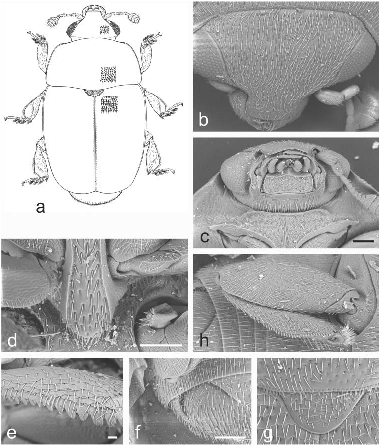
Fig. 19. Astylogethes Kirejtshuk, 1992: a–h – A. subrugosus (Gyllenhal, 1808). a – male habitus (length 2.4 mm); b – dorsal view of head; c – ventral view of head and anterior portion of prosternum; d – prosternal process and mesoventrite; e – outer margin of protibia; f – exposed portion of last visible abdominal ventrite; g – scutellum and microsetae on middle of posterior margin of pronotum; h – caudal marginal line of metacoxal cavity. Scale bars: Figs. c, d, f = 100 μm; Fig. e = 20 μm.

Fig. 23. Lamiogethes Audisio & Cline, gen. nov.: a – L. paschalis (Spornraft, 1975); b – L. convexus (Boheman, 1851); c–e, g–h, m – L. ruficollis (Reitter, 1872); k, f, n – L. difficilis (Heer, 1841). a, b – male habitus (a – length 3.0 mm; b – length 2.7 mm); c – ventral view of head and of anterior portion of prosternum; d – dorsal view of head; e–f – scutellum and microsetae on middle of posterior margin of pronotum; g – ventral view of body; h–k – prosternal process and mesoventrite; m – last tarsomere of middle leg; n – outer margin of mesotibia. Scale bars: Figs. c, d, e, f, h, n = 100 μm; Fig. g = 1 mm; Fig. m = 20 μm.

Fig. 24. Chromogethes Kirejtshuk, 1989: a – C. formosus (Kirejtshuk, 1989); b–h – C. mastax (Audisio & De Biase, 2004); k – C. splendidulus (Reitter, 1873). a – male habitus (length 2.0 mm); b, k – dorsal view of head; c – scutellum and microsetae on middle of posterior margin of pronotum; d – ventral view of head and anterior portion of prosternum; e – exposed portion of last visible abdominal ventrite; f – tarsal claws of middle leg; g – outer margin of mesotibia; h – prosternal process and mesoventrite. Scale bars: Figs. b, d, e, g, k = 100 μm; Fig. c, f = 30 μm; Fig. h = 200 μm.
Fig. 25. Cyclogethes Kirejtshuk, 1979: a–h, k, p–v – C. orientalis Kirejtshuk, 1979; i, o – C. abnormis Kirejtshuk, 1979; m – C. fuscipennis Jelínek, 2000; n – C. aldridgei Kirejtshuk, 1980. a – male habitus; b–c – punctation of pronotum and elytra; d – prosternal process, mesoventrite, and metaventrite; e – exposed portion of last visible abdominal ventrite; f – protibia; g – mesotibia; h – antennal club; k, i, m – dorsal view of head; n, o, p – prosternal process; q – labium and left palp; r–s – dorsal view of male genitalia; t – labrum; u – ovipositor; v – lateral view of male genitalia. Drawings a–h, k, q–v from KIREJTSHUK (1979a); drawings i, m–p from JELÍNEK (2000b); refer to KIREJTSHUK (1979a) and to JELÍNEK (2000b) for scale.
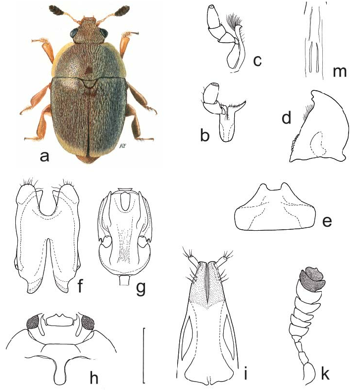
Fig. 26. Anthystrix Kirejtshuk, 1981: a, f–g, i – A.squamosa Kirejtshuk, 1981; b–e, h, k, m – A. longiclava Kirejtshuk & Easton, 1988. a – male habitus (length 2.5 mm); b – labium and right labial palpus; c – right maxilla and palpus; d – left mandible; e – labrum; f–g – male genitalia; h – ventral view of head and anterior portion of prosternum; i – distal portion of ovipositor; k – male antennal club; m – major sclerites of male endophallus. Drawings b–m: refer to AUDISIO et al. (2009a) for scale.

Fig. 27. Tarchonanthogethes Audisio & Cline, gen. nov.: a, c–i – T. rotundiclava (Kirejtshuk & Easton, 1988); b – T.sp.; k – T. martini (Grouvelle, 1899). a, b – male habitus (a – length 2.6 mm; b – length 2.0 mm); c–d – male genitalia (c – length 0. 43 mm; d – length 0.38 mm); e – major sclerites of male endophallus (length 0.20 mm); f – scutellum and microsetae on middle of posterior margin of pronotum; g – ventral view of body; h – male antenna (length 0.65 mm); i – dorsal view of head; k – distal portion of ovipositor (length 0.4 mm). Scale bars: Figs. f, i = 100 μm; Fig. g = 300 μm.

Fig. 28. Sebastiangethes Audisio, Kirk-Spriggs & Cline, 2008: a–i – S. anthystrixoides Audisio, Kirk-Spriggs & Cline, 2008. a – male habitus (length 2.7 mm); b – right maxilla and palpus; c – left mandible; d – labrum; e–f – male genitalia; g – major sclerites of male endophallus; h – distal portion of ovipositor; i – ventral view of head and anterior portion of prosternum. Drawings b–i: refer to AUDISIO et al. (2008) for scale.

Fig. 29. Xenostrongylogethes Audisio & Cline, gen. nov.: a–h – X. luculentus (Kirejtshuk & Easton, 1988). a – male habitus (length 2.5 mm); b – protibia (length 0.32 mm); c – male antenna (length 0.50 mm); d – ventral view of head and anterior portion of prosternum (pronotal width 1.22 mm); e–f – male genitalia (e – length 0.42 mm; f – length 0.47 mm); g – major sclerites of male endophallus (length 0.42 mm); h – distal portion of ovipositor (length 0.49 mm).

Fig. 31. Meligethinus Grouvelle, 1906: a, c–k – M. pallidulus (Erichson, 1843); b – M. muehlei Jelínek, 1992; m – M. humeralis Grouvelle, 1906. a – male habitus (length 1.8 mm); b – female habitus (length 2.4 mm); c – dorsal view of head; d – microsetae on middle of posterior margin of pronotum; e – ventral view of head and anterior portion of prosternum; f – prosternal process and mesoventrite; g – exposed portion of last visible abdominal ventrite; h – caudal marginal line of metacoxal cavity; k – mesotibia. Drawings b, m – refer to JELÍNEK (1992) for scale. Scale bars: Figs. c, e, f, g, h, k = 100 μm; Fig. d = 20 μm.

Fig. 32. Meligethes Stephens, 1830: a–e, g – M. atratus (A. G. Olivier, 1790); f – M. denticulatus (Heer, 1841). a, f – male habitus (a – length 3.6 mm; f – length 3.4 mm); b – dorsal view of head; c – exposed portion of last visible abdominal ventrite;d – scutellum and microsetae on middle of posterior margin of pronotum;e – ventral view of head and anterior portion of prosternum; g – prosternal process. Scale bars: Fig. b, e = 200 μm; Figs. c, d, g = 100 μm.
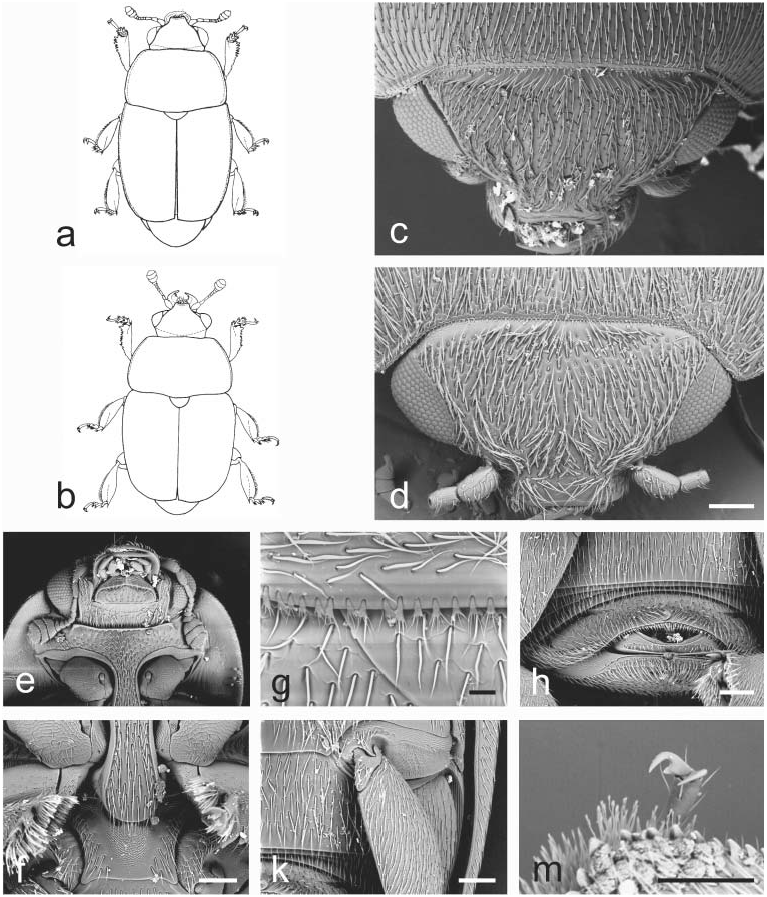
Fig. 17. Aristogethes Audisio & Cline, gen. nov.: a – A. translatus (Grouvelle, 1913); b – A. pecten (Audisio, Kirk-Spriggs & Kirejtshuk, 1998); c, e–k – A. pubescens (Reitter, 1872); d, m – A. marshalli (Grouvelle, 1914). a, b – male habitus (a – length 2.6 mm; b – length 2.4 mm); c, d – dorsal view of head; e – ventral view of head and anterior portion of prosternum; f – prosternal process and mesoventrite; g – anterior portion of scutellum and microsetae on middle of posterior margin of pronotum; h – exposed portion of last visible abdominal ventrite; k – caudal marginal line of metacoxal cavity; m – last tarsomere of middle leg. Scale bars: Figs. d, f, h, k, m = 100 μm; Fig. g = 20 μm.

Fig. 20. Stachygethes Audisio & Cline, gen. nov.: a, c, f, k – S. ruficornis (Marsham, 1802); b – S. variolosus (Easton, 1964); d, e, g, h – S. assimilis (Sturm, 1845). a, b – male habitus (a – length 2.5 mm; b – length 2.5 mm); c – dorsal view of head; d – ventral view of head and anterior portion of prosternum; e – scutellum and microsetae on middle of posterior margin of pronotum; f – caudal marginal line of metacoxal cavity; g – prosternal process and mesoventrite; h – exposed portion of last visible abdominal ventrite; k – outer margin of mesotibia. Scale bars: Figs. d, f, g, h = 100 μm; Fig. e = 30 μm; Fig. k = 20 μm.

Fig. 21. Paleogethes Audisio & Cline, gen. nov.: a–k – P. wollastoni (Easton, 1950). a – male habitus (length 1.9 mm); b – dorsal view of head; c – protibia; d – ventral view of head and anterior portion of prosternum; e – middle leg with outer margin of mesotibia; f – prosternal process and mesoventrite; g – anterior portion of scutellum and microsetae on middle of posterior margin of pronotum; h – exposed portion of last visible abdominal ventrite; k – ventral view of body. Scale bars: Figs. b, d, e, f, h = 100 μm; Fig. c = 20 μm; Fig. g = 10 μm; Fig. k = 200 μm.

Fig. 22. Rubiogethes Audisio & Cline, gen. nov.: a–k – R. newtoni (Kirejtshuk, 1990). a – female habitus; b – dorsal view of head; c – ventral view of head and anterior portion of prosternum; d – scutellum and microsetae on middle of posterior margin of pronotum; e – prosternal process and mesoventrite; f – exposed portion of last visible abdominal ventrite; g – caudal marginal line of metacoxal cavity; h – middle leg with outer margin of mesotibia; k – protibia. Scale bars: Figs. a, b = 200 μm; Figs. c, d, e, f, g = 100 μm; Figs. h, k = 20 μm.
No known copyright restrictions apply. See Agosti, D., Egloff, W., 2009. Taxonomic information exchange and copyright: the Plazi approach. BMC Research Notes 2009, 2:53 for further explanation.
|
Kingdom |
|
|
Phylum |
|
|
Class |
|
|
Order |
|
|
Family |