
Meligethinus Grouvelle, 1906
|
publication ID |
https://doi.org/10.5281/zenodo.5319334 |
|
persistent identifier |
https://treatment.plazi.org/id/03BE87CC-F605-FFF7-BAAB-FF2FFC27FE64 |
|
treatment provided by |
Felipe (2021-08-28 07:26:47, last updated by Plazi 2023-11-05 05:53:56) |
|
scientific name |
Meligethinus Grouvelle, 1906 |
| status |
|
31. Meligethinus Grouvelle, 1906
( Figs. 31 a–m View Fig )
Meligethinus Grouvelle, 1906: 202 . Prianella Reitter, 1919: 16 . Senior homonym of Prianella Lechanteur, 1955: 238 .
Type species. Meligethinus – Meligethinus humeralis Grouvelle, 1906: 202 (by monotypy); Prianella – Pria pallidula Erichson, 1843 (by monotypy).
Generic redescription and diagnosis. Inclusive species vary greatly in size (1.2–3.0 mm length), and share the following combination of characters.
Body color and pubescence: pubescence short and fine, silvery-whitish to golden, never obscuring the usually dull and densely isodiametrically reticulate, yellowish-ochraceous to partially blackish-brown ( Figs. 31a, b View Fig ) dorsal body surface; pronotal and elytral sides narrowly flattened, typically same color as disc or paler; lateral margin of pronotum and elytra with a series of faintly distinct, small, and short setae, each seta 0.3–0.5× as long as those on elytral disc; posterior margin of pronotum with moderately long, usually distally bifid or trifid microsetae ( Fig. 31d View Fig ), microsetae also uniformly distributed on middle region anterior to scutellum.
Dorsal habitus: body small, usually flatly convex, variably shaped ( Figs. 31a, b View Fig ; Figs. 1 View Fig , 12 View Fig , 24 View Fig in KIREJTSHUK 1980b; Figs. 33–34 View Fig View Fig in JELÍNEK 1992), pronotum as wide as or slightly more narrow than elytra; dorsal punctures on discal portion of pronotum and elytra usually fine, smaller than eye facet, moderately to shallowly impressed and densely distributed; anterior margin of clypeus usually truncate, rarely sinuate, narrowly but distinctly bordered, without small, faintly distinct, medial bulge, lateral angles obtuse ( Fig. 31c View Fig ; Figs. 2 View Fig , 13 View Fig , 25 View Fig in KIREJTSHUK 1980b); circum-ocular furrows (occipital sulci) on dorsal side of head absent; eyes moderately large and projecting laterally ( Figs. 31a, b View Fig ); pronotum with distinct and nearly right posterior angles, or slightly acute and more or less distinctly projecting posteriorly ( Figs. 31a, b View Fig ; Figs. 1 View Fig , 12 View Fig , 24 View Fig in KIREJTSHUK 1980b; Figs. 33–34 View Fig View Fig in JELÍNEK 1992); lateral area adjacent to posterior outer portions of pronotum normally punctate and setose; scutellum minutely and densely punctured on most of exposed portion; elytra usually finely and densely discretely punctured to almost completely and finely transversely strigose; elytral humeral angle faintly projecting laterally, frequently obscured by posterior corner of pronotum; elytral humeral striae scarcely distinct or indistinct; elytral pre-sutural striae visible, variable, originating at scutellar vertex or more posteriorly, terminating slightly before elytral apex, and delimiting on each elytron a flat sutural area, widest at posterior third, some species (i.e. the western Palaearctic M. pallidulus and M. gedrosiacus ) with sutural area faintly distinct and nearly as wide as distal width of 3 rd antennomere, in other species (i.e. the southern African M. dolosus ) the sutural area is markedly distinct at least posteriorly and much wider than distal width of third antennomere; elytral apices truncately rounded in both sexes ( Fig. 31a View Fig ), or slightly lobed in females; pygidium partially exposed, moderately convex, apically rounded in both sexes ( Fig. 31a View Fig ; Figs. 1 View Fig , 12 View Fig , 24 View Fig in KIREJTSHUK 1980b), rarely with obliquely outstanding predistal conical projections in females ( Fig. 31b View Fig ).
Ventral habitus: antennal furrows markedly delimited, parallel-sided anteriorly, strongly convergent posteriorly; mentum subpentagonal, moderately transverse, trapezoidal ( Fig. 31e View Fig ); prosternal antennal furrows on anterior margin of prosternum obliterated, indistinct ( Fig. 31e View Fig ); prosternal process variably shaped, usually wide, subapical dilated portion 2.5–3.5× as wide as maximum width of 1 st antennomere, apex convex ( Fig. 31f View Fig ; Figs. 5 View Fig , 16 View Fig in KIREJ- TSHUK 1980b; Fig. 44 in JELÍNEK 1992), posterior margin not microscopically crenulate; lateral borders of prosternal process not delimiting distinct furrows, distally terminating before or over predistal lateral expansions; posterior margin of mesoventrite never medially incised, transversely truncate ( Fig. 31f View Fig ); deep male impressions on metaventrite typically absent; first two visible abdominal ventrites simple in both sexes, without tufts of setae; caudal marginal lines of metacoxal cavities simple, subparallel and contiguous to posterior margin of metacoxal cavities, with shallow arched impression of outer ‘axillary’ line ( Fig. 31h View Fig ); ‘axillary’ space on first abdominal ventrite well developed, ‘axillary’ angle widely or broadly widely obtuse ( Fig. 31h View Fig ); variably impressed arched impressions on basal portion of last visible abdominal ventrite: small, short, shallow, and moderately close to lateral margins of ventrite, frequently largely or almost completely obscured by distal portion of penultimate visible abdominal ventrite ( Fig. 31g View Fig ) in the two W Palaearctic species; arched impressions much larger and deeper in some African and Oriental species (Fig. 40 in JELÍNEK 1992), or exceptionally large in the southern African M. dolosus ( COOPER 1980, AUDISIO unpublished); apex of last abdominal ventrite usually simple in males, rarely distinctly emarginate in females (Fig. 40 in JELÍNEK 1992), without shining tubercles or arcuate ridges in both sexes.
Appendages: male1 st antennomere small, 0.8–0.9× as long as width of protibiae excluding distal teeth ( Fig. 31a View Fig ); 3 rd antennomere usually long and slender in both sexes, 2.3–2.8× as long as wide, 1.2–1.5× longer and distinctly thinner than 2 nd antennomere; 4 th antennomere usually longer than 5 th antennomere in both sexes, moderately long, nearly 1.5–1.8× longer than wide; antennal club small or mid-sized, nearly as wide as width of protibiae, compact and rounded or loose and narrow, especially in males ( Fig. 31m View Fig ), simple, comprising last 3 antennomeres in both sexes (8 th antennomere scarcely widened, 0.4–0.5× as wide as 9 th antennomere), sexual dimorphism variably expressed; labial palpi moderately short in both sexes ( Fig. 31e View Fig ), terminal segment 1.3–1.6× as long as wide; maxillary palpi long and thin in both sexes, terminal segment 2.6–3.1× as long as wide ( Fig. 31e View Fig ); mandible mid-sized, apex moderately acuminate, no sexual dimorphism usually present; tarsal claws simple, not toothed at base ( Fig. 31g View Fig ); tarsi of normal size, 0.6–0.7× as long as corresponding tibiae ( Fig. 31a View Fig ); protibiae with a series of small and relatively blunt teeth on distal portion of lateral margin ( Fig. 31a View Fig ; Figs. 5 View Fig , 16 View Fig in KIREJTSHUK 1980b); lateral margin of meso- and metatibiae bearing a single and usually even row of short and thin pegs ( Figs. 31a, k View Fig ; Figs. 8 View Fig , 20 View Fig in KIREJTSHUK 1980b; Fig. 45 in JELÍNEK 1992), without U-shaped sinuosity at distal third; meso- and metatibiae variably shaped, flat, usually short and wide, rarely slender, frequently subtrapezoidal and axe-shaped ( Fig. 31a View Fig ; Figs. 8 View Fig , 20 View Fig in KIREJTSHUK 1980b; Fig. 45 in JELÍNEK 1992); sexual dimorphism variably expressed in metatibial shape in most species, rarely with marked projections on inner metatibial margin in males (Fig. 45 in JELÍNEK 1992); tarsal plates of prolegs usually distinctly wider in males; posterior margin of metafemora simple in both sexes, without tubercles or projections.
Male genitalia: variably shaped, processes along inner side of parameres absent ( Figs. 14–17 View Fig View Fig View Fig View Fig , 20– 23 View Fig View Fig View Fig View Fig in COOPER 1980; Figs. 9–10 View Fig View Fig , 21– 22 View Fig View Fig in KIREJTSHUK 1980b; Figs. 47–48 in JELÍ- NEK 1992; Figs. 114 a–b in AUDISIO 1993b), usually with narrow and deep incision on distal margin, without deep median longitudinal desclerotization from proximal portion of tegmen extending to medio-distal V-shaped excision; median lobe of aedeagus variably shaped, without lateral emargination, narrow and obtuse, distally acuminate or spatulate, without minute excisions or emarginations.
Female genitalia (ovipositor): variably shaped, relatively large; styli long or short, cylindrical, never darkly pigmented, inserted at apex of contiguous or markedly divergent gonostyloids ( Figs. 24–25 View Fig View Fig , 27– 28 View Fig View Fig in COOPER 1980; Figs. 11 View Fig , 23 View Fig in KIREJTSHUK 1980b; Figs. 41 View Fig , 49 in JELÍNEK 1992; Figs. 114 g in AUDISIO 1993b); each gonostyloid lightly sclerotized, never darkly pigmented distally, with a simple, never indentate outer portion of variably shaped basicoxites, and a single, narrow, pigmented and sclerotized arcuate area along outer subdistal portion of gonostyloids. ‘Central point’ of ovipositor usually located more proximad than middle, or centrally located, with or without proximad directed spicule.
Etymology. The generic name is a diminutive of Meligethes , which is indicative of the usually small and slender body sizes characterizing most of inclusive species. Gender masculine.
Biology. All true Meligethinus , whose larval biology is known, are strictly associated with male inflorescences of palms ( Arecaceae ) ( AUDISIO 1980, 1993b, and unpublished data; JELÍNEK 1992).
Phylogenetic position. Available molecular and morphological data provide strong combined evidence of a likely sister-group relationship of Meligethinus with the clade [ Meligethes + Brassicogethes gen. nov.] ( TRIZZINO et al. 2009, LAMANNA 2009); morphologically, Micropria should also be related to this clade ( KIREJTSHUK 1980b, JELÍNEK 1992). A marginal phylogenetic relationship may also be hypothesized with the small Oriental clade [( Cryptarchopria + Horakia ) + Kabakovia ].
Taxonomy and geographic distribution. This taxon includes 15 described species. Inclusive species exhibit a high degree of morphological differentiation, and there is a strong need to employ molecular phylogenetic protocols to clarify the taxonomic position of many of the derived species. Most species are distributed in tropical Africa and southeastern Asia, with a couple relictual species known from western Mediterranean and Irano-Arabic areas ( COOPER 1980; KIREJTSHUK 1980b; JELÍNEK 1981, 1988, 1992; AUDISIO 1993b; JELÍNEK & AUDISIO 2007). The two Mediterranean-Arabian species (i.e. M. pallidulus and M. gedrosiacus ) and a few Oriental species could possibly be attributed to a separate genus ( Prianella Reitter, 1919 ), however the entire group needs a complete phylogenetic and taxonomic revision prior to any further changes.
An additional species described by KIREJTSHUK (1989), i.e. Meligethinus larioides Kirejtshuk, 1989 , from South Africa: Cape Peninsula, is a member of Pria (AUDISIO unpublished data; holotype in BMNH). Likewise, the species described by KIREJTSHUK (1989) as Meligethinus formosus Kirejtshuk, 1989 , is a member of Chromogethes (see above). Finally, a few South African species described as Meligethinus by KIREJTSHUK & EASTON (1988), have been transferred here to Tarchonanthogethes gen. nov. (see above).
Meligethinus absonus Kirejtshuk, 1987 India: Uttar Pradesh; Vietnam
Meligethinus apicalis (Grouvelle, 1894) NE India, S China
Meligethinus bisignatus Kirejtshuk, 1980 Zaire, Rwanda
Meligethinus dolosus Grouvelle, 1919 E South Africa or S Zimbabwe?
Meligethinus gedrosiacus Jelínek, 1981 S Iran, Arabian Peninsula
Meligethinus grouvellei Kirejtshuk, 1980 India
Meligethinus humeralis Grouvelle, 1906 Zaire, Angola, Rwanda
Meligethinus kabakovi Kirejtshuk, 1980 Vietnam
Meligethinus muehlei Jelínek, 1992 Rwanda
Meligethinus pallidulus (Erichson, 1843) W Mediterranean
Meligethinus peringueyi ( Grouvelle, 1919) E South Africa or S Zimbabwe?
Meligethinus plagiatus (Grouvelle, 1894) India: Darjeeling; Vietnam, Taiwan
Meligethinus quadricollis Kirejtshuk, 1987 India: Uttar Pradesh
Meligethinus suffusus Kirejtshuk, 1980 Zaire
Meligethinus tschungseni Kirejtshuk, 1987 China: Fujian, Sichuan, Yunnan
AUDISIO P. 1980: Magyarorszag Allatvilaga (Fauna Hungariae), VIII. Kotet, Coleoptera III., 9 Fuzet: Nitidulidae. Fauna Hungarica. Vol. 140, Akademiai Kiado, Budapest, 171 + 6 pp (in Hungarian).
AUDISIO P. 1993 b: Coleoptera Nitidulidae - Kateretidae. Fauna d'Italia. Vol. 32. Calderini Edizione, Bologna, xvi + 971 pp.
COOPER M. C. 1980: Species of the genus Meligethinus Grouvelle (Coleoptera: Nitidulidae). Entomologica Scandinavica 11: 32 - 36.
GROUVELLE A. 1919: Description d'especes nouvelles de Coleopteres de l'Afrique australe. Pp. 47 - 61. In: Memoires Entomologique. Etudes sur les coleopteres. Vol. 2. Paris.
JELINEK J. 1981: Results of the Czechoslovak - Iranian entomological expeditions to Iran 1970 and 1973. Coleoptera: Nitidulidae. Acta Entomologica Musei Nationalis Pragae 40: 105 - 119.
JELINEK J. 1988: Coleoptera: Nitidulidae of Saudi Arabia (Part 2). Fauna of Saudi Arabia 9: 42 - 51.
JELINEK J. 1992: Nitidulidae (Coleoptera) associated with flowers of oil palm, Elaeis guineensis (Arecales, Arecaceae), in Rwanda. Acta Entomologica Bohemoslovaca 89: 409 - 427.
JELINEK J. & AUDISIO P. 2007: Family Nitidulidae. Pp. 459 - 491. In: LOBL I. & SMETANA A. (eds): Catalogue of Palaearctic Coleoptera. Vol. 4: Elateroidea - Derodontoidea - Bostrichoidea - Lymexyloidea - Cleroidea - Cucujoidea. Apollo Books, Stenstrup, 935 pp.
KIREJTSHUK A. G. 1980 b: New species of beetles of the subfam. Meligethinae (Coleoptera, Nitidulidae) from the Ethiopian Region. Revue de Zoologie Africaine 94: 249 - 294.
KIREJTSHUK A. G. 1987: Novye taksony zhukov-blestyanok (Coleoptera, Nitidulidae) vostochnogo polushariya (Chast' 1) Omosita nearctica sp. n., vikariruyushchii s palearkticheskim O. colon (L.). [New taxa of nitidulid beetles (Coleoptera, Nitidulidae) of eastern hemisphere (Part 1). Omosita nearctica sp. n., vicariant of Palaearctic O. colon (L.)]. Trudy Zoologicheskogo Instituta, Akademiya Nauk SSSR 164: 63 - 94 (in Russian).
KIREJTSHUK A. G. & EASTON A. M. 1988: Reviziya roda Anthystrix Kirejtshuk i novye vidy podsem. Meligethinae (Coleoptera, Nitidulidae) iz yuzhnoy Afriki. [Revision of the genus Anthystrix Kirejtshuk and new species of the subfamily Meligethinae (Coleoptera, Nitidulidae) from South Africa]. Trudy Vsesoyuznogo Entomologicheskogo Obshchestva 70: 41 - 55 (in Russian).
KIREJTSHUK A. G. 1989: Novye taksony zhukov-blestyanok (Coleoptera, Nitidulidae) vostochnogo polushariya (Chast' III). [New taxa of the Nitidulidae (Coleoptera) of the East hemisphere (Part III)]. Trudy Zoologicheskogo Instituta, Akademiya Nauk SSSR 208: 64 - 89 (in Russian).
LAMANNA F. 2009: Filogenesi molecolare della sottofamiglia Meligethinae (Coleoptera, Nitidulidae), attraverso l'utilizzo di marcatori molecolari (PEPCK, ITS 2). Master degree dissertation in Biological Sciences, Department of Human and Animal Biology, Sapienza Rome University, 107 pp (unpublished).
LECHANTEUR F. 1955: Description d'un genre nouveau et d'une espece de Meligethinae du Congo Belge (Cleoptera, Nitidulidae). Bulletin et Annales de la Societe Royale d'Entomologique de Belgique 91: 238 - 241.
REITTER E. 1919: Bestimmungs-Tabelle der Coleopterenfamilien: Nitidulidae und Byturidae aus Europa und den angrenzenden Landern. Verhandlungen des Naturforschenden Vereines in Brunn 56 (1918 - 1919): 1 - 104.
TRIZZINO M., AUDISIO P., ANTONINI G., DE BIASE A. & MANCINI E. 2009: Comparative analysis of sequences and secondary structures of the rRNA internal transcribed spacer 2 (ITS 2) in pollen-beetles of the subfamily Meligethinae (Coleoptera, Nitidulidae): potential use of slippage-derived sequences in molecular systematics. Molecular Phylogenetics and Evolution 51: 215 - 226.

Fig. 31. Meligethinus Grouvelle, 1906: a, c–k – M. pallidulus (Erichson, 1843); b – M. muehlei Jelínek, 1992; m – M. humeralis Grouvelle, 1906. a – male habitus (length 1.8 mm); b – female habitus (length 2.4 mm); c – dorsal view of head; d – microsetae on middle of posterior margin of pronotum; e – ventral view of head and anterior portion of prosternum; f – prosternal process and mesoventrite; g – exposed portion of last visible abdominal ventrite; h – caudal marginal line of metacoxal cavity; k – mesotibia. Drawings b, m – refer to JELÍNEK (1992) for scale. Scale bars: Figs. c, e, f, g, h, k = 100 μm; Fig. d = 20 μm.
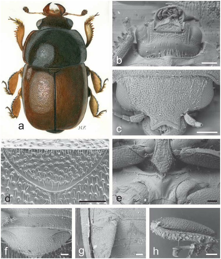
Fig. 1. Acanthogethes Reitter, 1871: a–h – A. fuscus (Olivier, 1790). a – male habitus (length 3.5 mm); b – ventral view of head and anterior portion of prosternum; c – dorsal view of head; d – scutellum and microsetae on posterior margin of pronotum; e – prosternal process and mesoventrite; f – exposed portion of last visible abdominal ventrite; g – caudal marginal lines of metacoxal cavities; h – middle leg illustrating outer margin of mesotibia. Scale bars: Figs. b, c = 200 μm; Figs. d, e, f, g, h = 100 μm.

Fig. 12. Bolbocerogethes Audisio & Cline, gen. nov.: a–e – B. pallipes (Boheman, 1851). a – male habitus (length 2.6 mm); b – ovipositor (modified from SPORNRAFT & KIREJTSHUK (1993); length 0.6 mm); c–d – male genitalia (c, d – length 0.3 mm); e – prosternal process (width 0.3 mm).

Fig. 24. Chromogethes Kirejtshuk, 1989: a – C. formosus (Kirejtshuk, 1989); b–h – C. mastax (Audisio & De Biase, 2004); k – C. splendidulus (Reitter, 1873). a – male habitus (length 2.0 mm); b, k – dorsal view of head; c – scutellum and microsetae on middle of posterior margin of pronotum; d – ventral view of head and anterior portion of prosternum; e – exposed portion of last visible abdominal ventrite; f – tarsal claws of middle leg; g – outer margin of mesotibia; h – prosternal process and mesoventrite. Scale bars: Figs. b, d, e, g, k = 100 μm; Fig. c, f = 30 μm; Fig. h = 200 μm.

Fig. 33. Brassicogethes Audisio & Cline, gen. nov.: a – B. longulus (Schilsky, 1894); b – B. salvan (Audisio, De Biase & Antonini, 2003); c–h – B. aeneus (Fabricius, 1775). a, b – male habitus (a – length 2.8 mm; b – length 2.6 mm); c – scutellum and microsetae on middle of posterior margin of pronotum; d – ventral view of head and anterior portion of prosternum; e – prosternal process and mesoventrite; f – exposed portion of last visible abdominal ventrite; g – caudal marginal line of metacoxal cavity; h – dorsal view of head. Scale bars: Fig. c = 20 μm; Fig. d = 200 μm; Figs. e, f, g, h = 100 μm.
Fig. 34. Kabakovia Kirejtshuk, 1979: a–s – K. latipes (Grouvelle, 1908). a – male habitus; b –anterior margin of clypeus; c – male antenna; d – female antennal club; e – elytral punctation; f – male protibia; g – prosternum, mesoventrite, and anterior portion of metaventrite; h – ventral view of head and prosternum; k – exposed portion of last visible abdominal ventrite; m – male mesotibia; n – labium and left palpus; p – labrum; q–r – male genitalia; s – ovipositor. Drawings a–g, m–s modified from KIREJTSHUK (1979a); drawings h–k modified from JELÍNEK (2000a)). Refer to KIREJTSHUK (1979a) and to JELÍNEK (2000a) for scale.
Fig. 2. Asterogethes Audisio & Cline, gen. nov.: a – A. endroedyi (Kirejtshuk & Audisio, 1995); b–d, f–n – A. arcuatus (Reitter, 1872); e – A. rufiventris (Reitter, 1872). a, b – male habitus (a – length 3.2 mm; b – length 2.4 mm); c – dorso-lateral view of head; d – ventral view of head and anterior portion of prosternum; e – outline of male metafemur (length 0.5 mm); f – caudal marginal lines of metacoxal cavities; g – exposed portion of last visible abdominal ventrite; h – middle leg with illustrating outer margin of mesotibia; k – antenna; m – pronotal setae and microsetae on posterior margin of pronotum; n – prosternal process and mesoventrite. Scale bars: Figs. c, h, m, n = 20μm; Figs. d, f, g = 100 μm.

Fig. 13. Genistogethes Audisio & Cline, gen. nov.: a – G. immundus (Kraatz, 1858); b–h – G. punctatus (C. N. F. Brisout de Barneville, 1863). a – male habitus (length 2.7 mm); b – dorso-lateral view of head; c – ventral view of head and anterior portion of prosternum; d – caudal marginal line of metacoxal cavity; e – anterior portion of scutellum and microsetae on middle of posterior margin of pronotum; f – exposed portion of last visible abdominal ventrite; g – prosternal process and mesoventrite; h – middle leg with outer margin of mesotibia. Scale bars: Figs. b, c, d, f, g, h = 100 μm; Fig. e = 30 μm.
Fig. 25. Cyclogethes Kirejtshuk, 1979: a–h, k, p–v – C. orientalis Kirejtshuk, 1979; i, o – C. abnormis Kirejtshuk, 1979; m – C. fuscipennis Jelínek, 2000; n – C. aldridgei Kirejtshuk, 1980. a – male habitus; b–c – punctation of pronotum and elytra; d – prosternal process, mesoventrite, and metaventrite; e – exposed portion of last visible abdominal ventrite; f – protibia; g – mesotibia; h – antennal club; k, i, m – dorsal view of head; n, o, p – prosternal process; q – labium and left palp; r–s – dorsal view of male genitalia; t – labrum; u – ovipositor; v – lateral view of male genitalia. Drawings a–h, k, q–v from KIREJTSHUK (1979a); drawings i, m–p from JELÍNEK (2000b); refer to KIREJTSHUK (1979a) and to JELÍNEK (2000b) for scale.
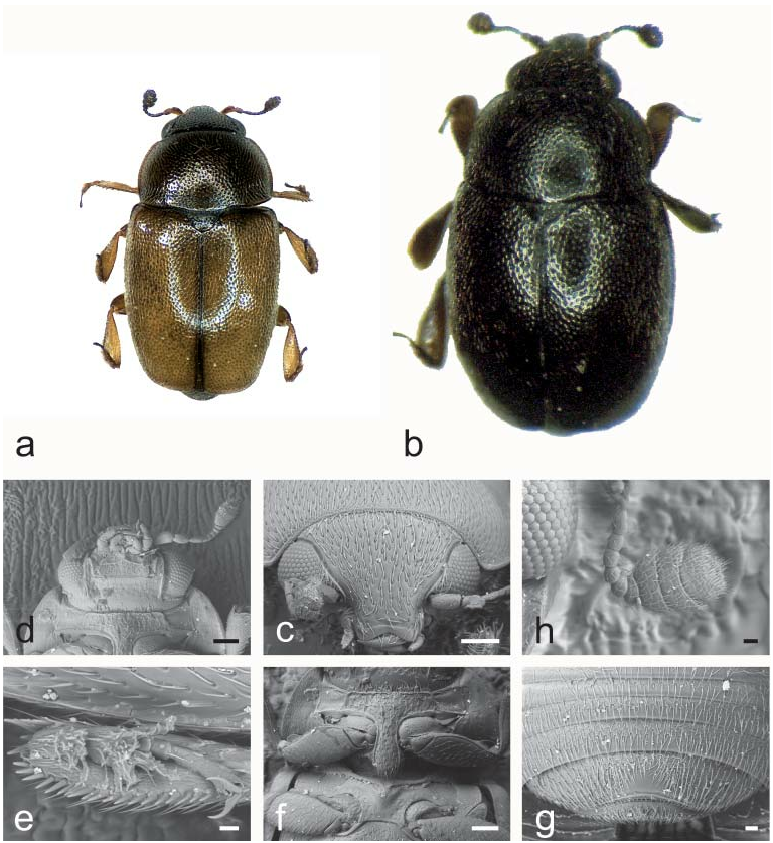
Fig. 5. Neolariopsis Audisio & Cline, gen. nov.: a, c–h – N. cercoides (Reitter, 1872); b – N. thalycroides (Kirejtshuk & Audisio, 1995). a, b – male habitus (a – length 2.1 mm, b – length 2.1 mm); c – dorsal view of head; d – ventral view of head and anterior portion of prosternum; e – middle leg with outer margin of mesotibia; f – prosternal process and mesoventrite; g – exposed portion of last visible abdominal ventrite; h – antenna. Scale bars: Figs. c, d, f = 100 μm; Figs. e, h = 20 μm; Fig. g = 30 μm.
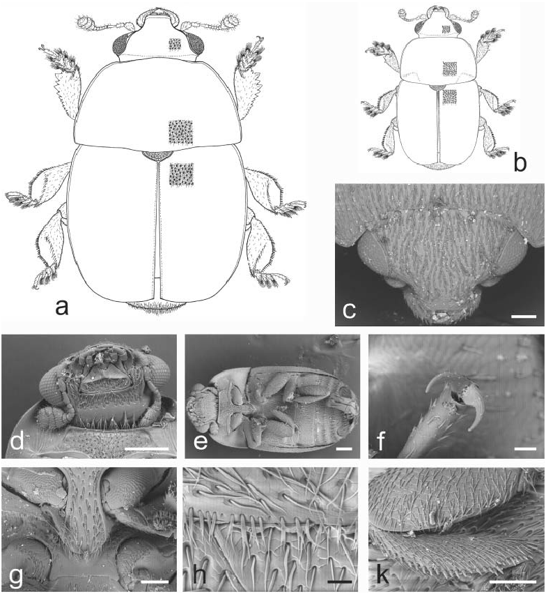
Fig. 16. Sagittogethes Audisio & Cline, gen. nov.: a – S. ater (C. N. F. Brisout de Barneville, 1863); b – S. lindbergi (Rebmann, 1940); c, f–g – S. maurus (Sturm, 1845); d–e, h–k – S. obscurus (Erichson, 1845). a, b – male habitus (a – length 2.9 mm; b – length 2.5 mm); c – dorsal view of head; d – ventral view of head and anterior portion of prosternum; e – ventral view of body; f – last tarsomere of a middle leg; g – prosternal process and mesoventrite; h – anterior portion of scutellum and microsetae on middle posterior margin of pronotum; k – middle leg with outer margin of mesotibia. Scale bars: Figs. c, g, k = 100 μm; Figs. d, e = 200 μm; Fig. f = 20 μm; Fig. h = 30 μm.

Fig. 8. Idiogethes Kirejtshuk, 1977: a–e – I. angustitarsus Kirejtshuk, 1977. a – male habitus (length 2.2 mm); b – dorsal view of head; c – antenna; d – anterior leg; e – mesotibia. Figs. b–e: refer to KIREJTSHUK (1977a) for scale.

Fig. 20. Stachygethes Audisio & Cline, gen. nov.: a, c, f, k – S. ruficornis (Marsham, 1802); b – S. variolosus (Easton, 1964); d, e, g, h – S. assimilis (Sturm, 1845). a, b – male habitus (a – length 2.5 mm; b – length 2.5 mm); c – dorsal view of head; d – ventral view of head and anterior portion of prosternum; e – scutellum and microsetae on middle of posterior margin of pronotum; f – caudal marginal line of metacoxal cavity; g – prosternal process and mesoventrite; h – exposed portion of last visible abdominal ventrite; k – outer margin of mesotibia. Scale bars: Figs. d, f, g, h = 100 μm; Fig. e = 30 μm; Fig. k = 20 μm.
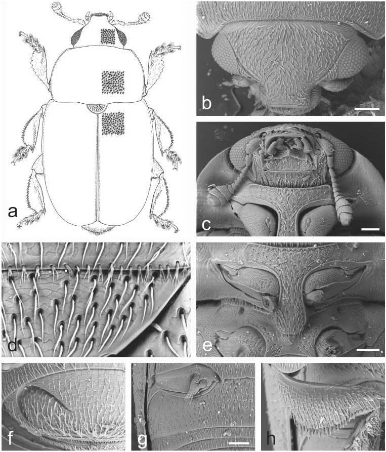
Fig. 14. Fabogethes Audisio & Cline, gen. nov.: a – F. opacus (Rosenhauer, 1856); b–h – F. nigrescens (Stephens, 1830). a – male habitus (length 2.7 mm); b – dorso-lateral view of head; c – ventral view of head and anterior portion of prosternum; d – anterior portion of scutellum and microsetae on middle of posterior margin of pronotum; e – prosternal process and mesoventrite; f – exposed portion of last visible abdominal ventrite; g – caudal marginal line of metacoxal cavity; h – middle leg with outer margin of mesotibia. Scale bars: Figs. b, c, e, g = 100 μm.

Fig. 15. Thymogethes Audisio & Cline, gen. nov.: a, h – T. egenus (Erichson, 1845); b – T. subfumatus (Ganglbauer, 1899); c–g, k – T. nigritus (Lucas, 1849). a, b – male habitus (a – length 2.5 mm; b – length 2.7 mm); c – ventral view of head and anterior portion of prosternum; d – anterior portion of scutellum and microsetae on middle of posterior margin of pronotum; e – dorsal view of head; f – prosternal process and mesoventrite; g – caudal marginal line of metacoxal cavity; h – exposed portion of last visible abdominal ventrite; k – middle leg with outer margin of mesotibia. Scale bars: Figs. c, e, f, g, h = 100 μm; Fig. d = 20 μm.
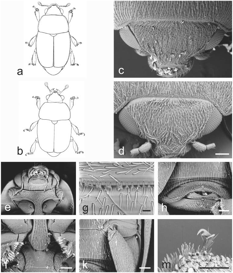
Fig. 17. Aristogethes Audisio & Cline, gen. nov.: a – A. translatus (Grouvelle, 1913); b – A. pecten (Audisio, Kirk-Spriggs & Kirejtshuk, 1998); c, e–k – A. pubescens (Reitter, 1872); d, m – A. marshalli (Grouvelle, 1914). a, b – male habitus (a – length 2.6 mm; b – length 2.4 mm); c, d – dorsal view of head; e – ventral view of head and anterior portion of prosternum; f – prosternal process and mesoventrite; g – anterior portion of scutellum and microsetae on middle of posterior margin of pronotum; h – exposed portion of last visible abdominal ventrite; k – caudal marginal line of metacoxal cavity; m – last tarsomere of middle leg. Scale bars: Figs. d, f, h, k, m = 100 μm; Fig. g = 20 μm.

Fig. 9. Boragogethes Audisio & Cline, gen. nov.: a, d, k, m – B. symphyti (Heer, 1841); b–c, e–h – B. rosenhaueri (Reitter, 1871). a, b – male habitus (a – length 3.0 mm; b – length 2.5 mm); c, d – dorsal view of head; e – microsetae on posterior margin of pronotum; f – ventral view of head and anterior portion of prosternum; g – prosternal process and mesoventrite; h – exposed portion of last visible abdominal ventrite; k – caudal marginal lines of metacoxal cavities; m – outer margin of mesotibia. Scale bars: Figs. c, d, f, g, h, m = 100 μm; Fig. e = 20 μm; Fig. k = 200 μm.
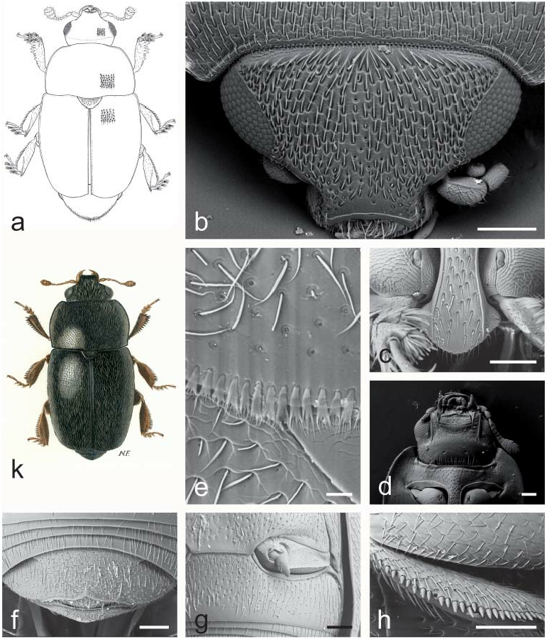
Fig. 10. Afrogethes Audisio & Cline, gen. nov.: a – A. tristis (Sturm, 1845); b–d, h – A. reticulatus (Reitter, 1872); e – A. alani (Kirejtshuk, 1988); f–g – A. planiusculus (Heer, 1841); k – A. isoplexidis (Wollaston, 1854). a, k – male habitus (a – length 2.6 mm; k – length 2.5 mm); b – dorsal view of head; c – prosternal process; d – ventral view of head and anterior portion of prosternum; e – microsetae on middle posterior margin of pronotum; f – exposed portion of last visible abdominal ventrite; g – caudal marginal lines of metacoxal cavities; h – outer margin of mesotibia. Scale bars: Figs. b, c, d, f, g, h = 100 μm; Fig. e = 20 μm.

Fig. 21. Paleogethes Audisio & Cline, gen. nov.: a–k – P. wollastoni (Easton, 1950). a – male habitus (length 1.9 mm); b – dorsal view of head; c – protibia; d – ventral view of head and anterior portion of prosternum; e – middle leg with outer margin of mesotibia; f – prosternal process and mesoventrite; g – anterior portion of scutellum and microsetae on middle of posterior margin of pronotum; h – exposed portion of last visible abdominal ventrite; k – ventral view of body. Scale bars: Figs. b, d, e, f, h = 100 μm; Fig. c = 20 μm; Fig. g = 10 μm; Fig. k = 200 μm.

Fig. 22. Rubiogethes Audisio & Cline, gen. nov.: a–k – R. newtoni (Kirejtshuk, 1990). a – female habitus; b – dorsal view of head; c – ventral view of head and anterior portion of prosternum; d – scutellum and microsetae on middle of posterior margin of pronotum; e – prosternal process and mesoventrite; f – exposed portion of last visible abdominal ventrite; g – caudal marginal line of metacoxal cavity; h – middle leg with outer margin of mesotibia; k – protibia. Scale bars: Figs. a, b = 200 μm; Figs. c, d, e, f, g = 100 μm; Figs. h, k = 20 μm.

Fig. 27. Tarchonanthogethes Audisio & Cline, gen. nov.: a, c–i – T. rotundiclava (Kirejtshuk & Easton, 1988); b – T.sp.; k – T. martini (Grouvelle, 1899). a, b – male habitus (a – length 2.6 mm; b – length 2.0 mm); c–d – male genitalia (c – length 0. 43 mm; d – length 0.38 mm); e – major sclerites of male endophallus (length 0.20 mm); f – scutellum and microsetae on middle of posterior margin of pronotum; g – ventral view of body; h – male antenna (length 0.65 mm); i – dorsal view of head; k – distal portion of ovipositor (length 0.4 mm). Scale bars: Figs. f, i = 100 μm; Fig. g = 300 μm.

Fig. 28. Sebastiangethes Audisio, Kirk-Spriggs & Cline, 2008: a–i – S. anthystrixoides Audisio, Kirk-Spriggs & Cline, 2008. a – male habitus (length 2.7 mm); b – right maxilla and palpus; c – left mandible; d – labrum; e–f – male genitalia; g – major sclerites of male endophallus; h – distal portion of ovipositor; i – ventral view of head and anterior portion of prosternum. Drawings b–i: refer to AUDISIO et al. (2008) for scale.
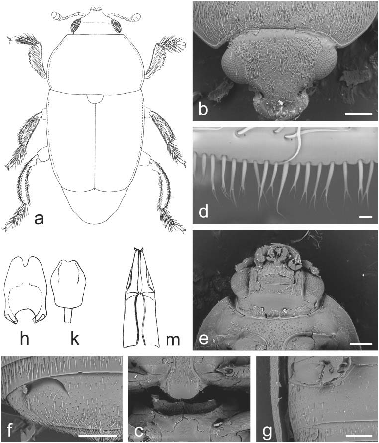
Fig. 11. Indogethes Audisio & Cline, gen. nov.: a–m – I. curvipes (Grouvelle, 1908). a – male habitus (pubescence and mandibles not illustrated; length 3.5 mm); b – dorsal view of head; c – prosternal process and mesoventrite; d – microsetae on middle of posterior margin of pronotum; e – ventral view of head and anterior portion of prosternum; f – exposed portion of last visible abdominal ventrite; g – caudal marginal line of metacoxal cavity; h–k – male genitalia (h – length 0.5 mm; k – 0.4 mm); m – ovipositor (length 0.7 mm). Scale bars: Figs. b, c, e, f, g = 200 μm; Fig. d = 10 μm.

Fig. 23. Lamiogethes Audisio & Cline, gen. nov.: a – L. paschalis (Spornraft, 1975); b – L. convexus (Boheman, 1851); c–e, g–h, m – L. ruficollis (Reitter, 1872); k, f, n – L. difficilis (Heer, 1841). a, b – male habitus (a – length 3.0 mm; b – length 2.7 mm); c – ventral view of head and of anterior portion of prosternum; d – dorsal view of head; e–f – scutellum and microsetae on middle of posterior margin of pronotum; g – ventral view of body; h–k – prosternal process and mesoventrite; m – last tarsomere of middle leg; n – outer margin of mesotibia. Scale bars: Figs. c, d, e, f, h, n = 100 μm; Fig. g = 1 mm; Fig. m = 20 μm.
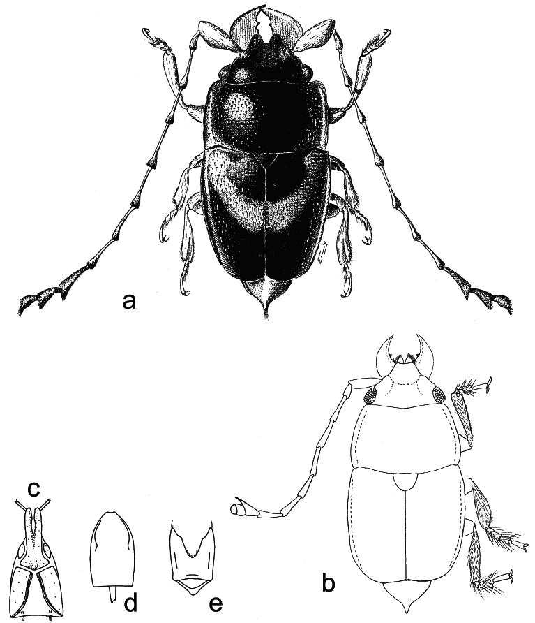
Fig. 41. Microporodes S. Endrödy-Younga, 1978 and Palmopria S. Endrödy-Younga, 1978: a – P. elaeidis S. Endrödy-Younga, 1978; b–e – M. dispar (Murray, 1864). a, b – male habitus (a – length 2.5 mm; b – length 2.6 mm); c – ovipositor; d–e – male genitalia. Drawing a modified from ENDRÖDY-YOUNGA (1978); drawings c–e modified from COOPER (1974). Drawings c–e: refer to COOPER (1974) for scale.
No known copyright restrictions apply. See Agosti, D., Egloff, W., 2009. Taxonomic information exchange and copyright: the Plazi approach. BMC Research Notes 2009, 2:53 for further explanation.
|
Kingdom |
|
|
Phylum |
|
|
Class |
|
|
Order |
|
|
Family |
Meligethinus Grouvelle, 1906
| Audisio, Paolo, Cline, Andrew Richard, Biase, Alessio De, Antonini, Gloria, Mancini, Emiliano, Trizzino, Marco, Costantini, Lorenzo, Strika, Sirio, Lamanna, Francesco & Cerretti, Pierfilippo 2009 |
Meligethinus
| LECHANTEUR F. 1955: 238 |
| REITTER E. 1919: 16 |