
Teruelius flavopiceus (Kraepelin, 1901), 2019
|
publication ID |
https://doi.org/10.18590/euscorpius.2019.vol2019.iss281.1 |
|
publication LSID |
lsid:zoobank.org:pub:FEBA0106-02A3-4465-8D39-9FF32634EEF |
|
DOI |
https://doi.org/10.5281/zenodo.7143698 |
|
persistent identifier |
https://treatment.plazi.org/id/03BB8789-FF8F-FF99-2722-D2F1FF4AF987 |
|
treatment provided by |
Felipe (2021-11-30 08:44:46, last updated 2024-11-26 05:52:46) |
|
scientific name |
Teruelius flavopiceus (Kraepelin, 1901) |
| status |
comb. nov. |
Teruelius flavopiceus (Kraepelin, 1901) View in CoL comb. n.
1♂ ( Figs. 16 View Figures 9–20 , 101, 173 View Figures 165–180 , 189 View Figures 181–195 , 230 View Figures 227–230 ), Toliara Province, Parc National de Bemaraha, Ankidrodroa , 2.5 km NE Bekopaka, 19°7.9’S 44°48.5’E, 100 m a. s. l., 25.XI.2001, secondary dry forest, leg. S. M. Goodman SMG12489 ( FMNH 73453 About FMNH ) GoogleMaps ; 1♀ ( Figs. 16 View Figures 9–20 , 47 View Figures 40–51 , 101, 129 View Figures 127–132 , 143 View Figures 133–144 , 153 View Figures 145–157 , 174 View Figures 165–180 ), Majunga, Melaky, Antsalova , Antsalova , Tsiandro , Bemaraha Plateau, Ambakoa forest, near Befanazava River , 18°47.838’S 44°52.904’E, 1400 ft a. s. l., 17.I.2006, valley marsh, pitfall 3, bucket 7, leg. H. A. Rakotondravony, HER 02557 ( FMNH 73428 About FMNH ) GoogleMaps ; 1♂ ( Fig. 75–76 View Figures 71–85 , 92 View Figures 90–93 ), No. 1196 ( FKCP, GLPC) ; 1♂ 6♀ 2♂ juvs. 1♀ juv., Montagne d`Ambre 30km south of Antseranana ( FKCP) ; 1♂ 3♀, no exact locality data, 2011 ( FKCP) ; 1juv. ( Figs. 597–599 View Figures 597–599 , with duplicated metasoma, dead during 2nd ecdysis) ( FKCP) ; 3♂ 6♀1♀ juv. ( Figs. 205 View Figures 196–210 , 217 View Figures 211–226 , 473–490 View Figures 473–476 View Figures 477–490 ), N Antsiranana Province, Tamatave, Plateau von Antsirana , Diego Suarez env., E of Ramena village, 12º15’9.95”S 49º21’31.05”E, ca 50 m a. s. l. ( FKCP) GoogleMaps .

Figures 9–20. Position of femur trichobothrium d 2in Grosphus and Teruelius gen. n. Dorsal surfaces of proximal pedipalp femur of adult females (♀, left panels) and adult males (♂, right panels), underUV fluorescence to highlightgranulation,carinae and trichobothrial areolae.Whitearrows indicate positions of trichobothrium d in each image. G. madagascariensis (9, 10; 2 samples from Anjiro (9) and Andasibe (10) show consistency of d 2 2 position),G. hirtus(11), G. voahangyae (12), T. ankarafantsika (13, 15; 2 samples fromAmpijoroa show consistency ofd 2position),T. ankarana (14), T. flavopiceus (16), T. grandidieri (17), T. limbatus (18), T. mahafaliensis (19), and T. olgae (20). Scale bars: 1 mm (9–20♀) or 500 μm (20 ♂).
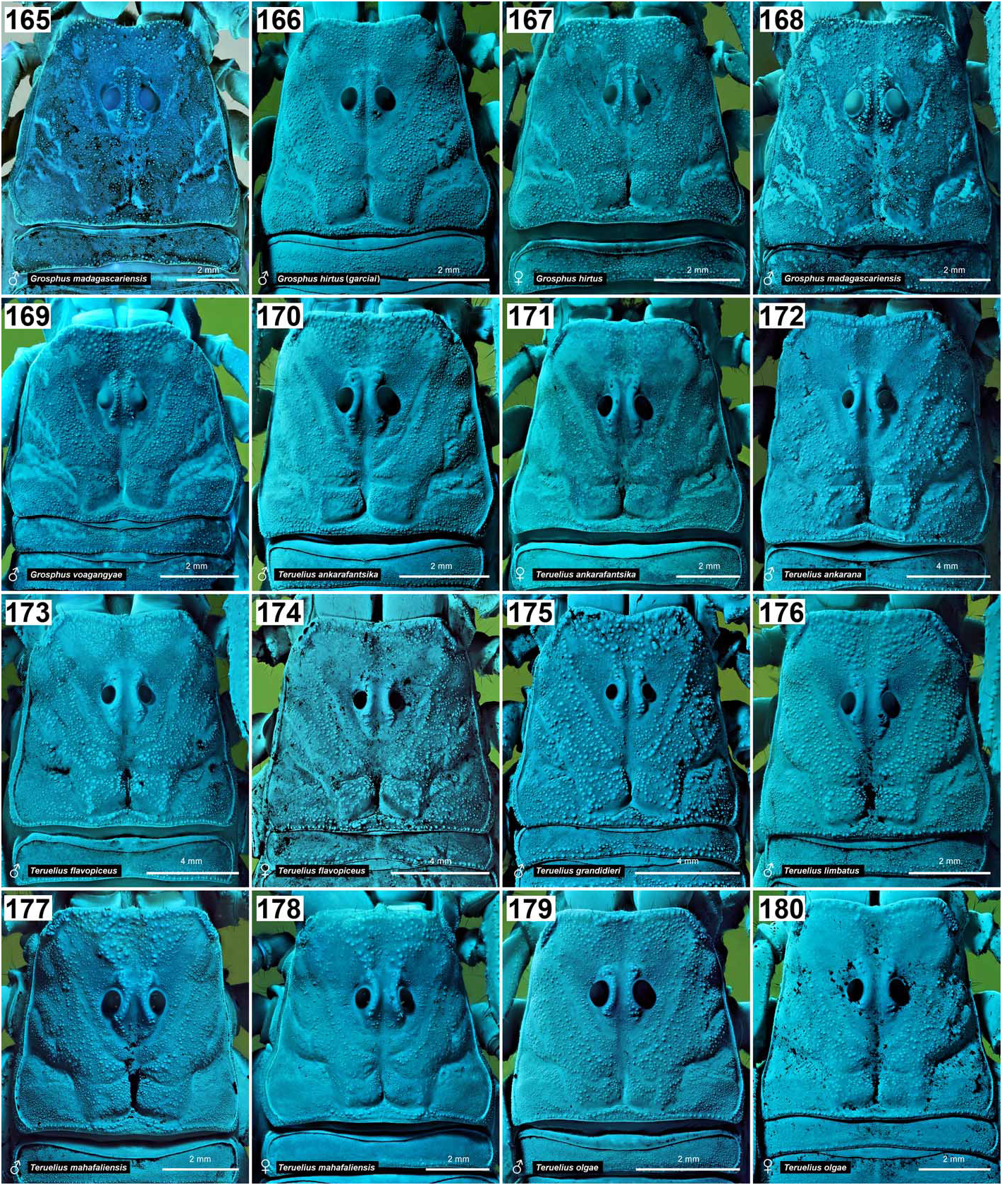
Figures 165–180. Carapace of representative Grosphus and Teruelius gen. n. G. madagascariensis (165, 168), G. hirtus (166–167), G. voahangyae (169), T. ankarafantsika (170–171), T. ankarana (172), T. flavopiceus (173–174), T. grandidieri (175), T. limbatus (176), T.mahafaliensis (177–178) andT. olgae (179–180).UV fluorescence,♂male, ♀female.Scale bars:2mm (165–171, 176–180),4 mm (172–175).
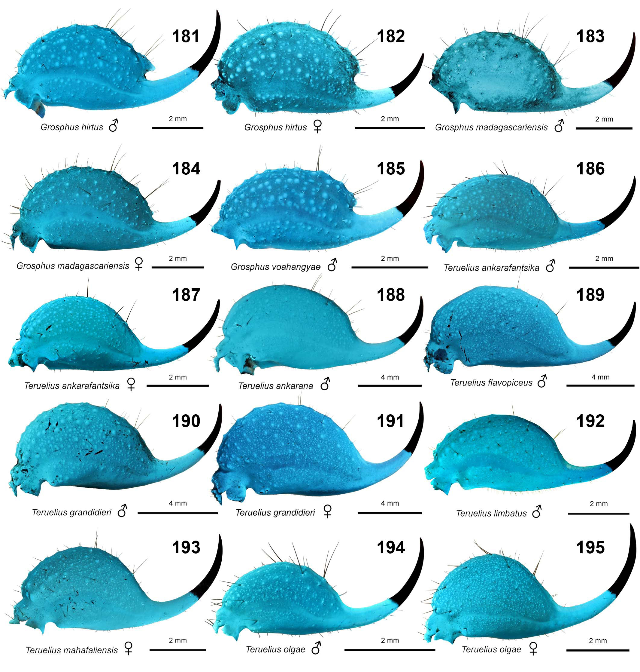
Figures 181–195. Telson of representativeGrosphus andTeruelius gen. n. G. hirtus (181–182), G. madagascariensis (183–184), G. voahangyae (185), T. ankarafantsika (186–187), T. ankarana (188), T. flavopiceus (189), T. grandidieri (190–191), T. limbatus (192), T. mahafaliensis (193) and T. olgae (194–195). UV fluorescence, ♂ male, ♀ female. Scale bars: 2 mm (181–187, 192–195), 4 mm (188–191).

Figures 227–230. Right lateral eyes of Grosphus and Teruelius gen. n. G. madagascariensis (227), G. voahangyae (228), T. limbatus (229) and T. flavopiceus (230). All species comply with the 5-eye buthid pattern with series of 3 larger ocelli in lower position, and two smaller ocelli in posterior and upper positions. UV fluorescence, males. Scale bars: 500 μm.

Figures 40–51. Female basal pectinal teeth in Grosphus and Teruelius gen. n. Ventral views of proximal left pectine of females shown under UV fluorescence to highlight cuticular surface texture, setation and absence of peg sensillae on basal tooth vs. their presence on other teeth. G. sp. nr hirtus (40), G. madagascariensis (41), G. hirtus (42), G. voahangyae (43), T. ankarafantsika (44–45; 2 samples fromAmpijoroa show variation in tooth shape), T. ankarana (46), T. flavopiceus (47), T. grandidieri (48), T. limbatus (49), T. mahafaliensis (50), and T. olgae (51). Scale bars: 1 mm.
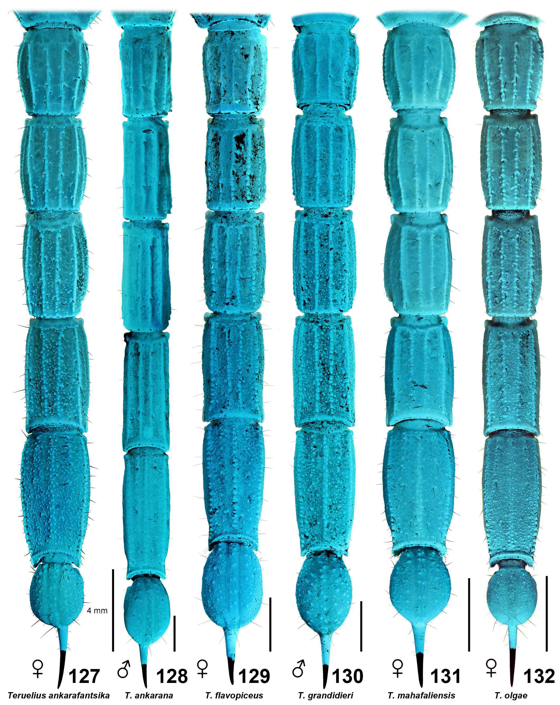
Figures 127–132. Ventral aspect of metasoma and telson of adult males (♂) or females (♀) of representative Teruelius gen. n under UV fluorescence to reveal carination and granulation. T. ankarafantsika (127), T. ankarana (128), T. flavopiceus (129), T. grandidieri (130), T. mahafaliensis (131), and T. olgae (132). Scale bars: 4 mm.

Figures 133–144. Ventral setation of telotarsus III in Grosphus and Teruelius gen. n. Ventral surfaces of right telotarsus III of adult males (♂) or females (♀), shown under UV fluorescence to highlight setation. Macrosetae appear dark with strongly fluorescent cuticular sockets at their base. Putative chemosensory microsetae appear bright. G. hirtus (= G. h. garciai) (133), G. hirtus (134), G. madagascariensis (135, from Anjiro, 136 from Andasibe), G. voahangyae (137), T. ankarafantsika (138), T. limbatus (139), T. mahafaliensis (140), T. ankarana (141), T. grandidieri (142), T. flavopiceus (143), and T. olgae (144). Scale bars: 500 μm (133–140, 144) and 1 mm (141–143).
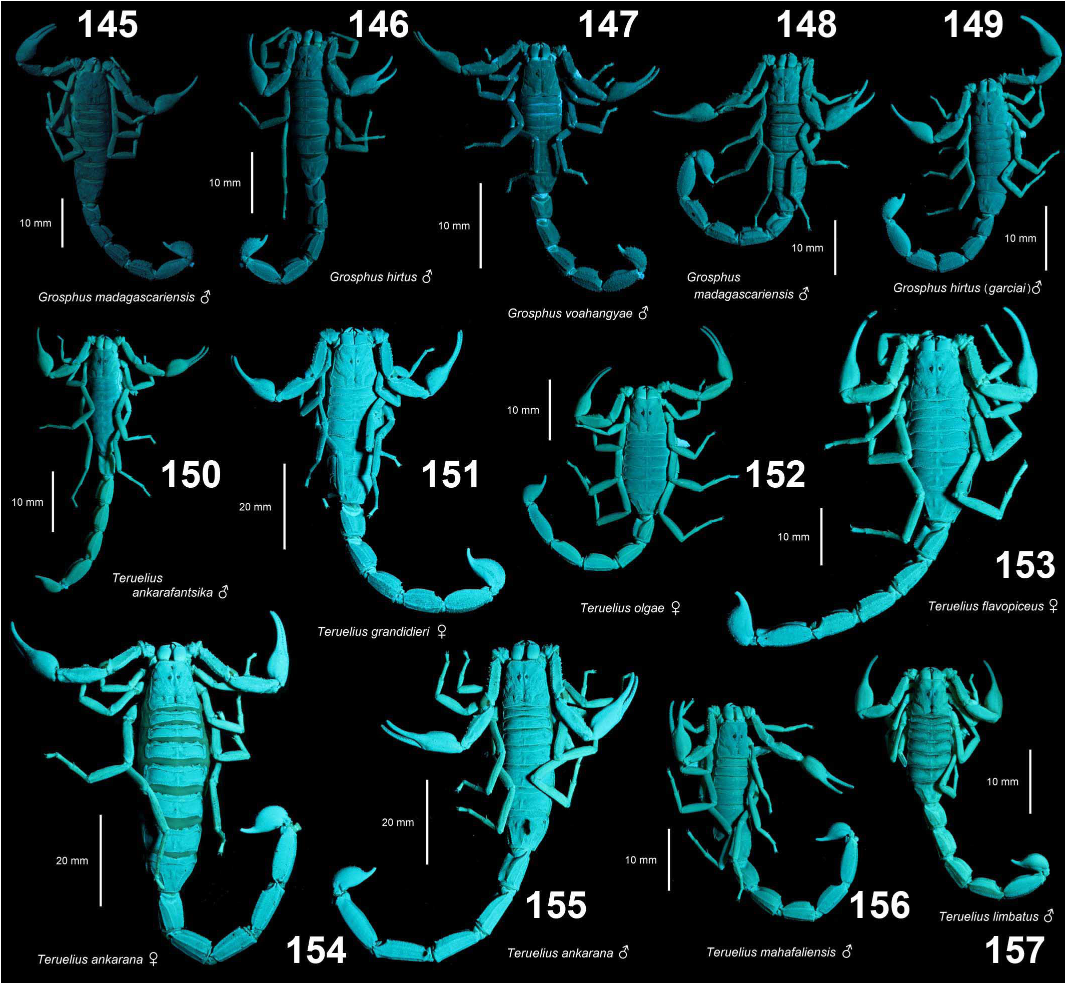
Figures 145–157. Comparative intensity of UV fluorescence in Grosphus and Teruelius gen. n. Photographic comparison of fluorescence emission intensities of representative species of each genus, including adult males (♂) or females (♀). G. madagascariensis (145 from Anjiro, 148 from Andasibe), G. hirtus (146), G. voahangyae (147), G. hirtus (= G. h. garciai) (149), T. ankarafantsika (150), T. grandidieri (151), T. olgae (152), T. flavopiceus (153), T. ankarana (154–155), T. mahafaliensis (156), and T. limbatus (157). Images acquired under identical intensities of UV excitation (395 nm LED source, 0.35 A current) and camera exposure (Canon EOS 7D Mark II, 100 mm f/13, 0.5 s, ISO 320), with 475 nm longpass filter to block excitation wavelengths. Scale bars: 10 mm (145–150, 152–153, 156–157), 20 mm (151, 154–155).
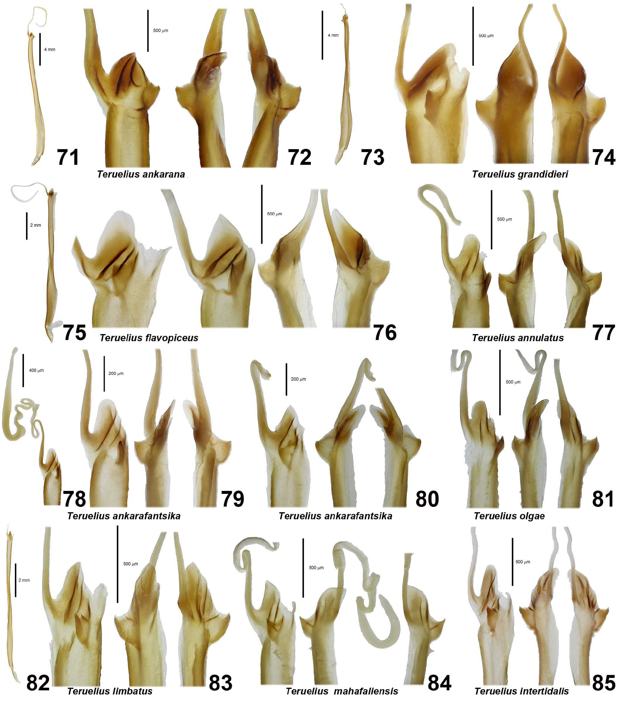
Figures 71–85. Hemispermatophores and capsule regions of Teruelius gen n. Multi-panel figures show: whole hemispermatophore; whole hemispermatophore and/or capsule with flagellum; capsule region in convex (or convex compressed), anterior and posterior views (panels in left to right sequence). Right hemispermatophores. Figure 71. T. ankarana, whole hemispermatophore. Scale bar: 4 mm. Figure 72. T. ankarana, capsule, left mirrored, Forêt d’Ankavanana, FMNH. Scale bar: 500 μm. Figure 73. T. grandidieri, whole hemispermatophore (flagellum truncated). Scale bar: 4 mm. Figure 74. T. grandidieri, capsule, Antsakabe River, FMNH. Scale bar: 500 μm. Figure 75. T. flavopiceus, whole hemispermatophore. Scale bar: 2 mm. Figure 76. T. flavopiceus, capsule, Madagascar, GLPC, FKCP. Scale bar: 500 μm. Figure 77. T. annulatus, capsule, Tsimanampetsotsa National Park, GLPC, FKCP. Scale bar: 500 μm. Figure 78. T. ankarafantsika, capsule and flagellum. Scale bar: 400 μm. Figure 79. T. ankarafantsika, capsule, Forêt d’Ankavanana, FMNH. Scale bar: 200 μm. Figure 80. T. ankarafantsika, capsule, Réserve Forestière de l’Ankarafantsika, FMNH. Scale bar: 200 μm. Figure 81. T. olgae, capsule, Itampolo village, FMNH. Scale bar: 500 μm. Figure 82. T. limbatus, whole hemispermatophore (flagellum truncated). Scale bar: 2 mm. Figure 83. T. limbatus, capsule, Forêt d’Ianasana, FMNH. Scale bar: 500 μm. Figure 84. T. mahafaliensis, capsule views, Zombitse-Vohibasia National Park, GLPC, FKCP. Scale bar: 500 μm. Figure 85. T. intertidalis, capsule, Madagascar, GLPC, FKCP. Scale bar: 500 μm.
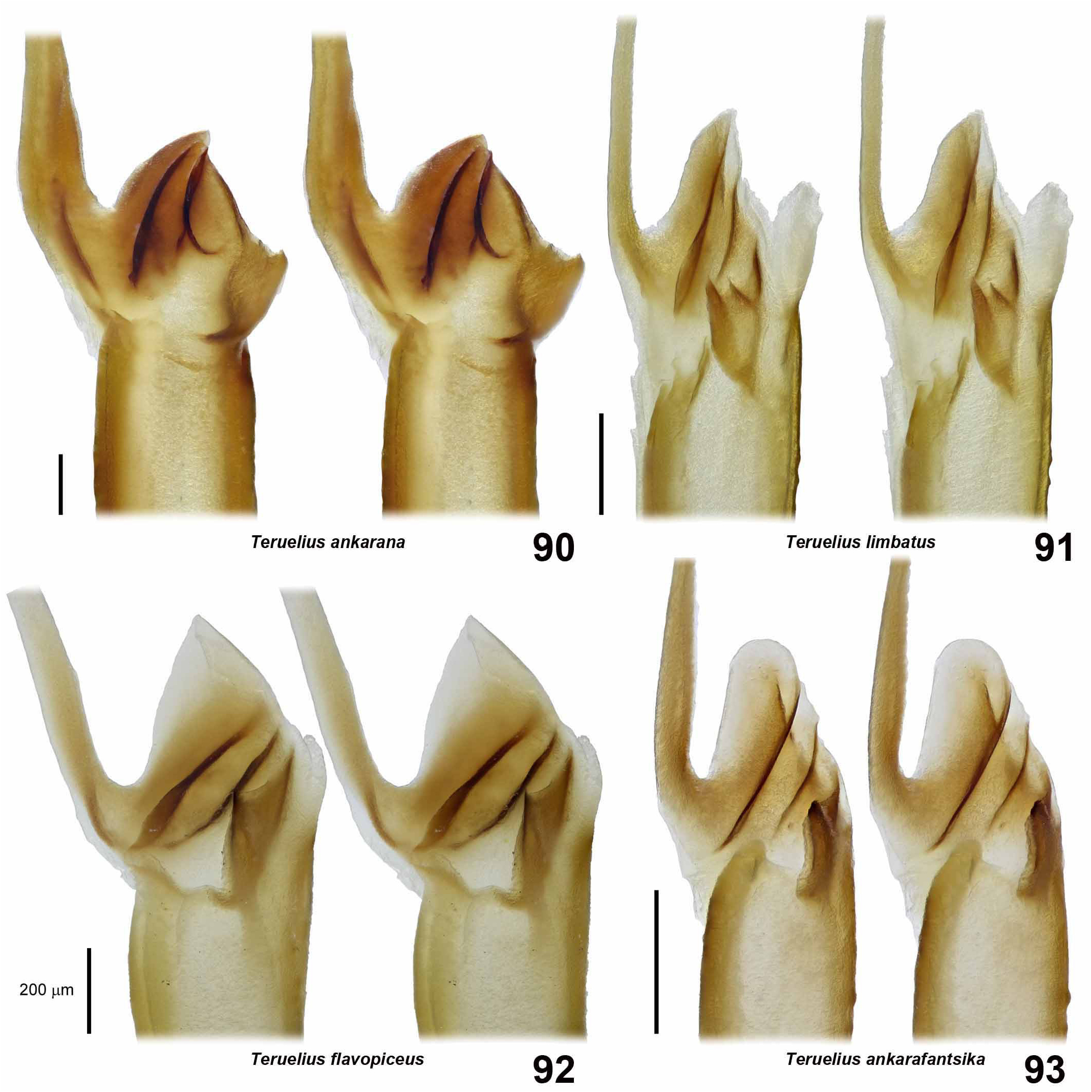
Figures 90–93. Hemispermatophores and capsule regions of Teruelius gen. n. Cross stereoscopic convex views. Figure 90. T. ankarana, left mirrored, Forêt d’Ankavanana, FMNH. Figure 91. T. limbatus, Forêt d’Ianasana, FMNH. Figure 92. T. flavopiceus, Madagascar, GLPC, FKCP. Figure 93. T. ankarafantsika, Forêt d’Ankavanana, FMNH. Scale bars: 200 μm.

Figures 597–599. Teruelius flavopiceus. Juvenile with duplicated metasoma (597, 599) and its unsuccesful second ecdysis (598).
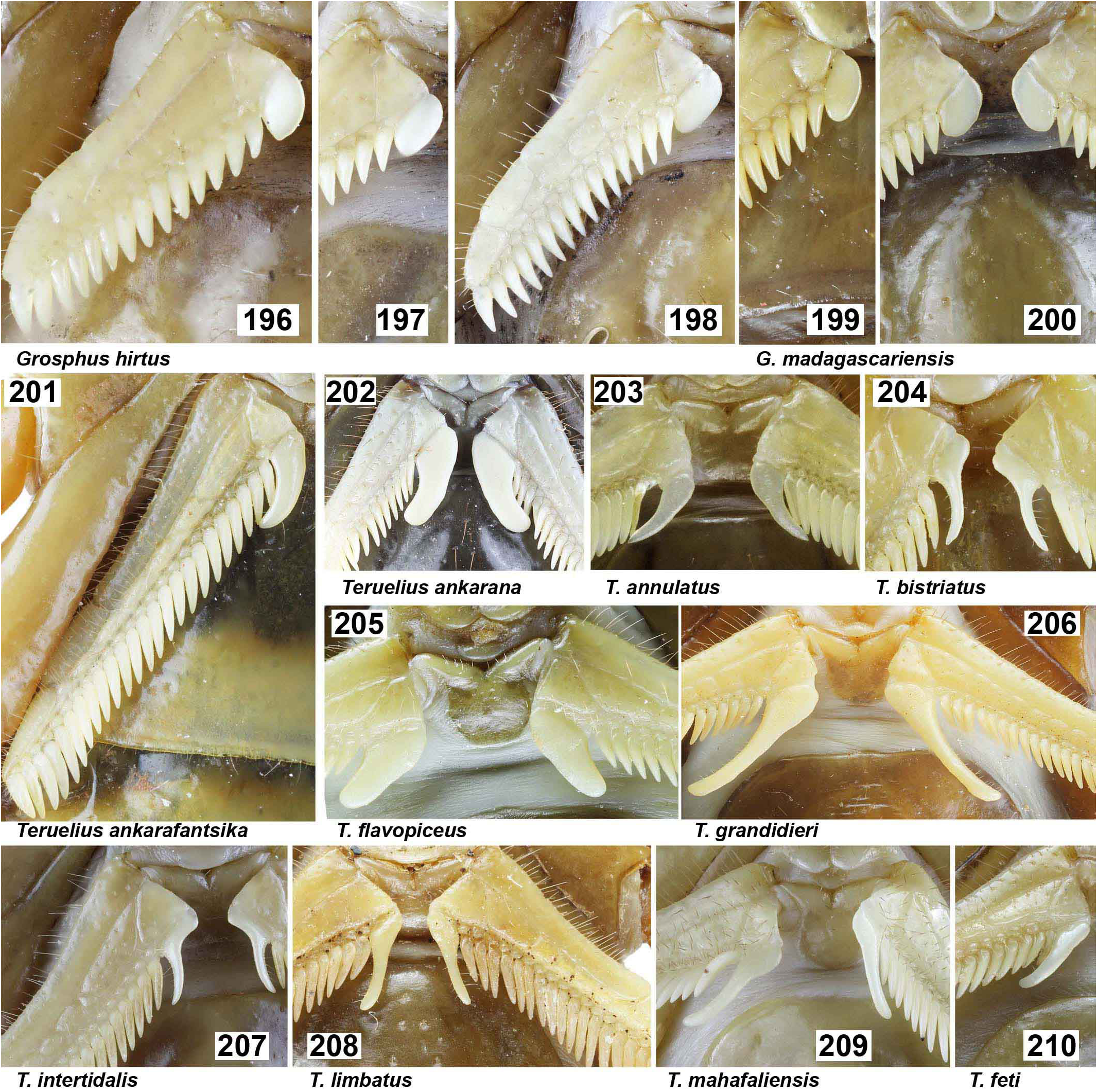
Figures 196–210. Female basal pectinal teeth of representative Grosphus and Teruelius gen. n. G. hirtus (196–198), G. madagascariensis (199), G. madagascariensis, paratype of G. mandena(200), T. ankarafantsika (201),T. ankarana (202), T. annulatus (203),T. bistriatus (204), T. flavopiceus (205), T. grandidieri (206), T. intertidalis (207), T. limbatus (208), T. mahafaliensis (209), T. feti (210).

Figures 211–226. Ventral tarsal setation of legs III or IV in Grosphus and Teruelius gen. n. G. goudoti (211), G. hirtus (212), G. hirtus (G. garciai) (213), G. madagascariensis (214), G. madagascariensis (G. mandena) (215), Teruelius ankarana (216), T. flavopiceus (217), T. annulatus (218), T. ankarafantsika (219–220), T. bistriatus (221), T. intertidalis (222), T. grandidieri (223), T. mahafaliensis (224), T. limbatus (225), T. feti (226).
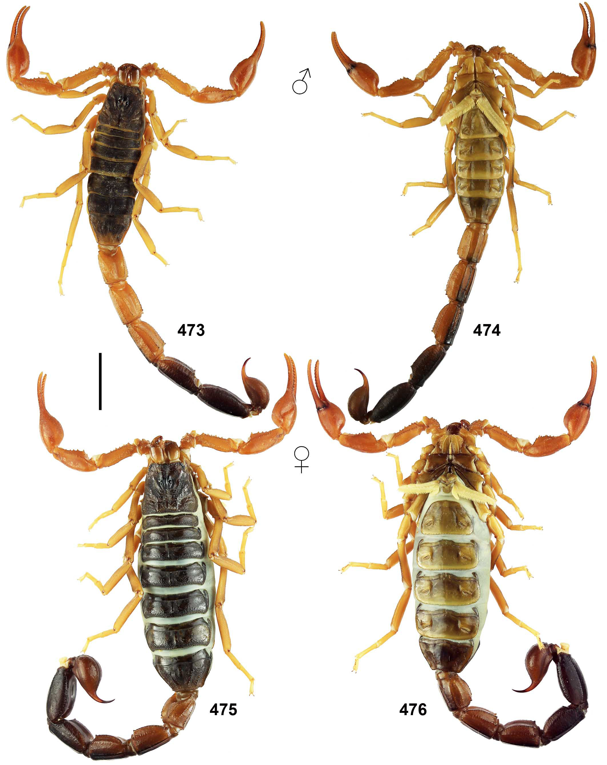
Figures 473–476. Teruelius flavopiceus. Habitus. Male (473–474) and female (475–476), dorsal (473, 475) and ventral (474, 476) views. Scale bar: 10 mm.

Figures 477–490. Teruelius flavopiceus, male. Figures 477–486. Pedipalp chela, dorsal (477), external (478) and ventrointernal (479) views; pedipalp patella, dorsal (480), external (481) and ventral (482) views; pedipalp femur and trochanter, internal (483) and dorsal (484) views; pedipalp chela, movable (485) and fixed (486) finger dentate margin. The trichobothrial pattern is indicated by white circles in Figures 477a–484a. Figures 487–490. Right legs tibia, basitarsus and telotarsus, retrolateral views. Leg IV (487), leg III (488), leg. II (489), leg I (490).
| HER |
Felix d'Herelle Reference Center for Bacterial Viruses |
No known copyright restrictions apply. See Agosti, D., Egloff, W., 2009. Taxonomic information exchange and copyright: the Plazi approach. BMC Research Notes 2009, 2:53 for further explanation.
|
Kingdom |
|
|
Phylum |
|
|
Class |
|
|
Order |
|
|
Family |
|
|
Genus |